ANATOMY EXAM 3 UIOWA
1/99
There's no tags or description
Looks like no tags are added yet.
Name | Mastery | Learn | Test | Matching | Spaced | Call with Kai |
|---|
No analytics yet
Send a link to your students to track their progress
100 Terms
excitability
ability to respond to stimuli
conductivity
The ability to transmit electrical events along the cell membrane
contractility
ability to generate tension and shorten cell length
elasticity
ability to return to resting length after shortening or lengthening
extensibility
ability to be stretched beyond resting length
(skeletal) muscle characteristics
each muscle is considered an organ
- each muscle contains all 4 tissue types: epitheleal, connective, muscle, nervous
- muscles usually attach to bones
- muscles and muscle cells usually vary in shape and size
skeletal muscle functions
- body movement
- maintenance of posture
- protection and support
- regulating elimination of materials
- heat production
skeletal muscle structure hierarchy
- whole muscle
- muscle fascicle
- muscle fiber
- myofibrils
- myofilaments
CT overview
muscles have multiple layers of connective tissue within and around them
- connective tissue layers are made mostly of collagen and elastic fibers
CT functions
- protection
- sites for blood vessel and nerve distribution
- attachment to the skeleton
endomysium
connective tissue surrounding a muscle fiber
perimysium
connective tissue surrounding a fascicle
epimysium
connective tissue covering the entire muscle
tendon
connects muscle to bone
ligment
Connects bone to bone
aponeurosis
strong sheet of tissue that acts as a tendon to attach muscles to bone
muscle attachments
most muscles extend over a joint and attach to bones on either side of the joint
- muscle contraction usually causes one bone to move while the other bone remains fixed
muscle fiber orientation
muscle fibers are organized into fascicles
four patterns of muscle fascicle arrangements:
1) circular
2) parallel
3) convergent
4) pennate (unipennate, bipennate, multipennate)
circular muscle fiber orientation
fibers arranged concentrically around an opening
- functions as a sphincter to close a passageway or opening
- EX: orbits, mouth, anus
parallel muscle fiber orientation
fascicles are parallel to the long axis of the muscle
- body of muscle increases in diameter with contraction
- high endurance, not very strong
convergent muscle fiber orientation
triangular muscle with common attachment site
- direction of pull of muscle can be changed
- does not pull as hard as equal-sized parallel
pennate muscle fiber orientation
muscle body has one or more tendons
- fascicles at oblique angle to tendon
- pulls harder than a parallel muscle of equal size
- unipennate, bipennate, multipennate
unipennate (pennate) muscle fiber orientation
all muscle fibers on the same side of the tendon
bipennate (pennate) muscle fiber orientation
muscle fibers on both sides of the tendon
multipennate (pennate) muscle fiber orientation
tendon branches within the muscle
naming muscle compounds
skeletal muscle fibers have many of the same components of a typical cell, but some are named differently
EX:
- sarcolemma: cell membrane of a skeletal muscle cell
- sarcoplasm: cytoplasm of a skeletal muscle cell
- sarcoplasmic reticulum: endoplasmic reticulum of a skeletal muscle cell
two main structures are unique to muscle fibers
- transverse tubules (T-tubules)
- sarcoplasmic reticulum
transverse tubules (T-tubules)
deep invaginations of the sarcolemma that extend into the sarcoplasm
- carry impulses from sarcolemma to help stimulate muscle contraction
- unique to muscle fibers
sarcoplasmic reticulum
specialized endoplasmic reticulum of muscle cells
- stores calcium
- internal membrane complex
myofibrils
Cylindrical structures within muscle fibers that run the length of the cell
- make up 80% of fiber volume
- have the ability to shorten, resulting in contraction of the muscle fiber
- contain myofilaments: two types (thick and thin)
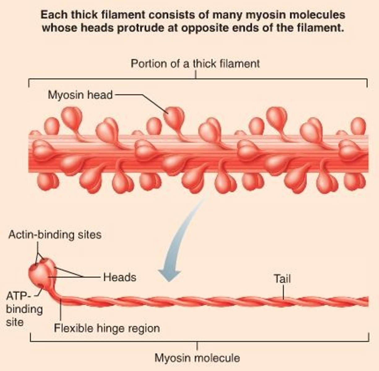
thick filaments
myosin
- contains many myosin molecules whose heads protrude on opposite ends of the filament
- heads bind to active sites on actin molecules
- heads pull on thin filaments, sliding them over thick filaments toward center of sarcomere
thin filaments
actin
two regulatory proteins are also part of the thin filament:
- Tropomyosin: regulatory protein, covers the binding sites on the actin and prevents myosin cross bridge binding
- Troponin: aids in exposure of the binding sites
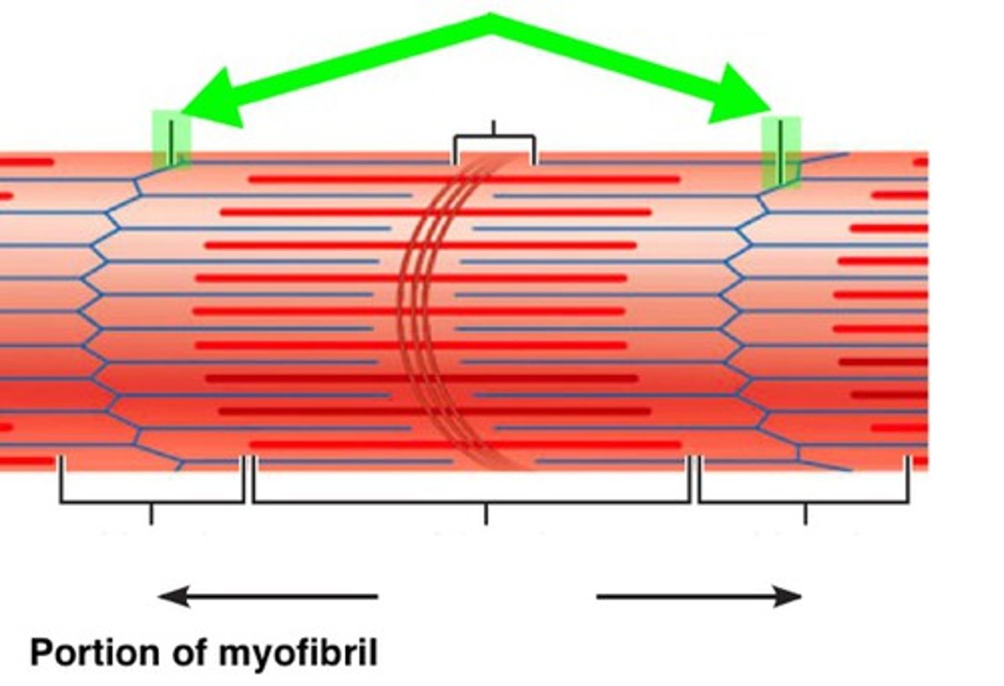
sarcomeres
contractile units within myofibrils, the smallest contractile unit of a muscle
- contain overlapping thick and thin filaments
- one sarcomere spans from one Z disc to the next

Z disc (line)
coin-shaped sheet of proteins on midline of light I band that anchors thin filaments and connects myofibrils to one another
- a sarcomere extends from z disc to z disc
M line
middle of sarcomere
- myosin
- dark line in the center of the H band of a sarcomere
- structural proteins that anchor myosin during contraction
I band
thin filaments only (actin)
- bisected by the Z line
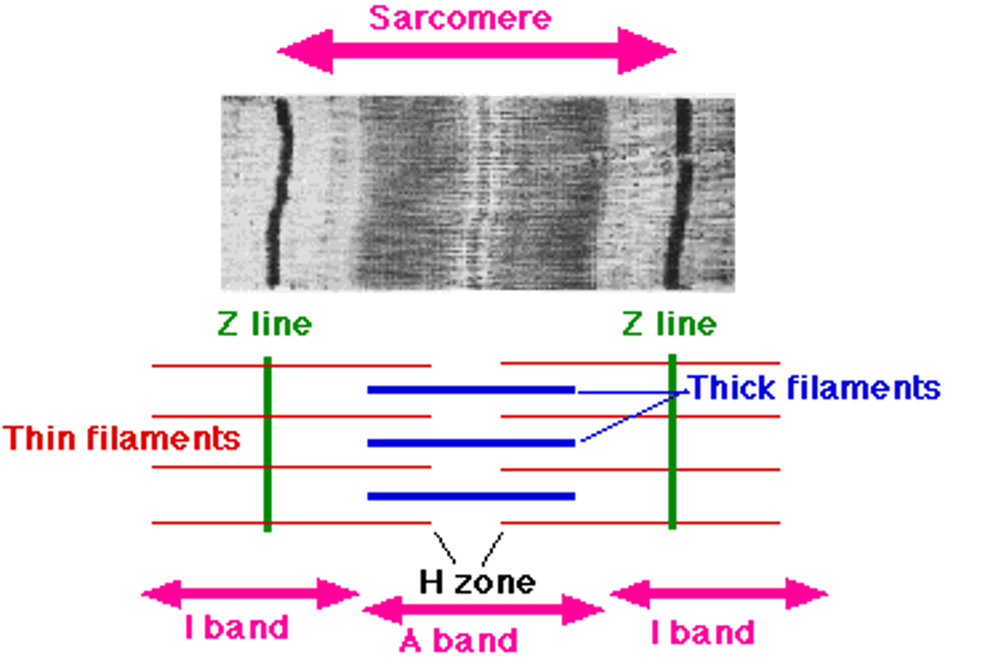
A band
dark area; extends length of the thick filaments (myosin)
- made up of H zone and M line
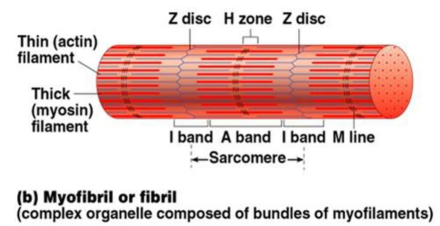
h zone
thick filaments only (myosin)
- the region of a striated muscle fibre that contains only thick (myosin) filaments. the H zone appears as a lighter band in the middle of the dark A band at the centre of a sarcomere

contraction of skeletal muscle fibers
Contracting muscles pull on tendons to produce movement, contraction begins when a motor neuron impulse stimulates an impulse in a muscle fiber
- muscles develop tension as sarcomeres shorten
- for sarcomeres to shorten, thick filaments attach to thin filaments and pull them toward the centers of the sarcomeres
- sliding filament theory
sliding filament theory
During contraction, thin filaments slide past thick filaments
- Z discs move closer together so sarcomeres shorten
- widths of A bands remain constant, but H zones disappear
- I bands narrow
lengths of filaments never change whether muscle is contracted or relaxed, only their relative positions change
sliding filament theory steps
1. Contraction Cycle Begins
2. Active-Site Exposure
3. Cross-Bridge Formation
4. Myosin Head Pivoting
5. Cross-Bridge Detachment
6. Myosin Reactivation
neuromuscular junction (NMJ)
synapse between the axon terminal of a motor neuron and the section of the membrane of a muscle fiber with receptors for the acetylcholine released by the terminal
components of neuromuscular junction (NMJ)
- Synaptic knob: expanded tip of neuron axon
- Synaptic vesicles: membrane sacs in synaptic knob, filled with acetylcholine (ACh)
- Synaptic cleft: narrow space separating synaptic knob and motor end plate
- Motor end plate: region of sarcolemma with many folds (increased surface area) under the synaptic knob
- ACh receptors: proteins that bind Ach on the motor end plate
- Acetylcholinesterase (AChE): enzyme in synaptic cleft that breaks down Ach (prevents continuous stimulation of muscle)
muscle contraction step 1
A nerve impulse causes acetylcholine (Ach) release into the synaptic cleft
- ACh binds to receptors on the motor end plate of sarcolemma, initiating a muscle fiber impulse
muscle contraction step 2
Spread of the impulse down T-tubules causes calcium to leak into the sarcoplasm
muscle contraction step 3
Calcium ions bind to troponin, and troponin changes shape
- Troponin moves tropomyosin, exposing active sites on actin
- Myosin heads bind to actin's active sites and form cross-bridges
muscle contraction step 4
Myosin pulls actin toward center of sarcomere
- Repeating cycle of attach-pivot-detach-return shortens sarcomere
- Requires ATP (energy)
muscle contraction step 5
impulse stops (power stroke)
- Calcium ions are actively transported into the sarcoplasmic reticulum
- Tropomyosin re-covers active sites
- Filaments passively slide back to their relaxed state
motor unit
a motor neuron and all of the muscle fibers it innervates
- a motor unit contains only some of the muscle fibers in an entire muscle
- when a motor unit is stimulated, all muscle fibers within it contract
- movements that require more force recruit more motor units
Skeletal muscles consist of a mixture of 3 fiber types
- Slow oxidative (SO) fibers, Type I
- Fast oxidative (FO) fibers, Type IIa
- Fast glycolytic (FG) fibers, Type IIb
Slow oxidative (SO) muscle fibers, Type I
endurance, maintaining posture → marathon running
- ATP use: slow
- capacity to make ATP: high, aerobic
- concentration of capillaries: extensive
- color of fibers: dark red
- contractile velocity: slow
- resistance to fatigue: highest
- fiber diameter: smallest
- number of mitochondria: many
- amount of myoglobin: large
- muscles with a large abundance of fiber type: trunk and lower libs
Fast oxidative (FO) fibers, Type IIa
medium duration, moderate movement → walking, biking
- ATP use: fast
- capacity to make ATP: moderate, aerobic
- concentration of capillaries: moderately extensive
- color of fibers: lighter red
- contractile velocity: fast
- resistance to fatigue: high
- fiber diameter: intermediate
- number of mitochondria: many
- amount of myoglobin: medium
- muscles with a large abundance of fiber type: lower limbs
Fast glycolytic (FG) fibers, Type IIb
short duration, intense movement → sprinting, lifting weights
- ATP use: fast
- capacity to make ATP: limited, anaerobic
- concentration of capillaries: sparse
- color of fibers: white (pale)
- contractile velocity: fast
- resistance to fatigue: low
- fiber diameter: largest
- number of mitochondria: few
- amount of myoglobin: small
- muscles with a large abundance of fiber type: upper limbs
distribution of fiber types
- skeletal muscle usually contains all three fiber types
- a single motor unit contains only muscle fibers of the same type
- slow fibers dominate postural muscles, such as those in the back and calf, which contract almost continually
- there are no slow muscle fibers in muscles that require swift but brief contractions, such as those in the eye and hand
muscle hypertrophy
muscle growth from heavy training
- building muscle increases fiber size but not number of fibers
- results from repetitive, exhaustive stimulation of muscle
muscle atrophy
loss of muscle size due to muscle disease, nervous system disease, or lack of use; commonly called muscle wasting
- reduced stimulation results in reduced muscle size, tone, and power
muscle tone
the state of balanced muscle tension that makes normal posture, coordination, and movement possible
- Some muscle fibers activated and some in resting state
two types of muscle contraction
Isometric contraction: muscle tension is less than the resistance, although tension is generated, the muscle does not shorten (no movement occurs) → static
Isotonic contraction: muscle tension equals or is greater than the resistance, the muscle shortens, and movement occurs
- concentric (isotonic) contraction: causes muscles to shorten, generating force as the tension in the muscle is great enough to overcome the resistance
- eccentric (isotonic) contraction: cause muscles to elongate in response to a greater opposing force
agonist
the muscle that contracts to cause a movement
- also called the prime mover
- EX: triceps brachii is the agonist for forearm extension
antagonist
the muscle that yields to the agonist yet aides in regulating movement; muscle acts in opposition to the agonist and is responsible for returning the limb to its original position
- EX: biceps brachii is the antagonist for forearm extension; it is antagonistic to the triceps brachii
synergist
muscle that aids a prime mover in a movement and helps prevent rotation
- muscles that contract to stabilize intermediate joints in a system
fibromyalgia
unexplainable chronic muscle pain
- treatment: antidepressants, exercise, pain relievers
musclular dystrophy
inherited disease characterized by progressive deterioration of muscle tissue, usually resulting in winged scapulae and scoliosis
- results in atrophy of the affected muscle
- muscle fibers are replaced by fibrous connective and fatty tissue
- no cure, but experimenting with stem cell treatment
myasthenia gravis
autoimmune disease in which antibodies are produced that attach to the acetylcholine receptors on the sarcolemma, thus blocking or reducing the stimulatory effect of the neurotransmitter
- symptoms: ptosis, muscle weakness, double vision, difficulty swallowing
- treatments: steroids, immunosuppressants, surgery
Amytropic Lateral Sclerosis (ALS)
neurodegenerative disease affecting various motor neurons
- also known as Lou Gehrigs Disease
- ogliodendrocytes
- loss of function leads to muscle weakness, atrophy, and spastic paralysis
- death usually occurs from respiratory failure within 5 years of diagnosis
cramps
involuntary painful, sustained contractions of a muscle
- cause unknown, but may be due to lactic acid build-up, dehydration, or calcium deficiencies
- can also be caused by a severe blow to the muscle
- treatment: stretching
muscle nomenclature
Muscles are named according to several criteria:
- Muscle action
- Specific body regions
- Muscle attachments
- Orientation of muscle fibers
- Muscle shape and size
- Muscle heads/tendons of origin
axial muscle overview
axial muscles have both their attachments on parts of the axial skeleton
- formerly: "origin and insertion"
- currently: "superior and inferior attachments" or "proximal and distal attachments"
axial muscle functions
- support the head and spinal column
- used in facial expression, chewing, and swallowing
- aid in breathing
- support and protect abdominal and pelvic organs
axial muscle groups
organized into five groups based on their location:
- head and neck
- vertebral column
- respiration
- abdominal wall
- pelvic floor
muscles of the head and neck
separated into several groups based on:
- location
- general functions
- most attach to skull or hyoid bone
muscles of facial expression
attach to superficial fascia or to skull bones
- since fascia is connected to skin, contraction moves skin and changes expression
muscles of mastication
muscles involved in chewing
- move mandible at TMJ
- temporalis: Elevates and retracts mandible
- masseter: Elevates and protracts mandible
muscles that move the head and neck
- anterolateral neck muscles: flex the head and/or neck
- posterior neck muscles: extend head and/or neck
- arise from: vertebrae, thoracic cage, pectoral girdle
- attach to: cranial bones
erector spinae
erector spinae muscles help determine posture
- bilateral contraction extends vertebral column
- unilateral contraction flexes vertebral column towards active muscles
- erector spinae organized into three groups of muscles (iliocostalis, longissimus, spinalis)
erector spinae muscles
- iliocostalis: most lateral group; composed of cervical, thoracic, and lumbar parts
- longissimus: composed of capitis, cervical, and thoracic parts
- spinalis: most medial group; attach to spinous processes of vertebrae; composed of cervical and thoracic parts
muscles of respiratoin
muscles involved with inhalation and exhalation
- external intercostals: elevates ribs during inhalation
- internal intercostals: depresses ribs during forced exhalation
- diaphragm: enlarges thoracic cavity during inhalation
diaphagm
the diaphragm is the most important muscle for breathing
- partition between thorax and abdomen
- dome shaped with a central tendon
- contraction pulls central tendon inferiorly, enlarging thorax
diaphragm contraction also increases intra-abdominal pressure
- important effect for urination, defecation, childbirth, movement of venous blood
muscles of abdominal wall
anterolateral wall of abdomen contains sheets of muscles
- hold organs in place
- together they flex and stabilize vertebral column
- unilateral contraction laterally flexes vertebral column
Four pairs of abdominal muscles
- external oblique: superficial, lateral muscle; fibers directed inferomedially
- internal oblique: deep to external oblique; fibers directed superomedially
- transverse abdominis: deepest of lateral muscles; fibers directed horizontally
- rectus abdominis: long, anterior muscle connecting sternum to pubic bone; divided into four muscle segments
posterior muscles
muscles that function to stabilize or move the scapula
- pectoralis minor
- serratus anterior
- subclavius
- trapezius
- levator scapulae
- rhomboid minor
- rhomboid major
Muscles That Move the Glenohumeral Joint/Arm
nine arm muscles attach to the scapula
- biceps brachii
- triceps brachii
- deltoid
- coracobrachialis
- teres major
rotator cuff muscles:
- subscapularis
- supraspinatus
- infraspinatus
- teres minor
muscles that move the elbow
the anterior compartment contains elbow flexors:
- biceps brachii
- brachialis
- brachioradialis
the posterior compartment contains elbow extensors:
- triceps brachii
- anconeus
muscles moving elbow / forearm
some forearm muscle pronate or supinate the forearm
two muscles on the anterior forearm that pronate the forearm are:
- pronator teres
- pronator quadratus
the muscle on the posterior forearm that supinates the forearm is:
- supinator
muscles that move wrist, hand, and fingers
most muscles in forearm move hand and fingers
- called extrinsic muscles of the wrist and hand
most anterior compartment muscles attach to the medial epicondyle of the humerus
- they flex the wrist, hand, and fingers
most posterior compartment muscles attach to the lateral epicondyle of the humerus
- they extend the wrist, hand, and fingers
forearm anterior compartment muscles
superficial layer
- pronator teres
- Flexor carpi radialis
- Palmaris longus
- Flexor carpi ulnaris
intermediate layer
- Flexor digitorum superficialis
Deep layer
- Flexor pollicis longus
- Flexor digitorum profundus
- Pronator quadratus
forearm posterior compartment muscles
Superficial layer
- Extensor carpi radialis longus
- Extensor carpi radialis brevis
- Extensor digitorum
- Extensor digiti minimi
- Extensor carpi ulnaris
Deep layer
- Abductor pollicis longus
- Extensor pollicis brevis
- Extensor pollicis longus
- Extensor indicis
- Supinator
carpal tunnel
tendons of anterior compartment muscles pass over anterior surface of carpal bones
- along with median nerve, they are held in place by flexor retinaculum
- carpal tunnel: space between carpal bones and flexor retinaculum
pelvic girdle and lower limbs
include the largest and most powerful muscles in the body
organized into specific groups:
- muscles that move the hip joint/thigh
- muscles that move the knee joint/leg
muscles of anterior compartment of leg
muscles of anterior compartment of the thigh flex the hip, most of them proximally attach to os coxae and distally attach to femur
thigh (hip) flexors include:
- Iliacus
- Psoas Major
- Sartorius
- Rectus femoris
muscles of medial compartment of leg
six muscles in medial compartment of thigh
- Adductor longus
- Pectineus
- Adductor brevis
- Gracilis
- Adductor magnus
muscles of lateral compartment of leg
Only one muscle in the lateral compartment of the thigh: tensor fasciae latae
- Abducts and medially rotates the thigh
- Attaches to the iliotibial tract (band), which extends from the iliac crest to the lateral condyle of the tibia
gluteal group
Gluteal group and deep muscles of gluteal region extend, abduct, and rotate hip joint/thigh, most of them attach proximally to os coxae and distally to femur
- Gluteus maximus
- Gluteus medius
- Gluteus minimus
- Piriformis
muscles of posterior compartment of thigh
muscles of the posterior compartment of the thigh extend the hip joint/thigh and flex the leg
hamstring muscles
- biceps femoris
- semimembranosus
- semitendinosus
muscles of anterior compartment of knee/leg
contains muscles that extend the knee joint/leg, ollectively called quadriceps femoris and consist of:
- Rectus femoris
- Vastus lateralis
- Vastus medialis
- Vastus intermedius
crural muscles
The muscles that move the ankle, foot, and toes
muscles of anterior compartment of the leg → foot and toes
muscles in the anterior compartment primarily dorsiflex the foot and extend the toes
- Extensor digitorum longus
- Extensor hallucis longus
- Tibialis anterior
- Fibularis tertius
muscles of lateral compartment of the leg → foot and toes
Muscles in the lateral compartment evert and plantar flex the foot
- Fibularis longus
- Fibularis brevis
muscles of posterior compartment of the leg → foot and toes
Muscles in the posterior compartment primarily plantar flex the foot
- Gastrocnemius
- Soleus
- Plantaris
- Flexor digitorum longus
- Flexor hallucis longus
- Tibialis posterior
nervous system organization
nervous system function: whole-body communication
two models to categorize information flow across the body
- Structural Organization
- Functional Organization