Histo Slides Exam 3
1/210
Earn XP
Description and Tags
Slides from both lecture and self labs
Name | Mastery | Learn | Test | Matching | Spaced | Call with Kai |
|---|
No analytics yet
Send a link to your students to track their progress
211 Terms
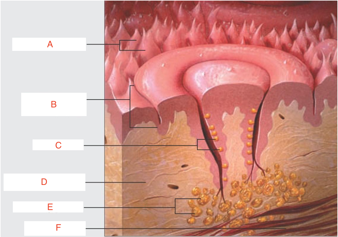
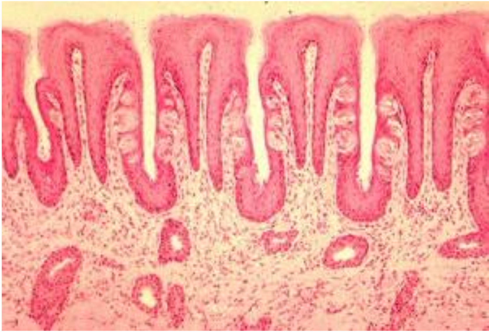
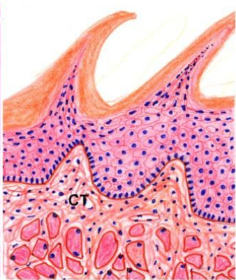
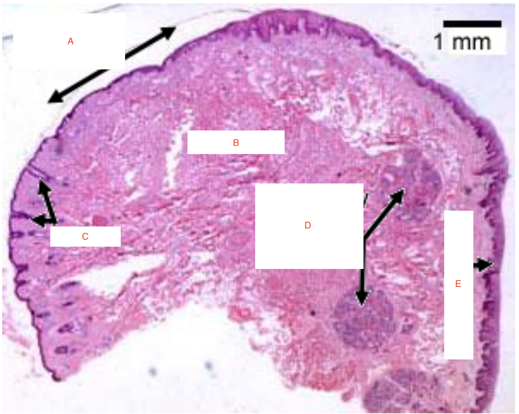
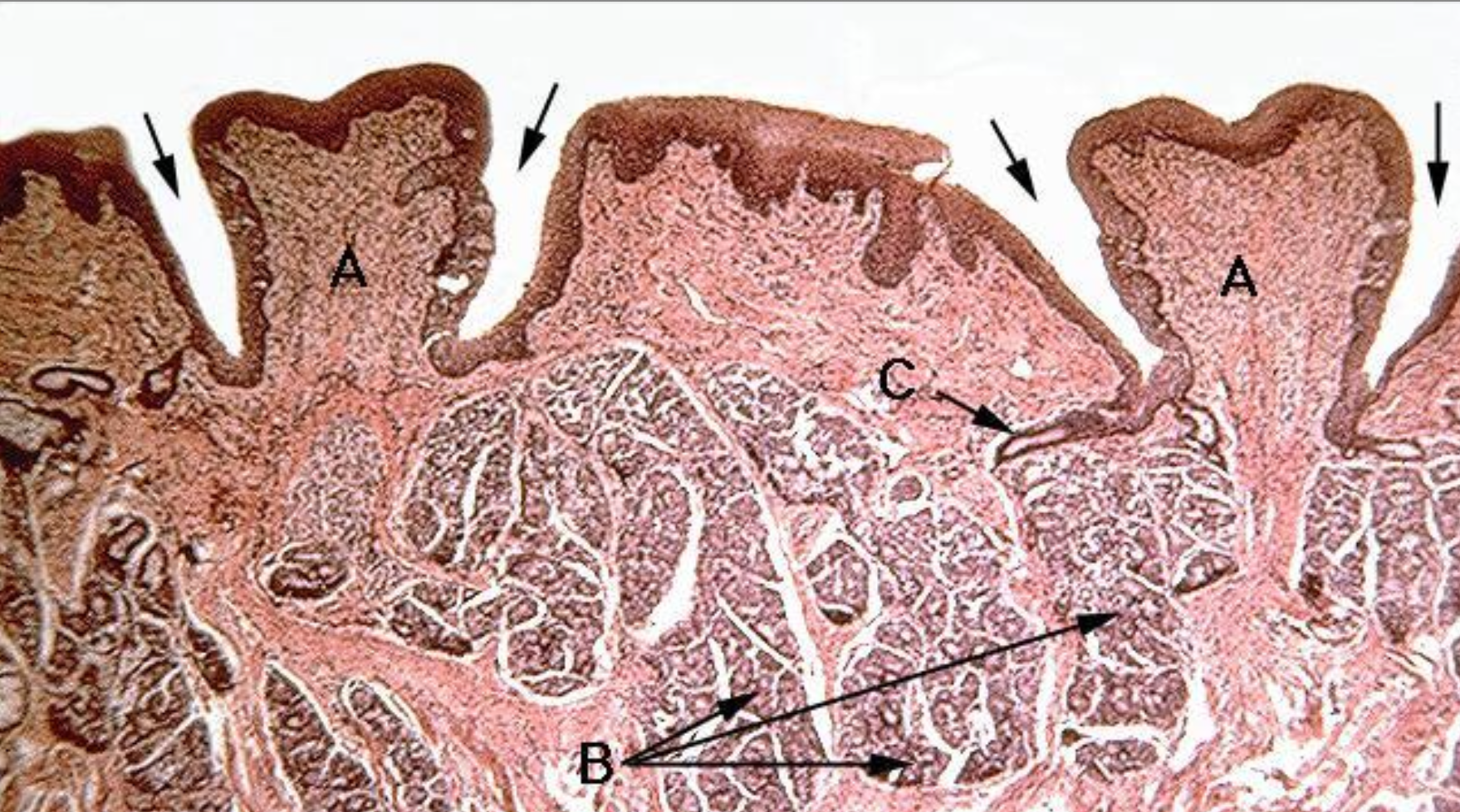
a) filiform papillae
b) circumvallate papilla
c) taste buds
d) connective tissue
e) salivary glands
f) muscle layer
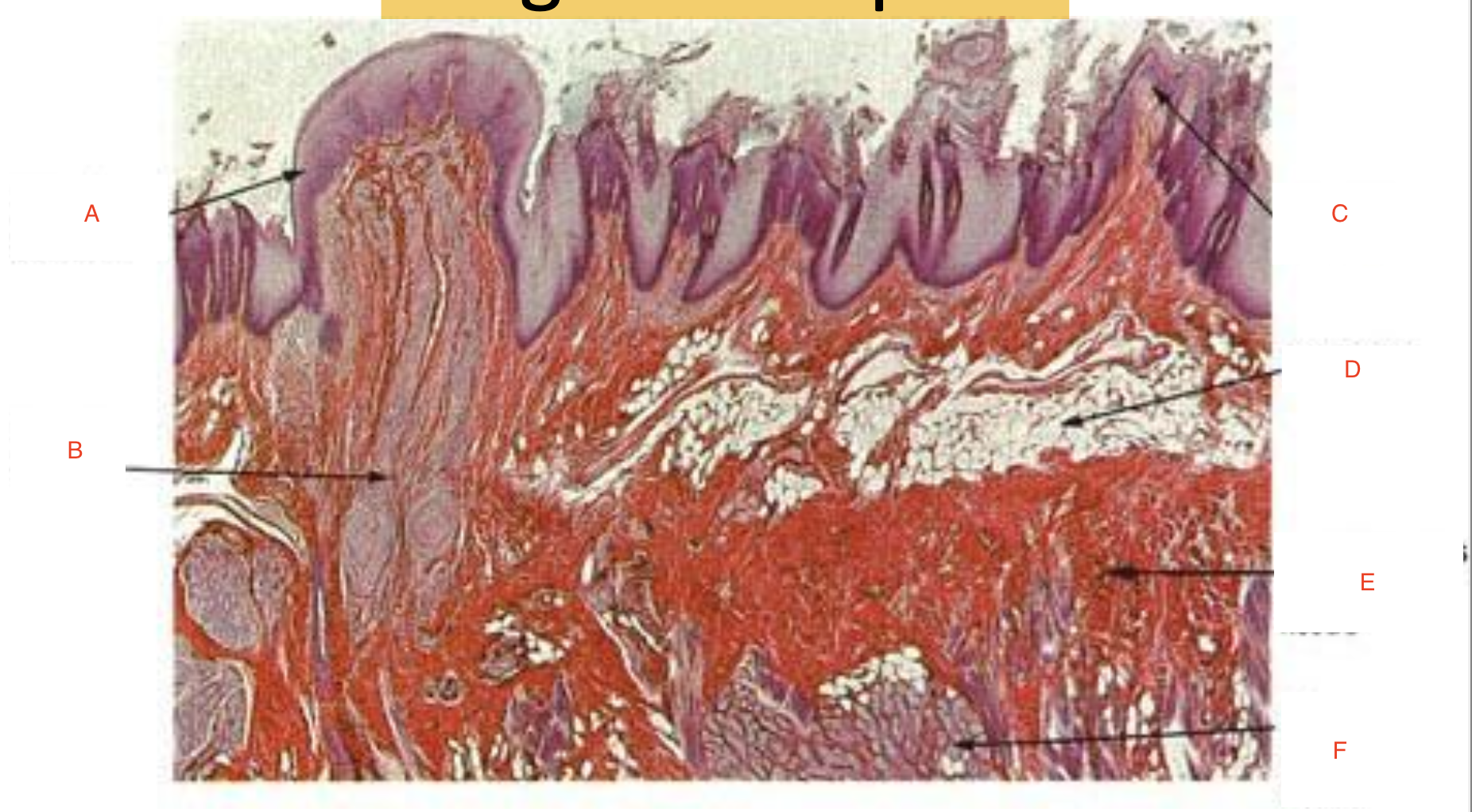
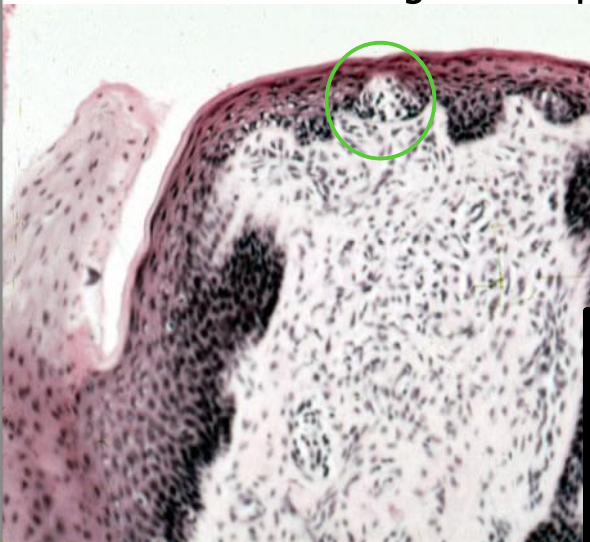
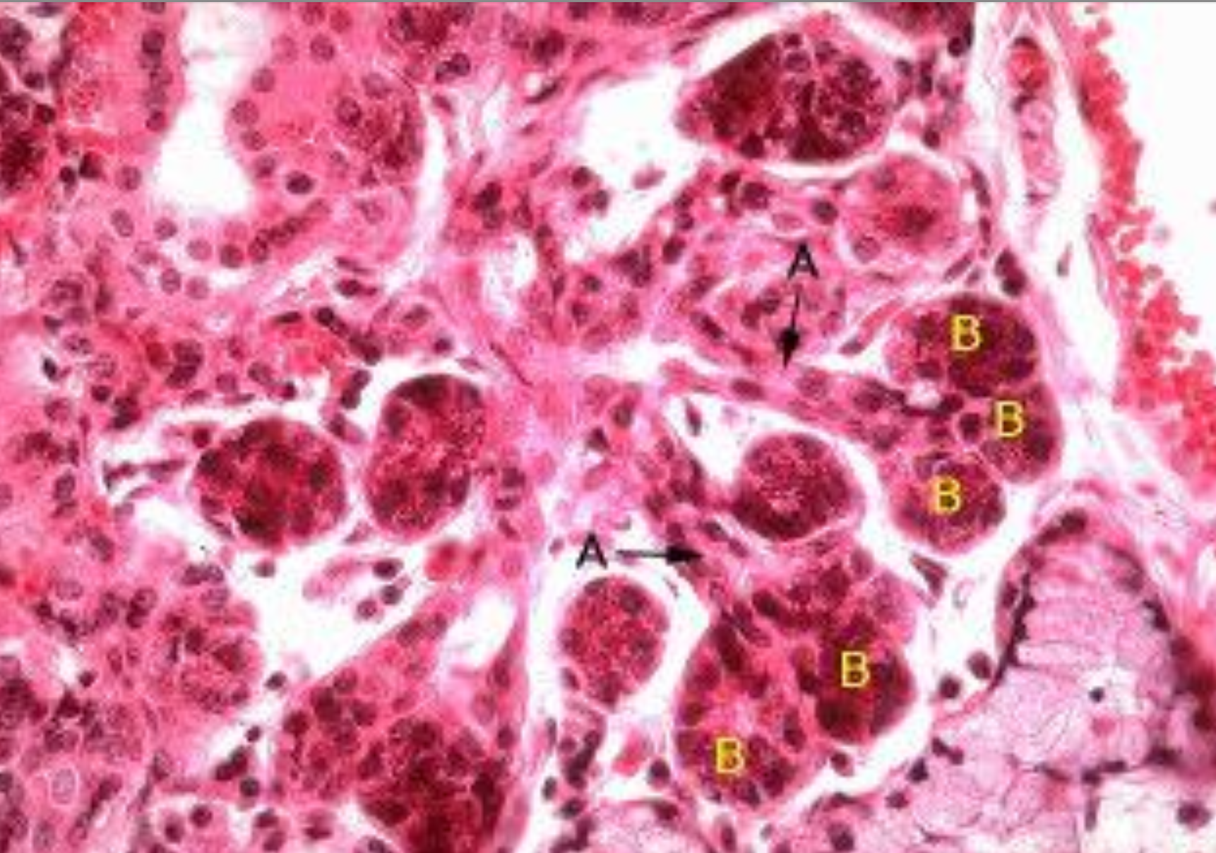
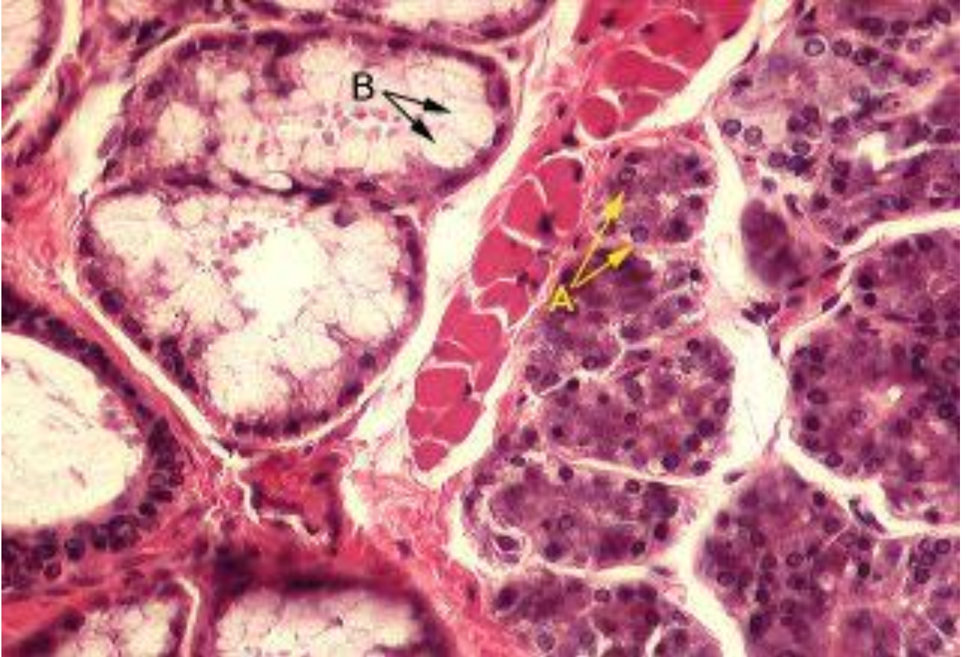
a) Fungiform papilla
b) nerve fibers
c) filiform papilla
d) fat cells
e) connective tissue
f) serous gland acini
ID the image. BE SPECIFIC
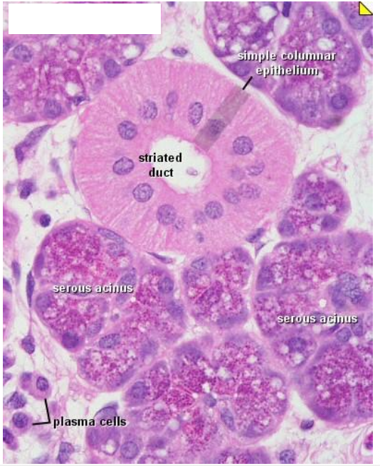
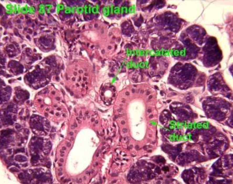
Parotid gland
ID the image. BE SPECIFIC
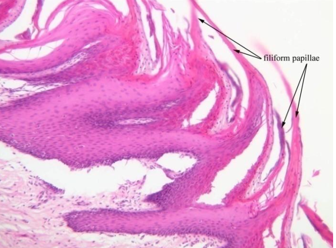
Filiform papillae
ID the image. BE SPECIFIC
Filiform papillae
ID the image. BE SPECIFIC
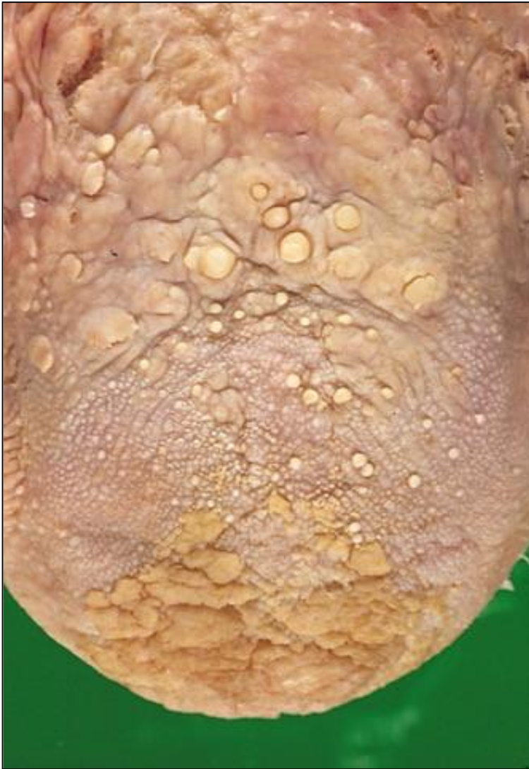
Foliate papillae
ID the image. BE SPECIFIC
Fungiform papilla
ID the image. BE SPECIFIC
Lingual papilla
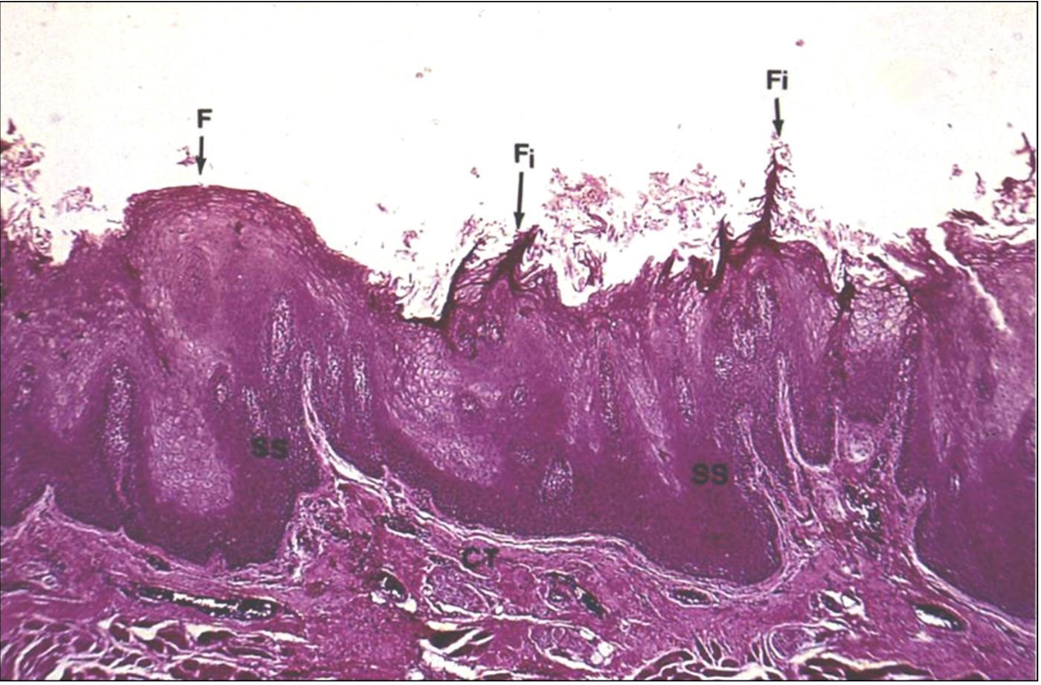
ID the image. BE SPECIFIC
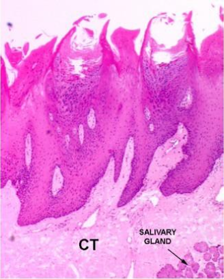
Masticatory mucosa
ID the image. BE SPECIFIC
Filiform papillae
ID the image. BE SPECIFIC
Parotid gland
ID the image. BE SPECIFIC
Parotid gland
ID the image. BE SPECIFIC
Parotid gland
ID the image. Bonus if you’re specific
Salivary gland (parotid)
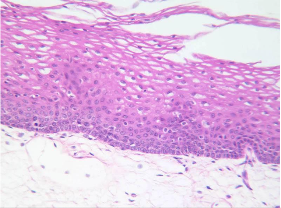
ID the image. BE SPECIFIC
Filiform papillae
ID the image. BE SPECIFIC
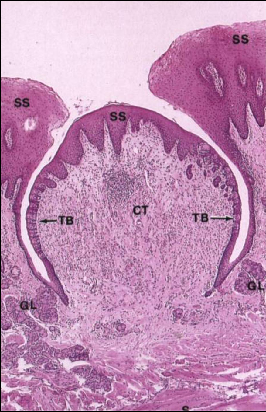
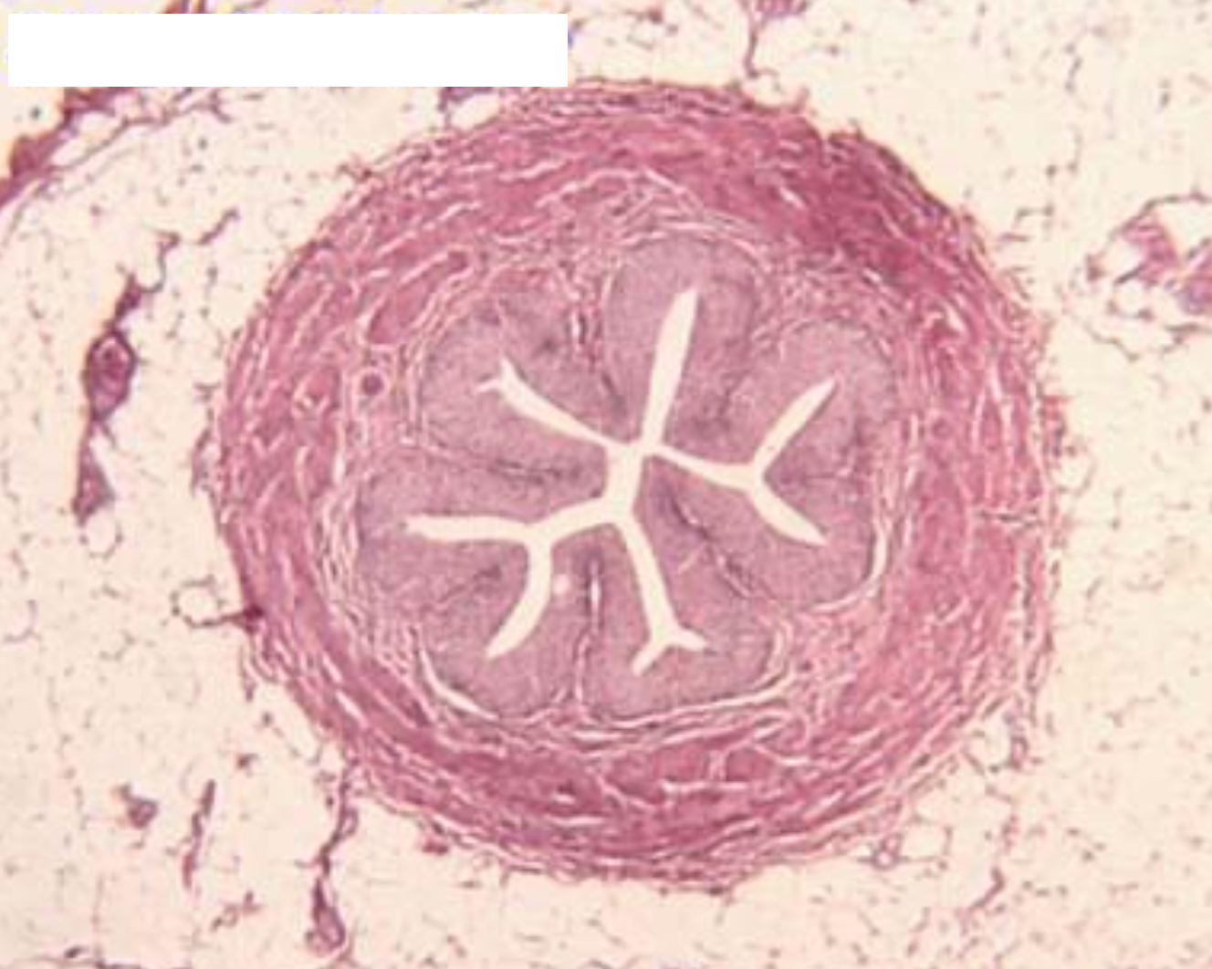
circumvallate papilla
ID the image. BE SPECIFIC
Sublingual gland
ID the image. BE SPECIFIC
Sublingual gland
ID the image. BE SPECIFIC
Submandibular gland
ID the image. BE SPECIFIC
Submandibular gland
ID the image
Circumvallate papilla
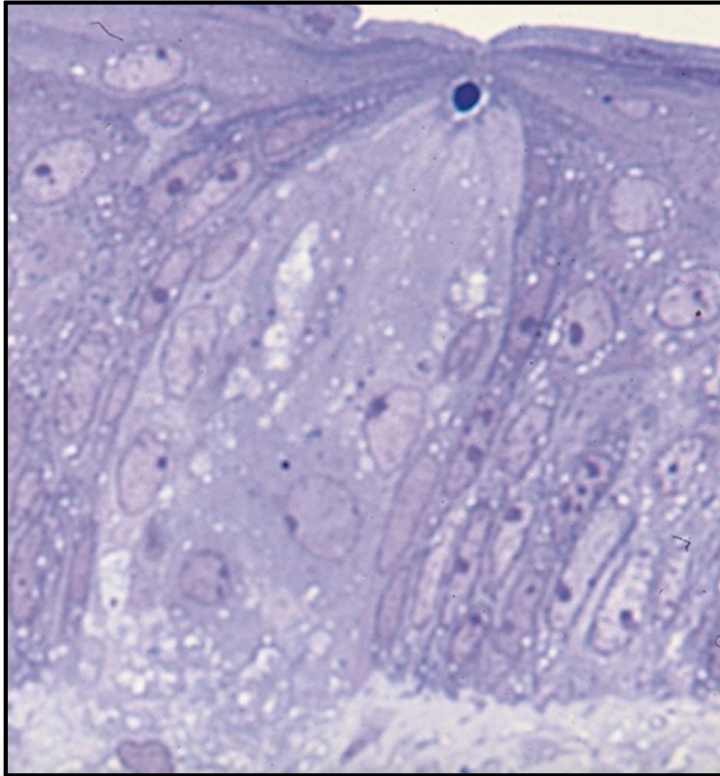
ID the image
Taste bud
ID the image
Taste bud
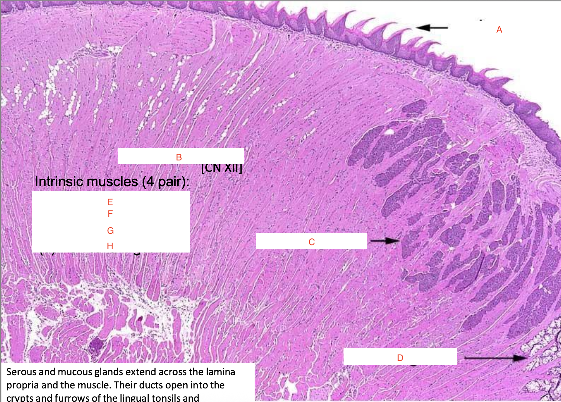
a) vermillion border
b) muscle
c) hairs
d) salivary glands
e) oral mucosa
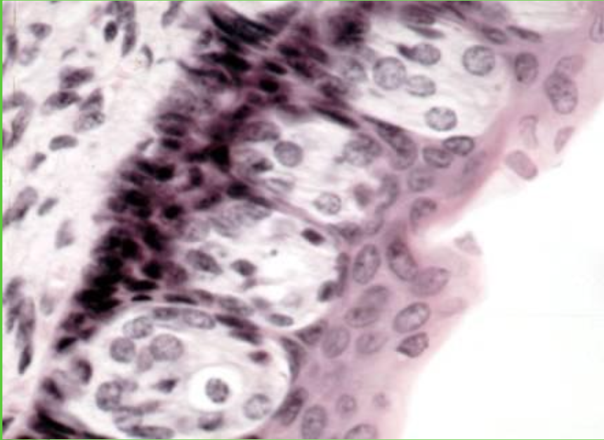
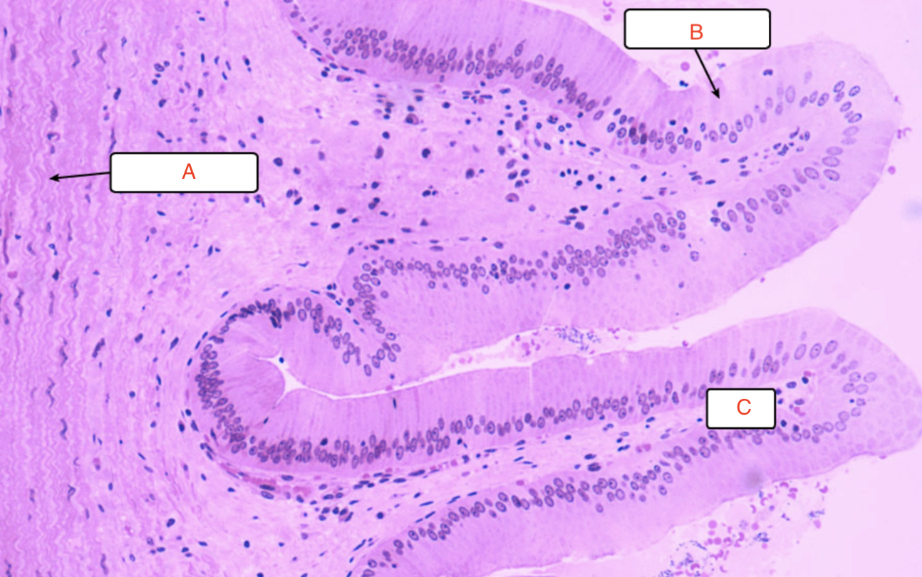
a) Foliate papilla
b) taste bud
c) seromucus gland
d) lamina propria
e) muscle layer
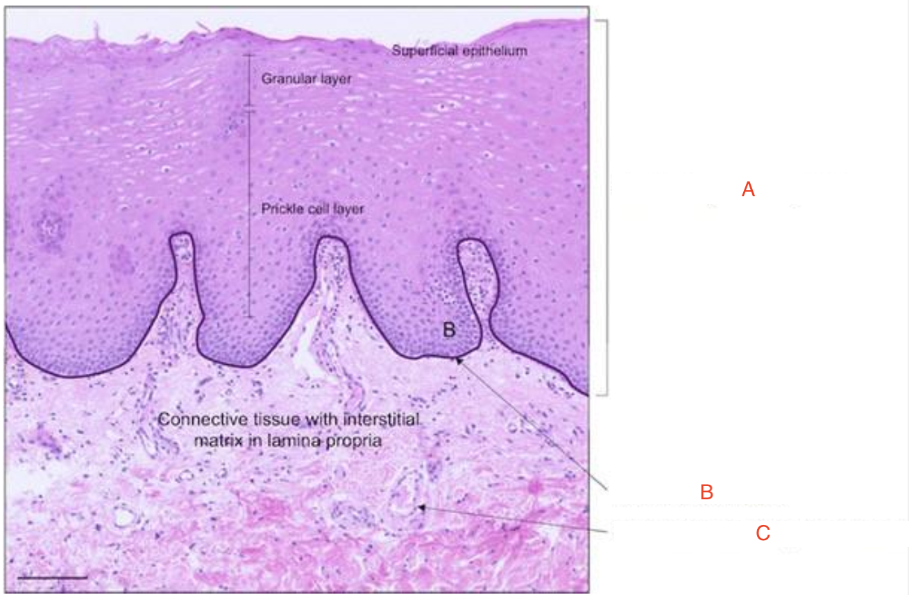
a) Stratified squamous
b) basement membrane
c) endothelial cells (blood vessels)
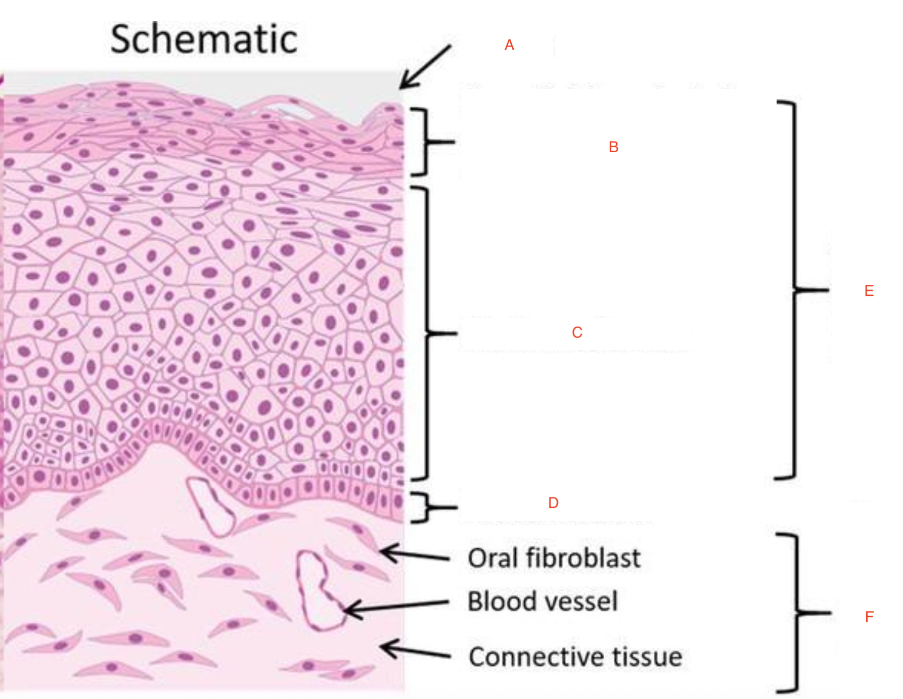
a) mucus
b) non-keratinized epithelium
c) stratum spinosum
d) stratum basale
e) stratified squamous
f) lamina propria
forget e-h
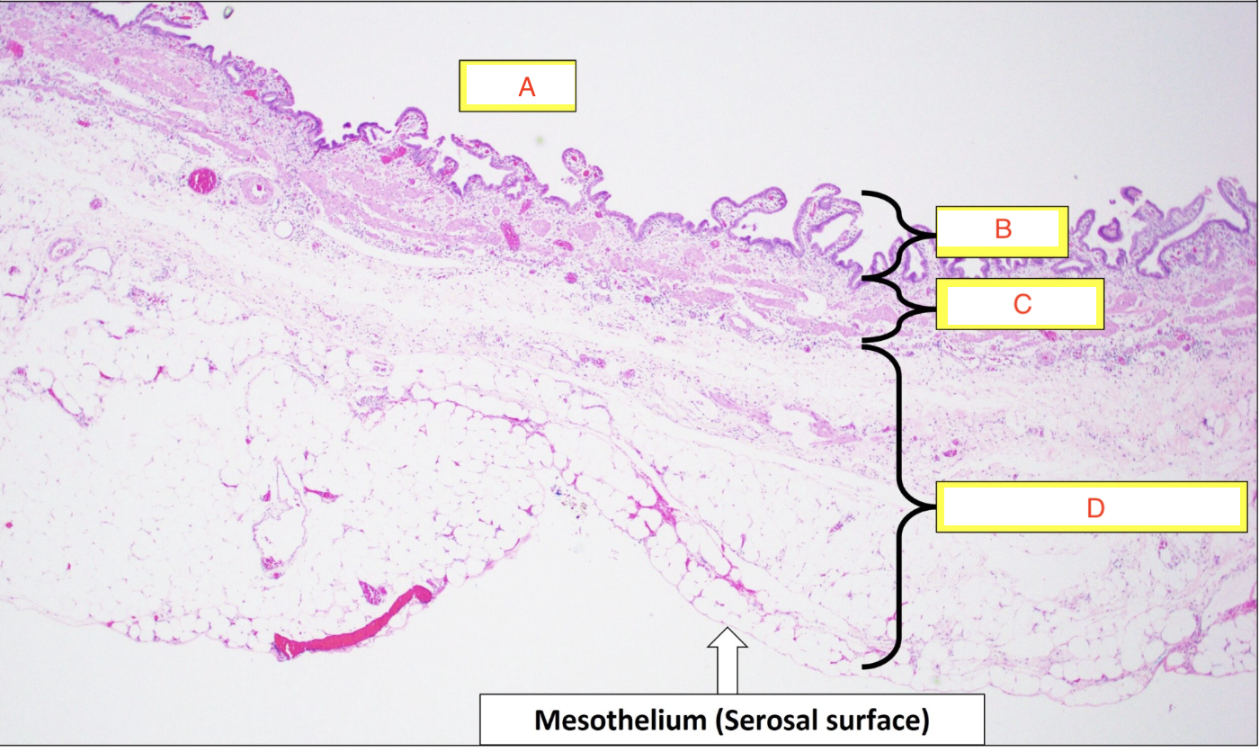
a) oral mucosa
b) muscle bundles
c) serous glands
d) mucous glands
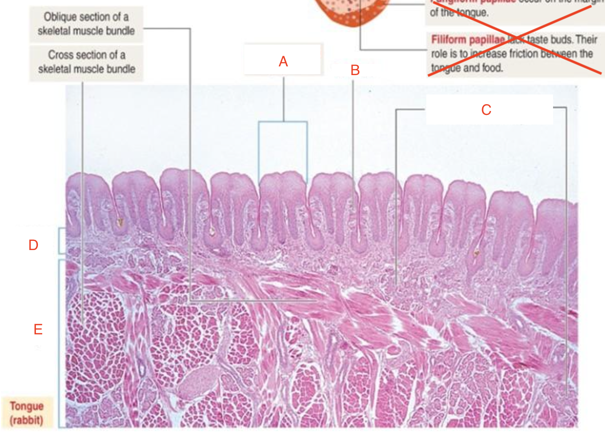
a) filiform papilla
b) adipose tissue
c) lingual salivary glands
d) skeletal muscle
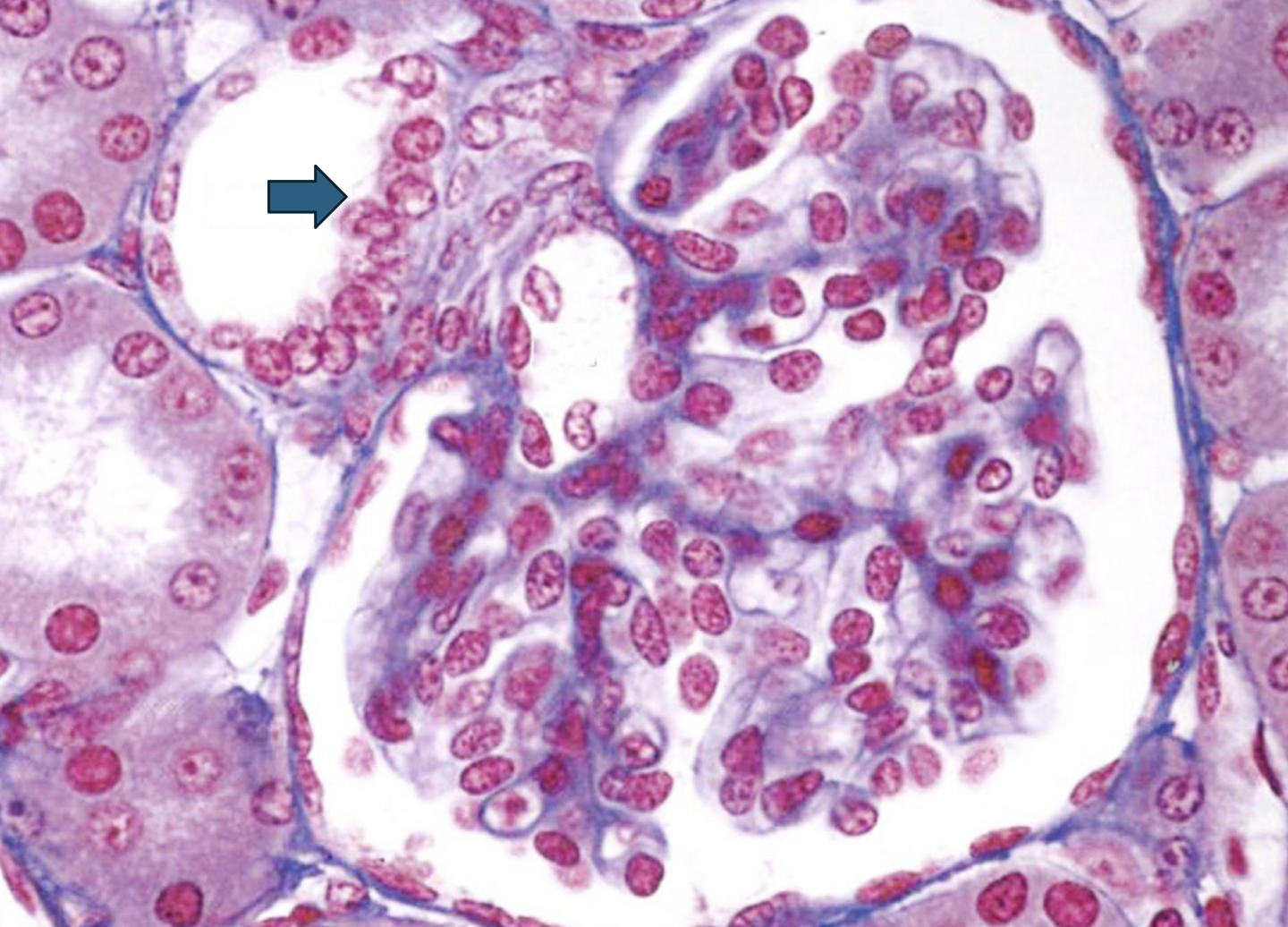
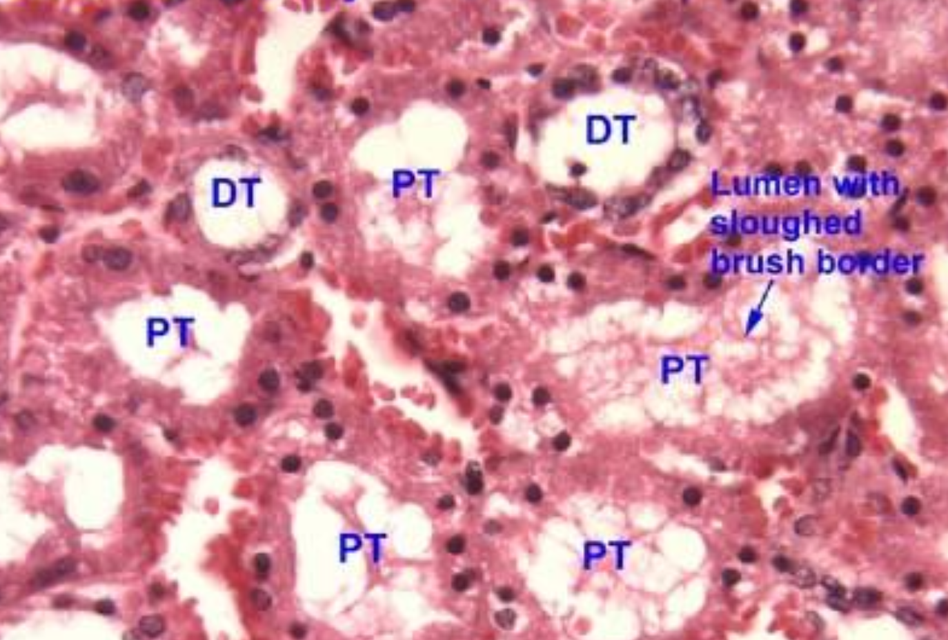
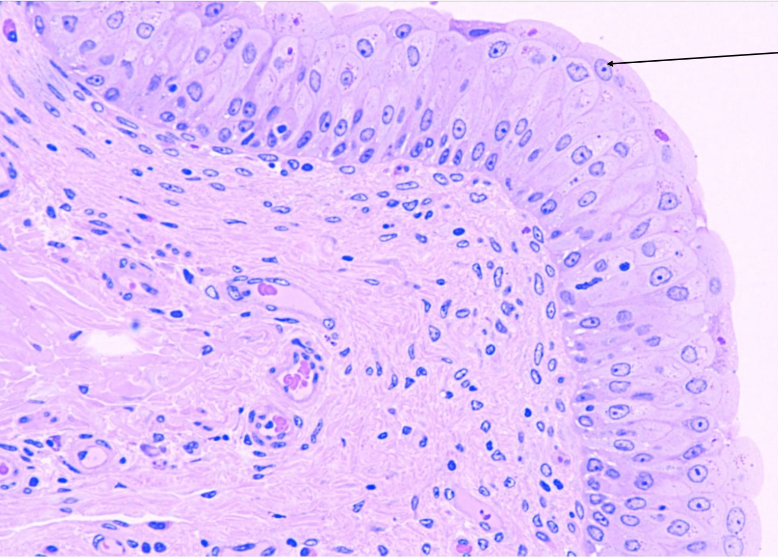
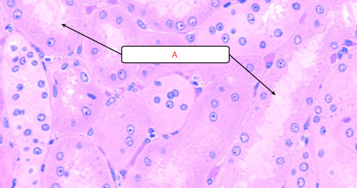
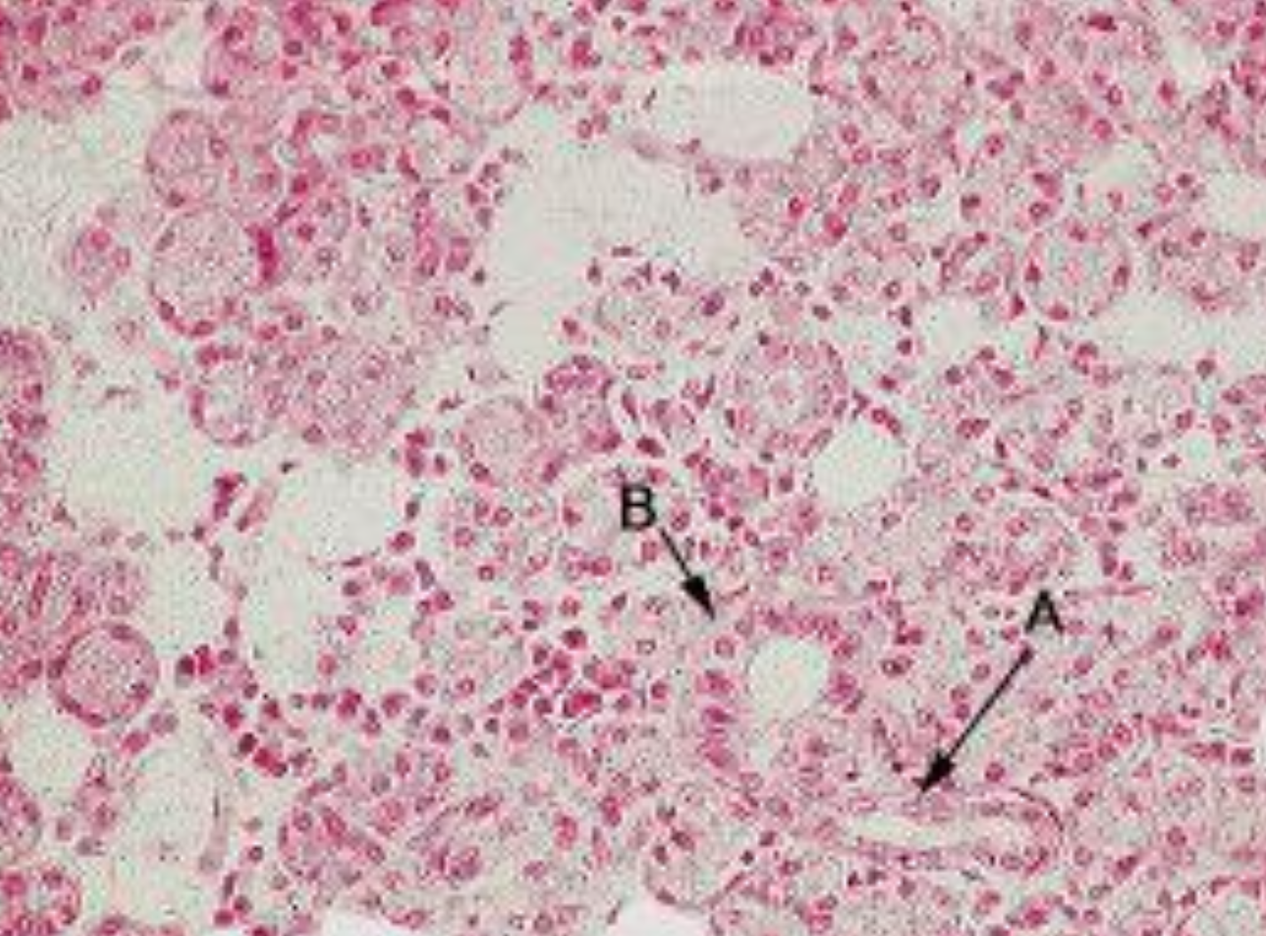
What is the arrow pointing to?
macula densa
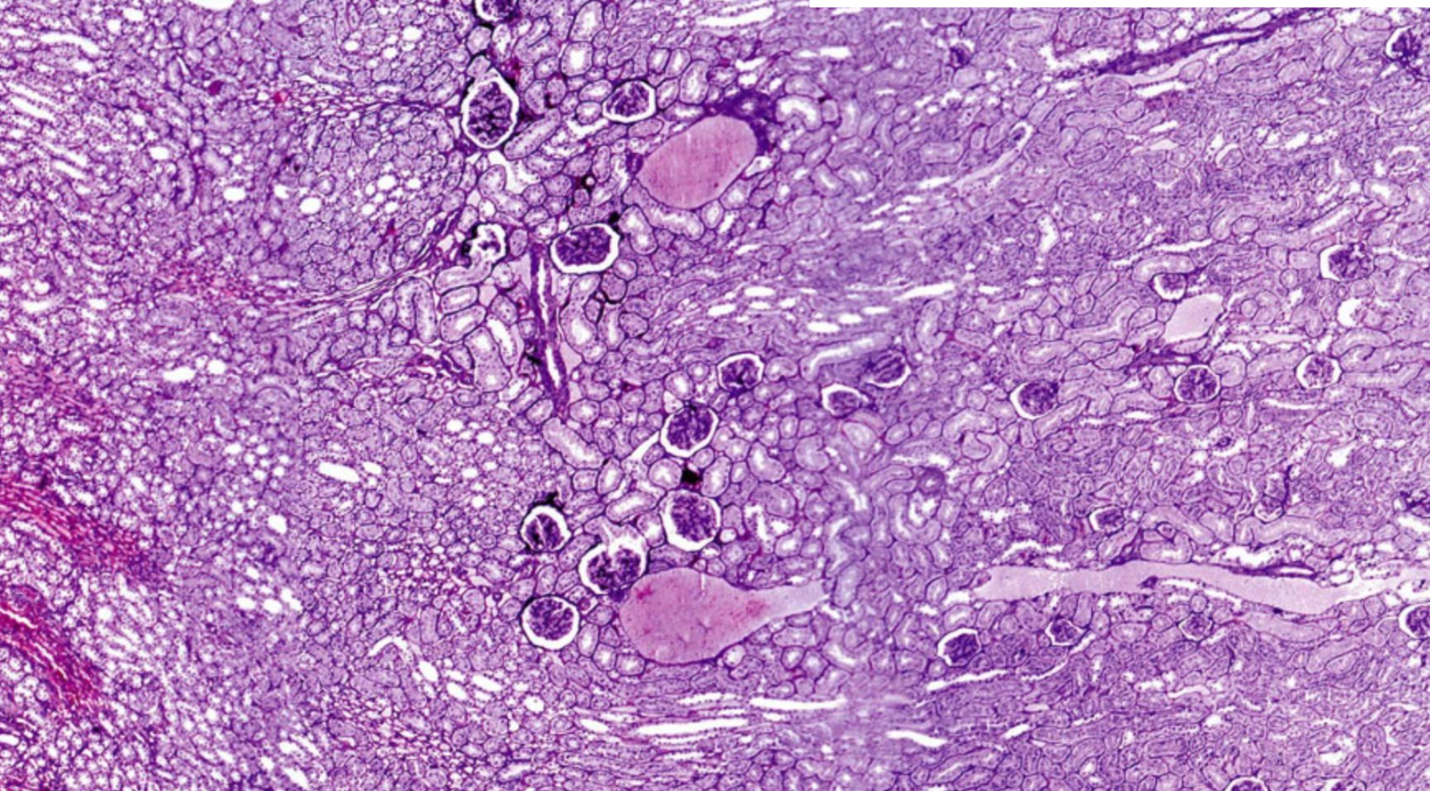
ID the image
Kidney
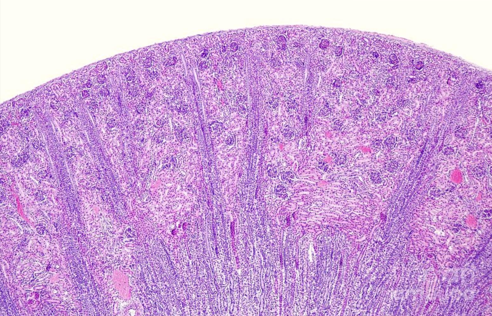
ID the image
Kidney
ID the image
Kidney
ID the image
Kidney
ID the image
Loop of Henle
ID the image
Loop of Henle
ID the image
Podocyte
ID the image
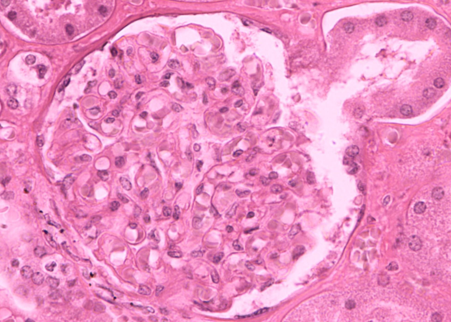
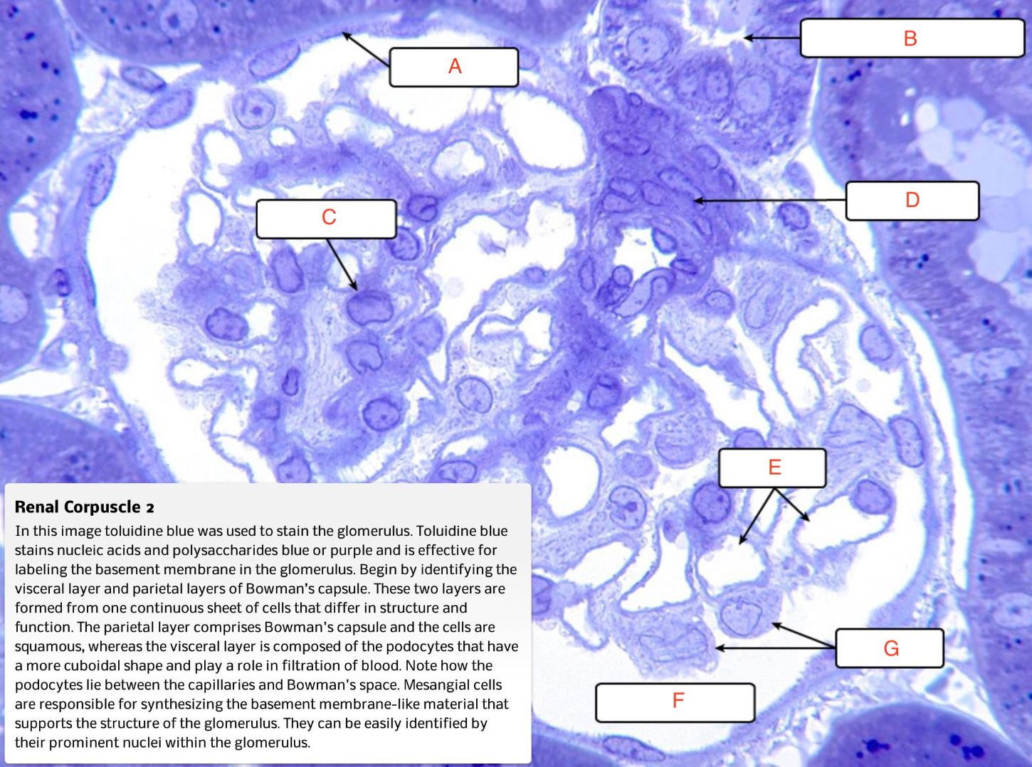
Renal corpuscle
ID the image
Renal corpuscle
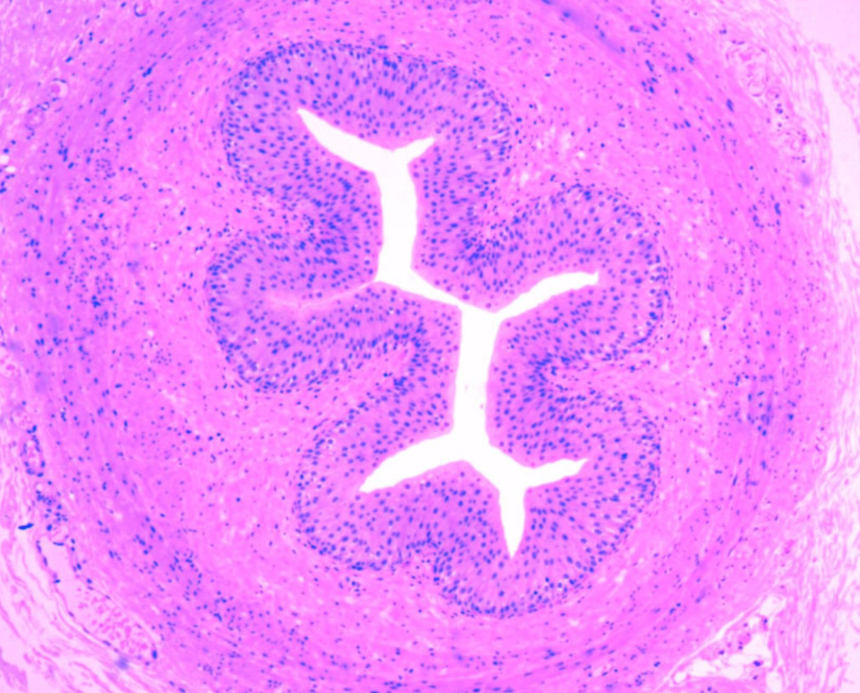
ID the image
Ureter
ID the image
Ureter
ID the image
Urinary bladder
ID the image
Urinary bladder
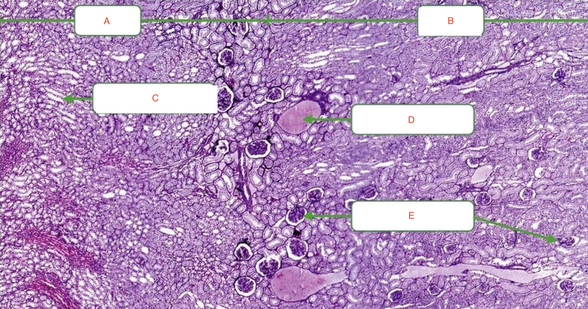
a) Medulla
b) cortex
c) collecting duct
d) arcuate artery
e) renal corpuscle
ignore f
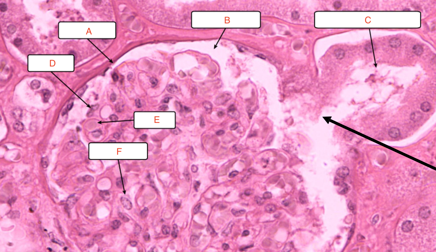
a) Parietal layer
b) Bowman’s space
c) proximal convoluted tubule
d) podocyte
e) capillary
ignore c
a) Parietal layer
b) distal convoluted tubules
d) vascular pole
e) capillaries
f) Bowman’s space
g) podocytes
ignore a
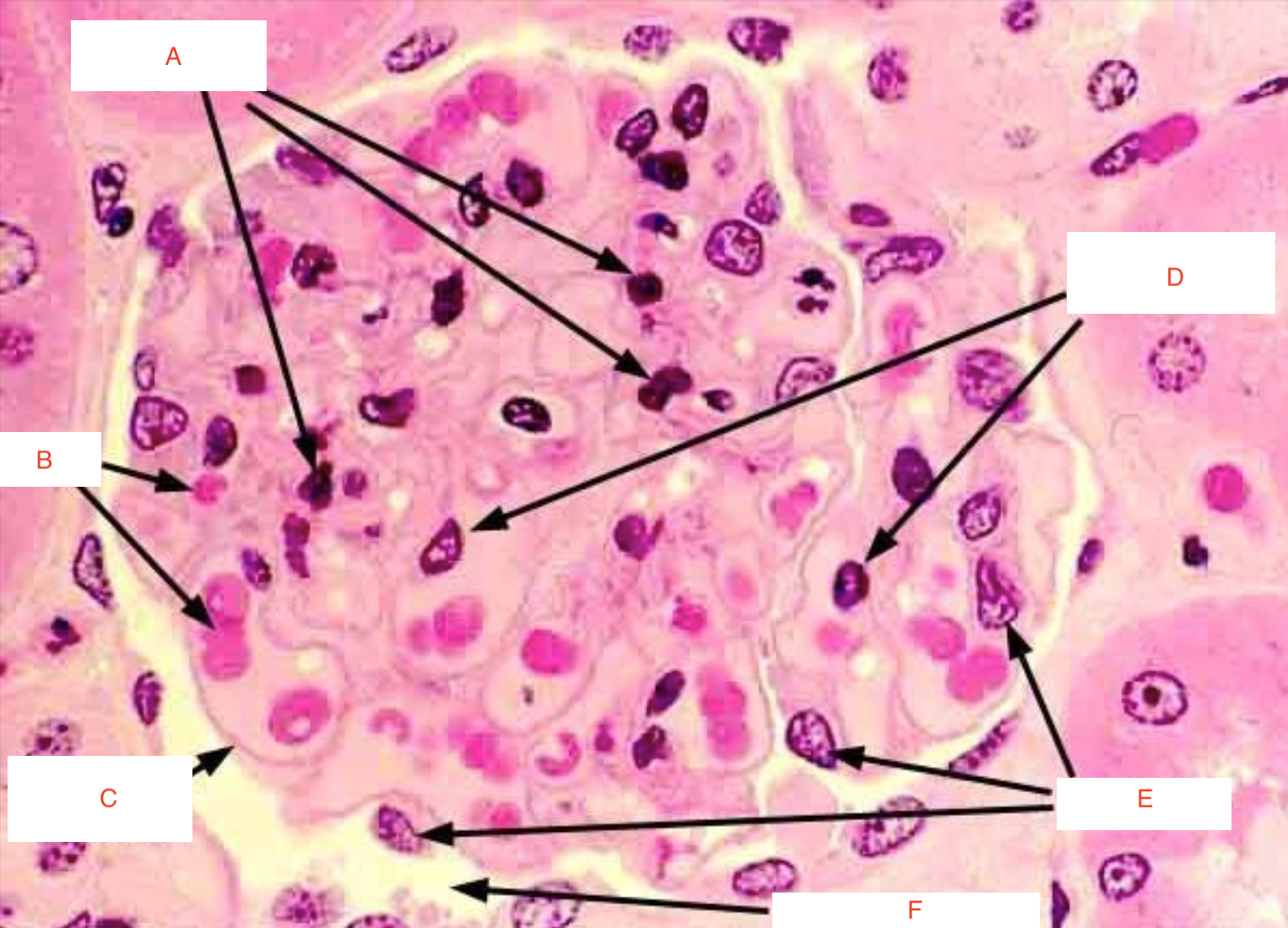
b) RBCs
c) filtration membrane
d) capillary endothelium
e) podocyte
f) Bowman’s space
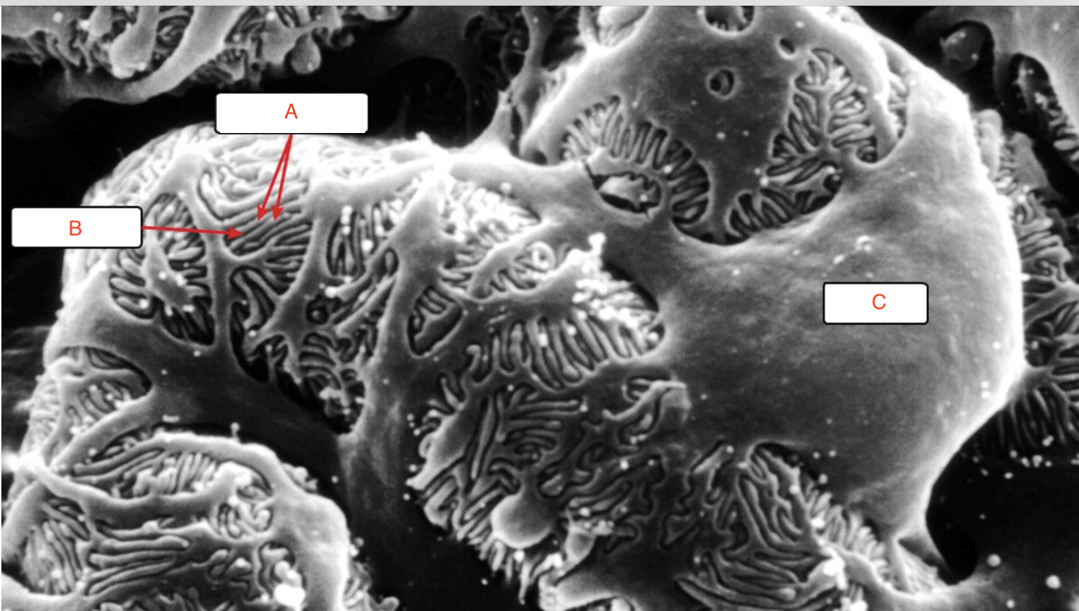
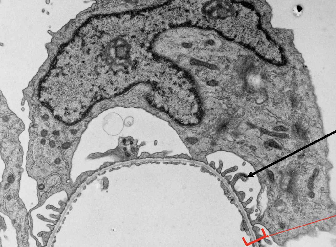
a) Foot processes
b) filtration slit
c) podocyte
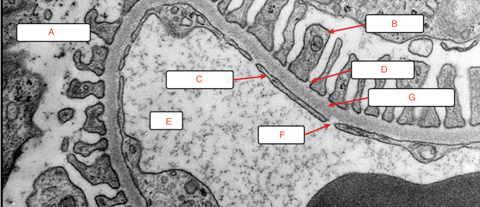
a) Bowman’s space
b) Foot process
c) endothelial cell
d) slit diaphragm
e) blood
f) fenestrae
g) basement membrane
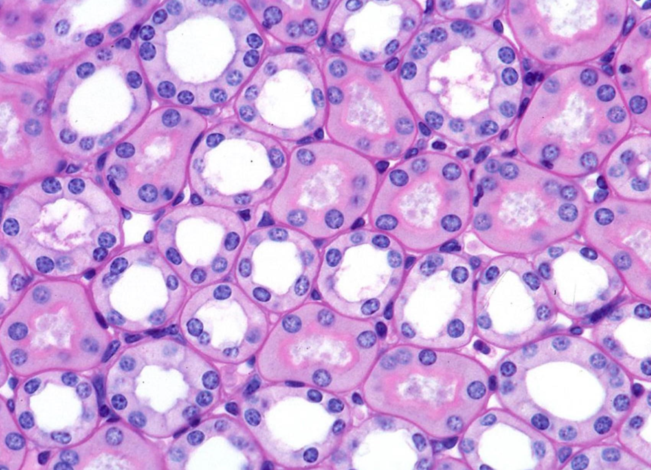
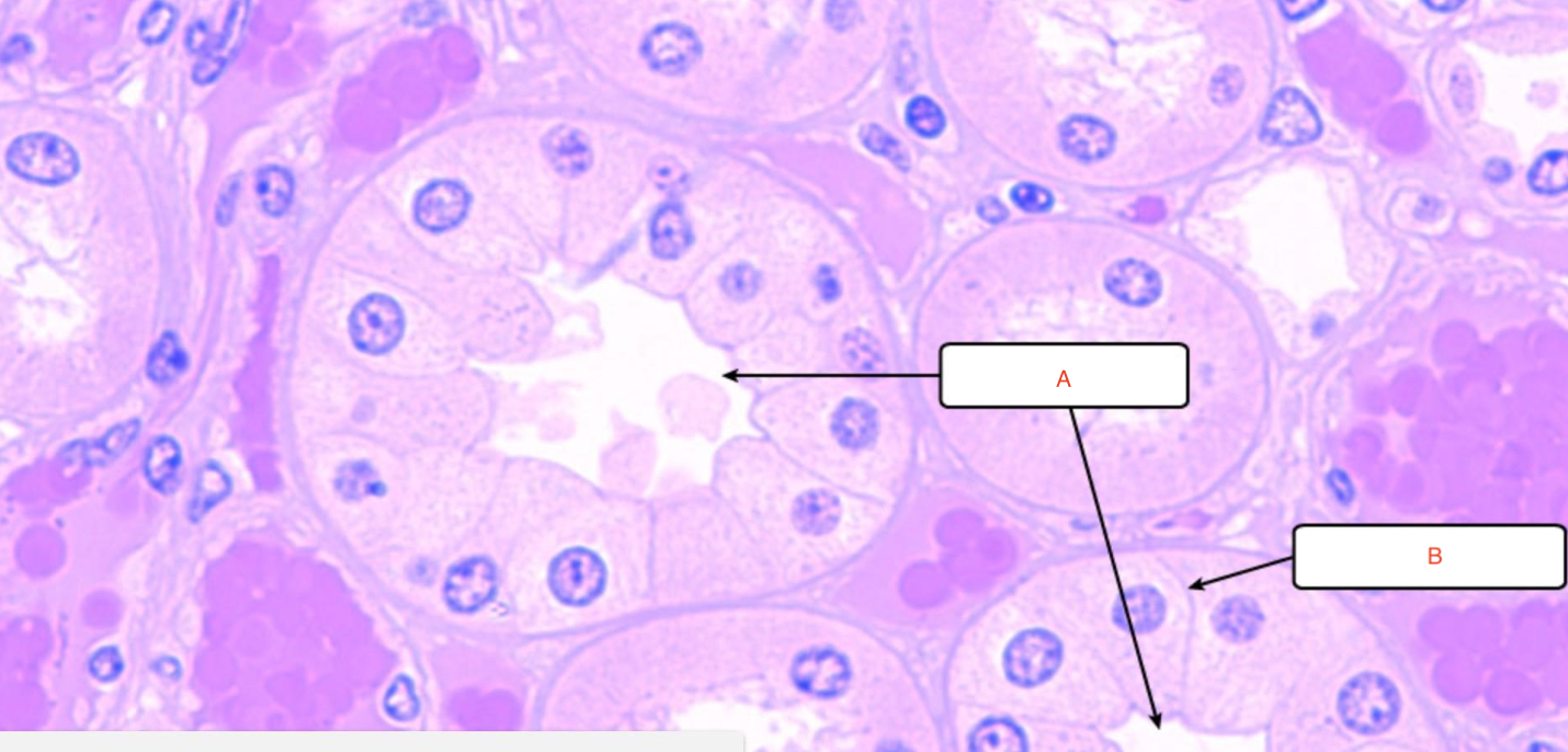
Proximal convoluted tubules
ID this structure
Loop of Henle
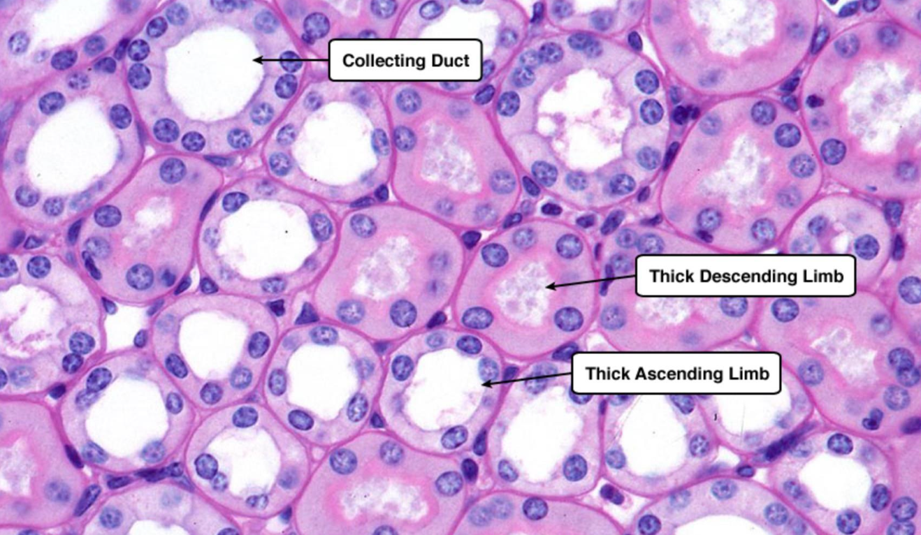
a) Collecting duct
b) thick descending limb
c) thick ascending limb
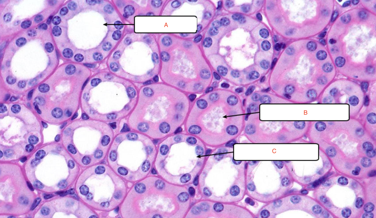
a) thin segment of loop of Henle
b) thick segment of loop of Henle
a) thick segment
b) thin segment
c) collecting duct
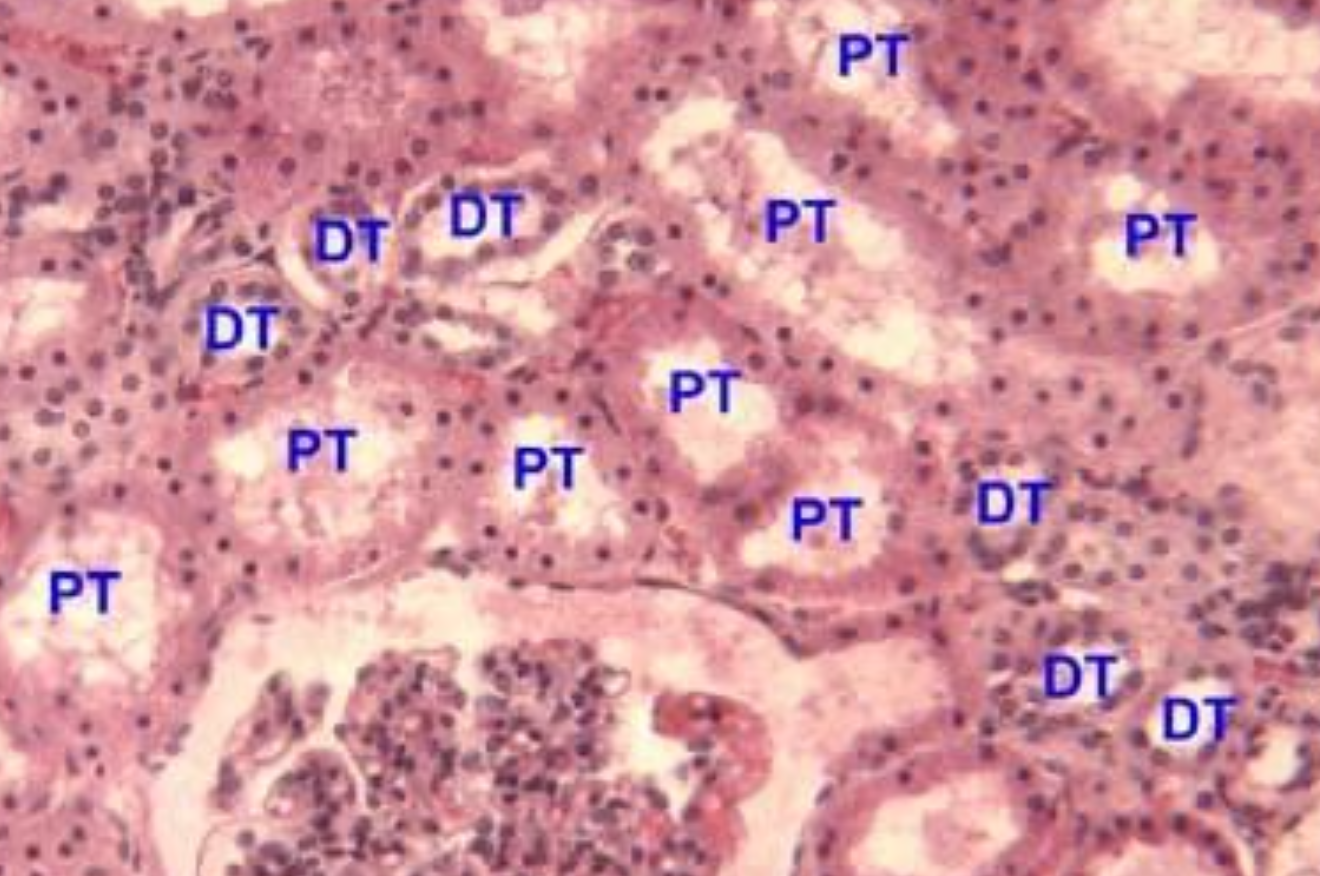
a) Distal convoluted tubules
b) proximal convoluted tubule
c) glomerulus
Bowman’s capsule
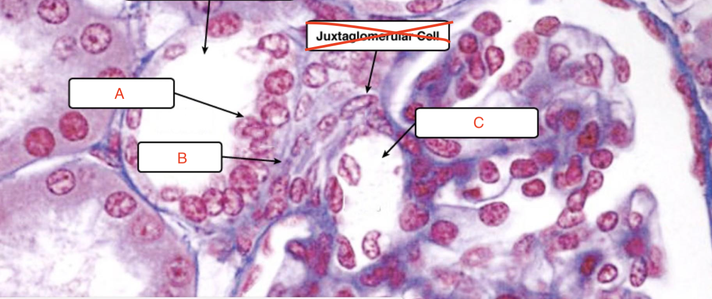
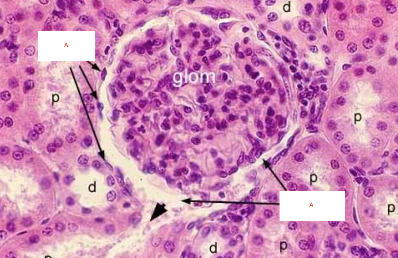
a) Macula densa
b) Lacis cells
c) afferent arteriole
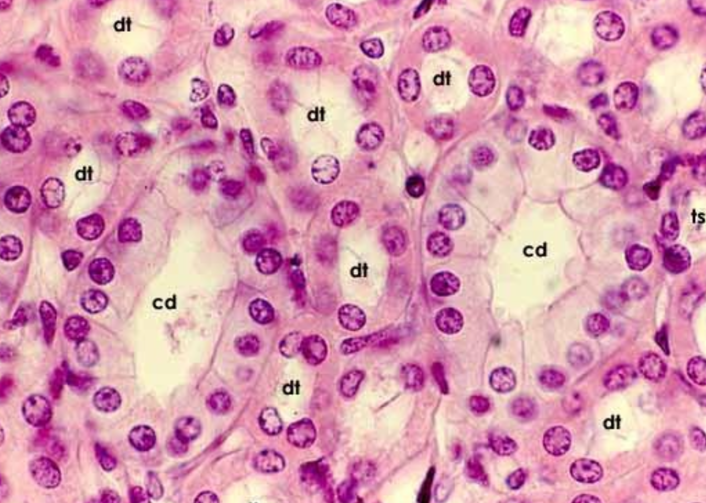
a) collecting duct
b) plasma membrane
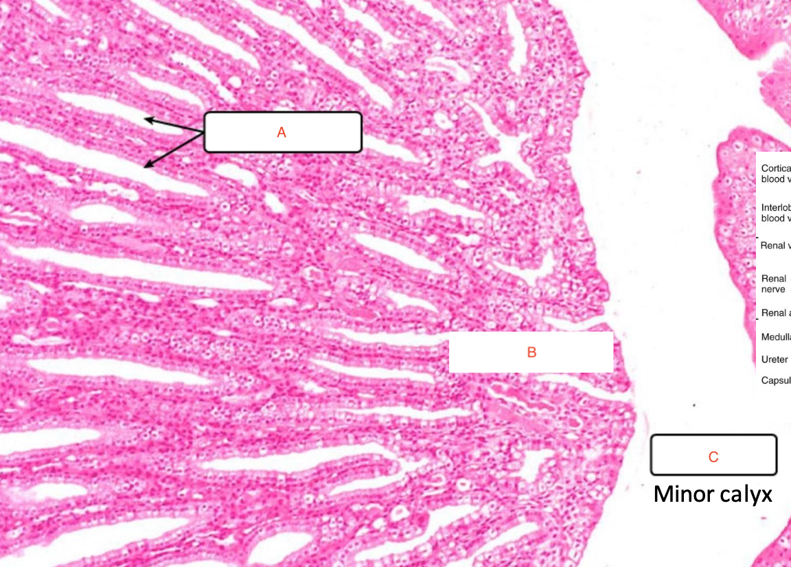
a) collecting ducts
b) renal papilla
c) renal pelvis
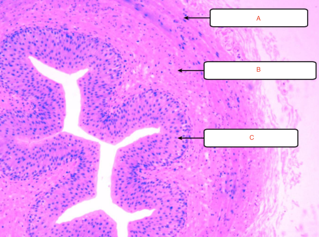
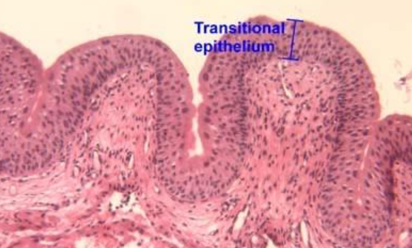
a) circular smooth muscle
b) longitudinal smooth muscle
c) transitional epithelium
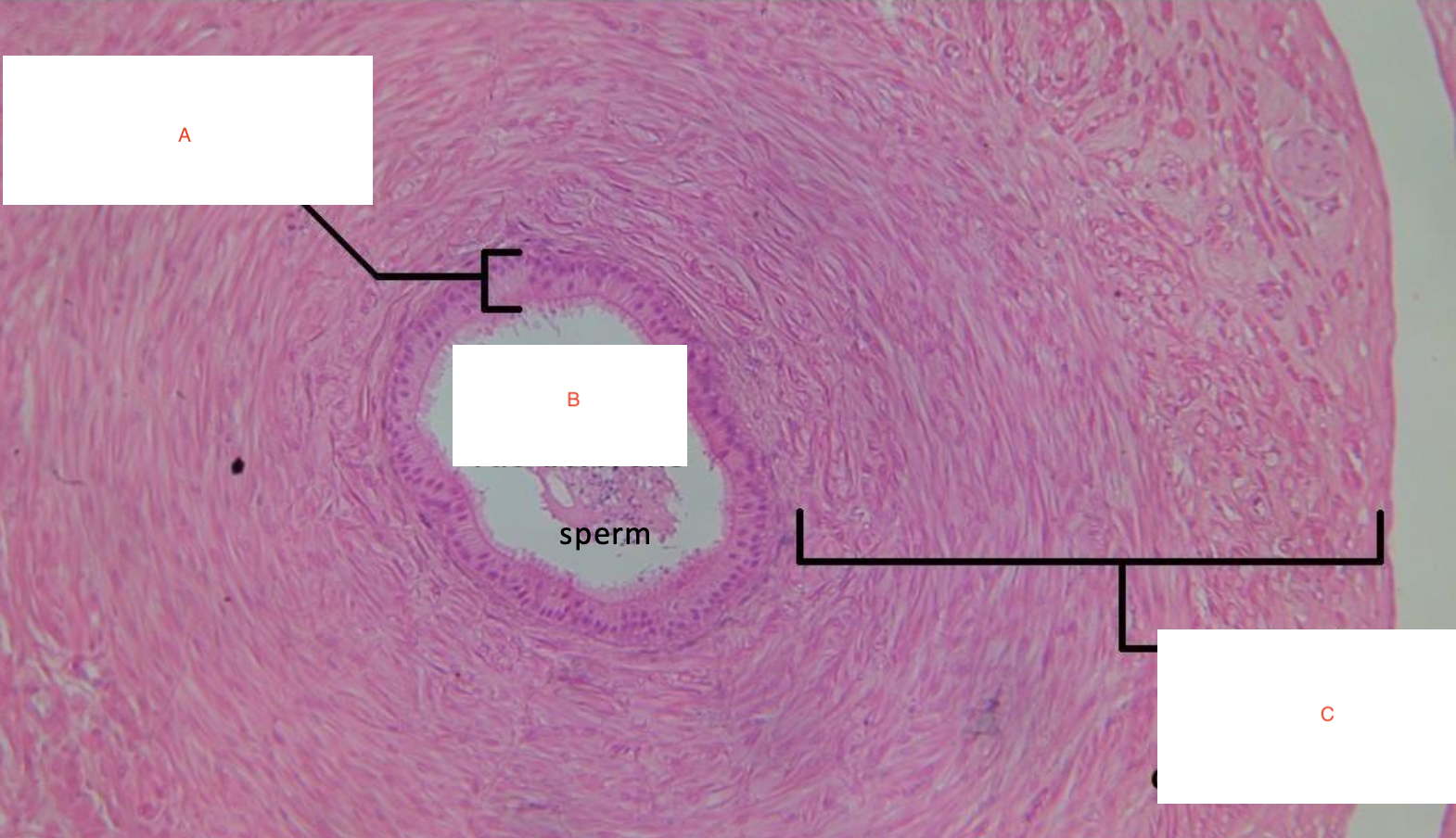
a) pseudostratified ciliated columnar epithelium
b) lumen of vas deferens
c) smooth muscle and connective tissue
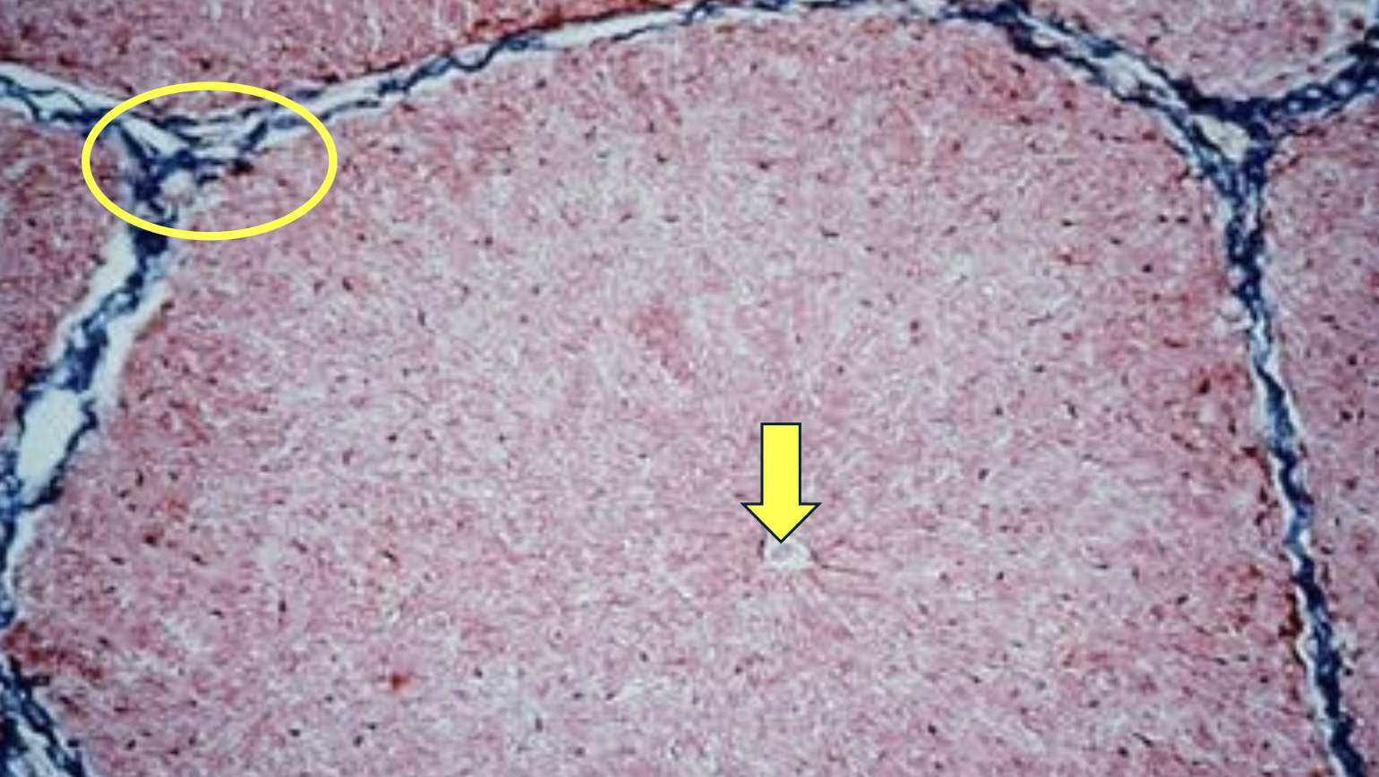
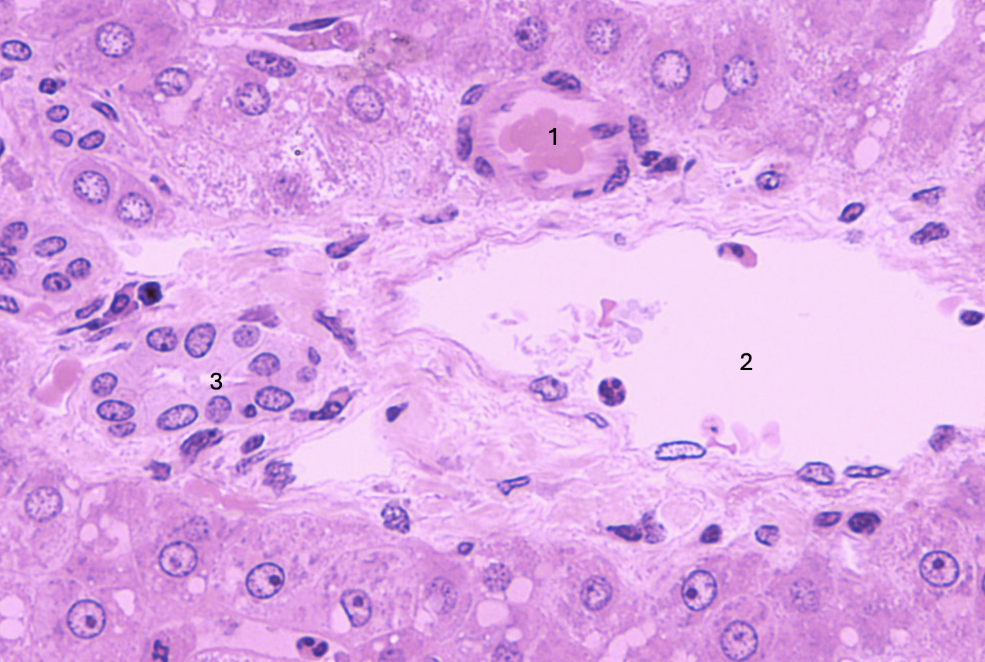
What is the yellow arrow pointing to?
Portal triad
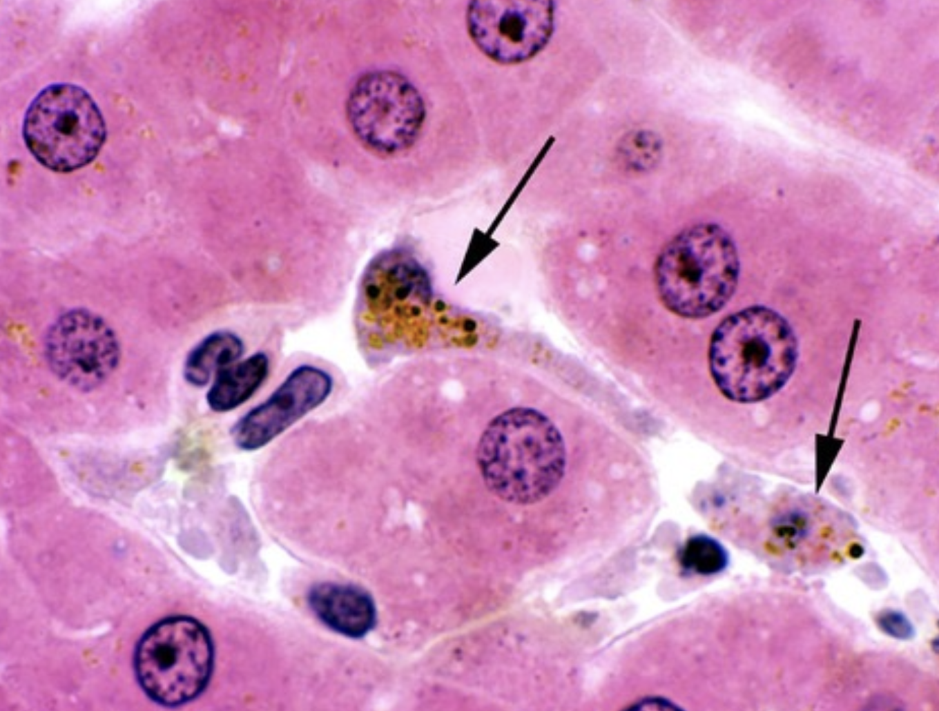
What are the arrows pointing to?
Kupffer cells
ID the structure
Gallbladder
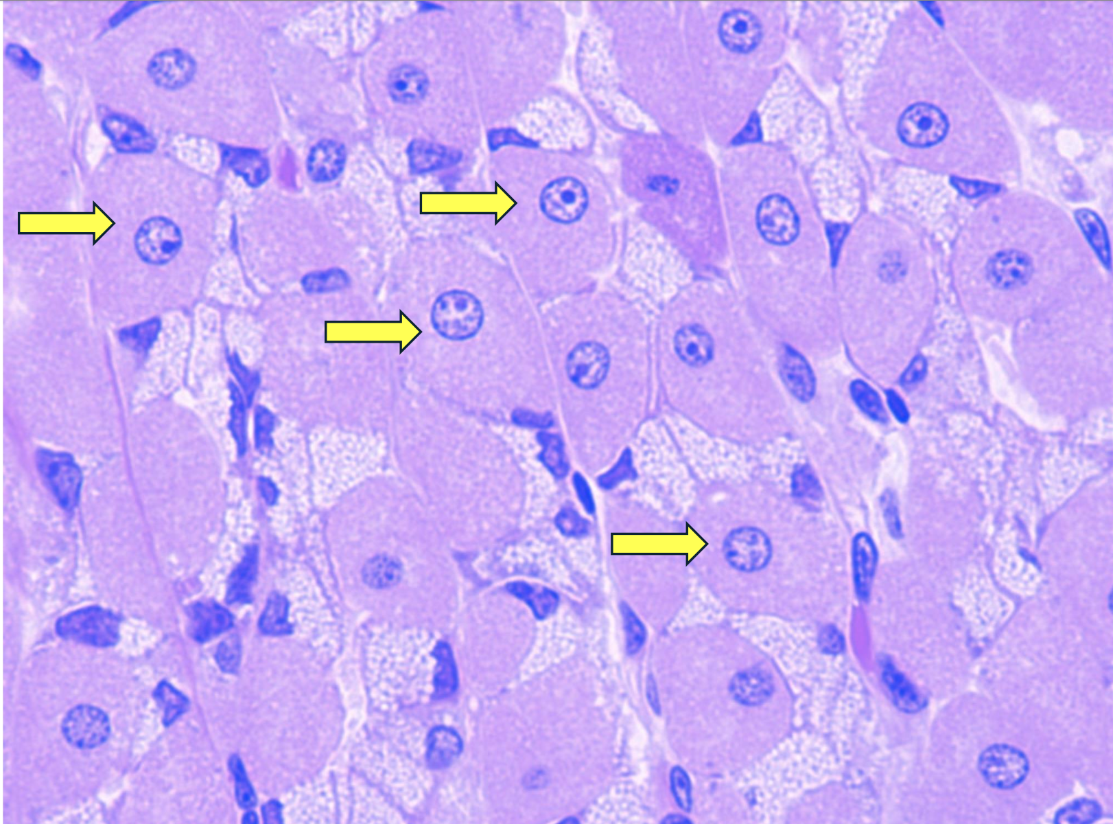
ID this structure
Hepatocytes
ID the structure
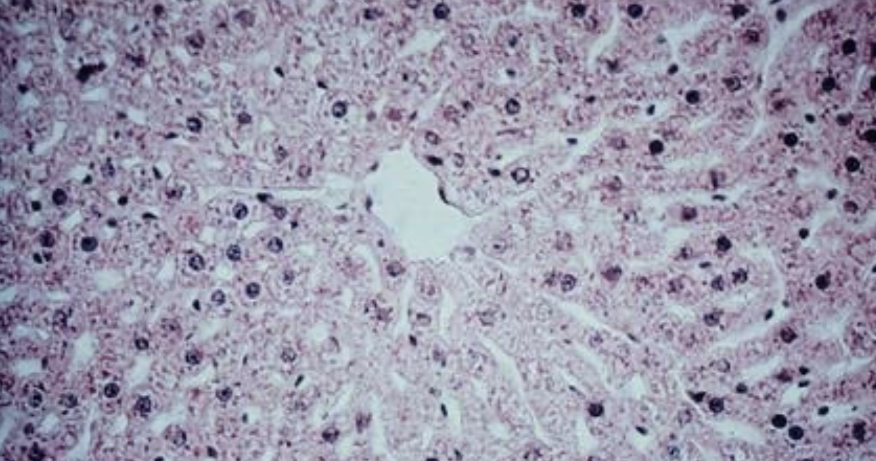
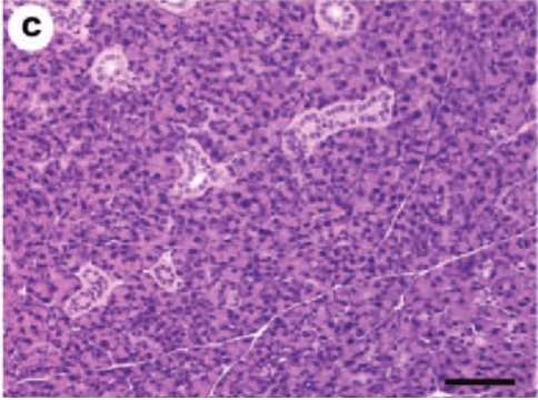
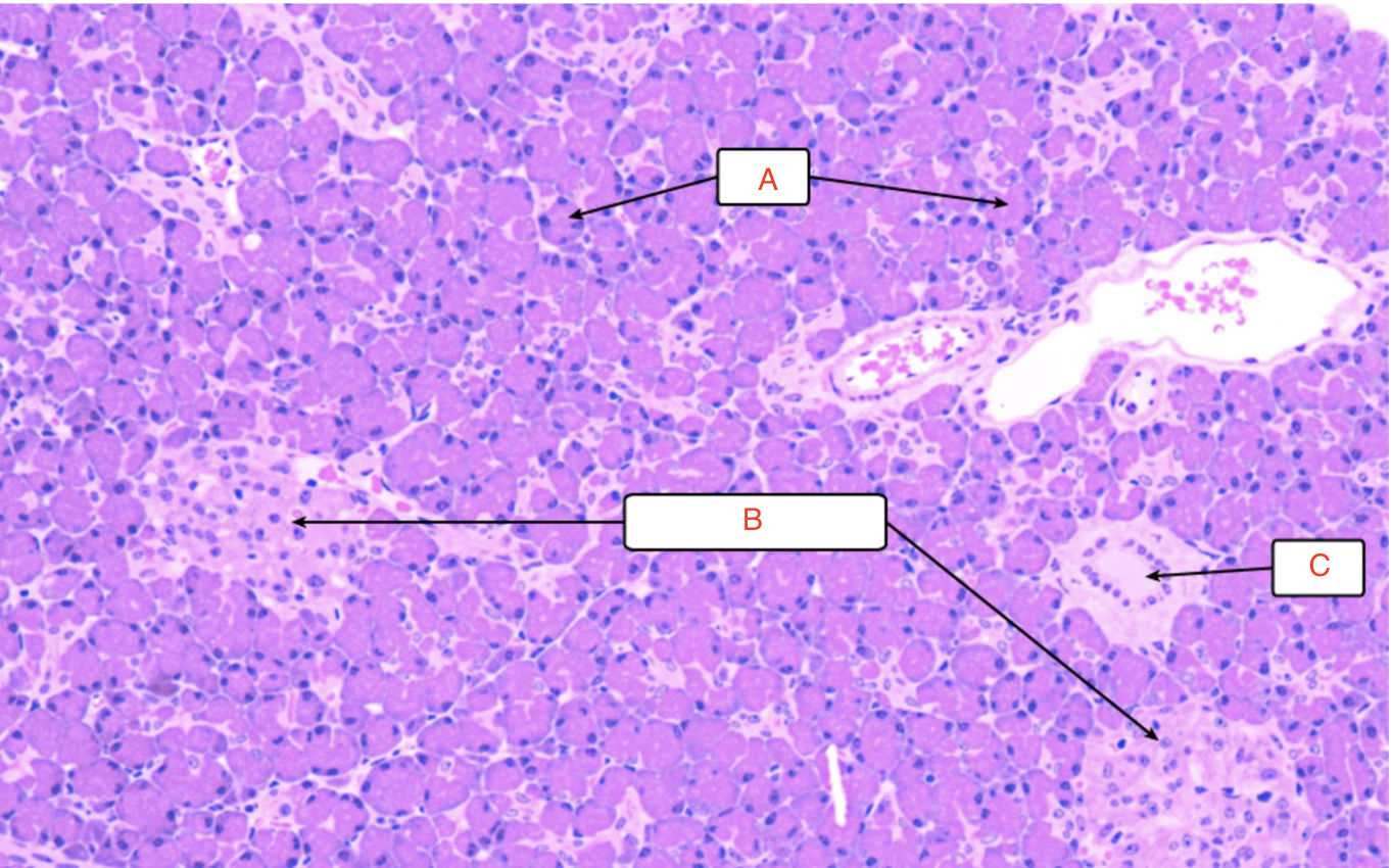
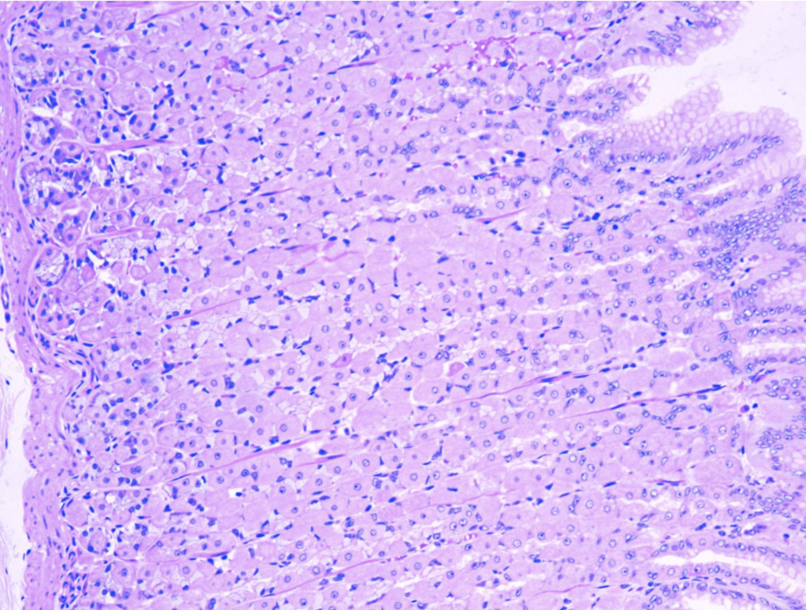
Liver
ID the structure
Liver
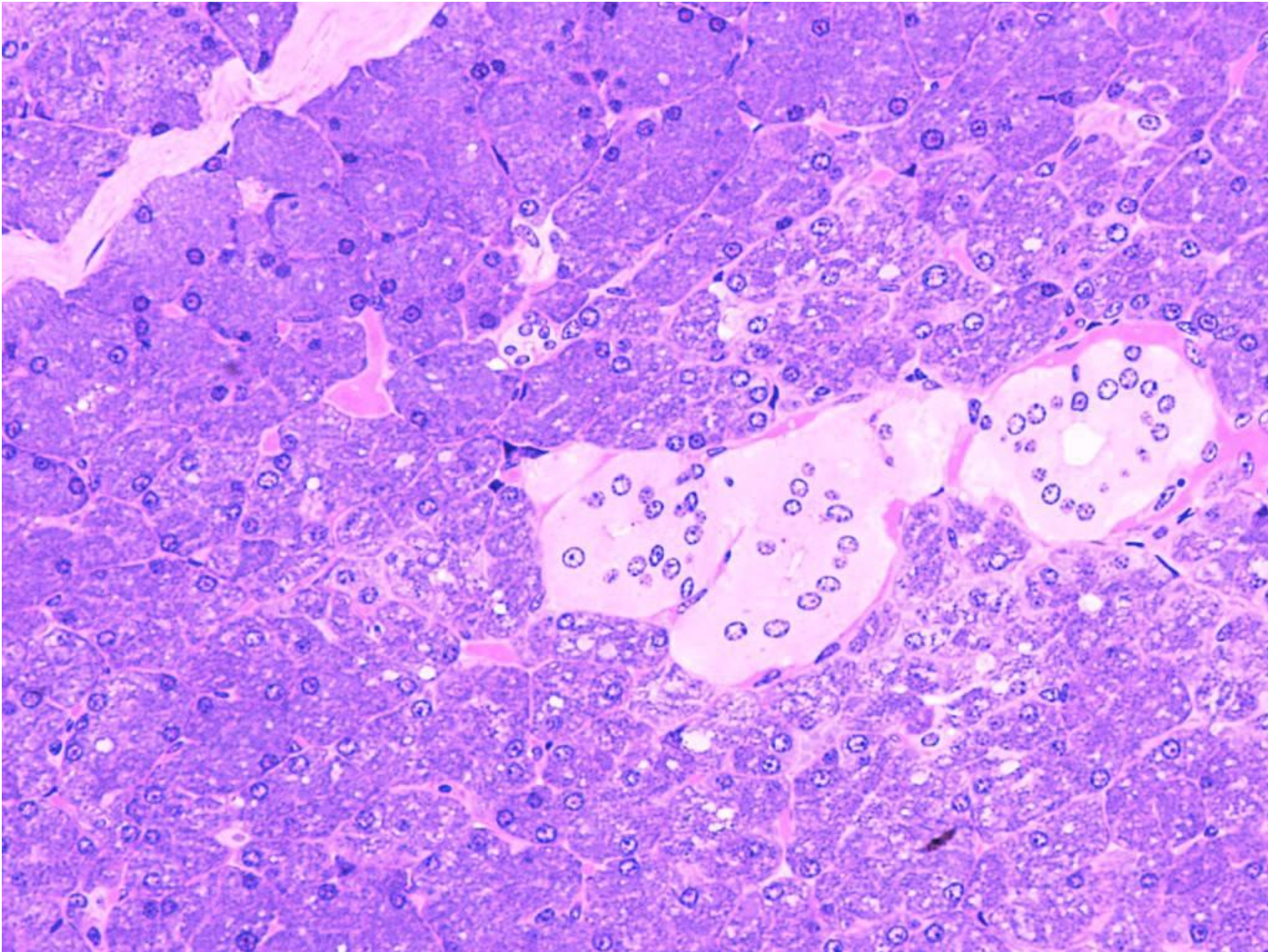
ID the structure
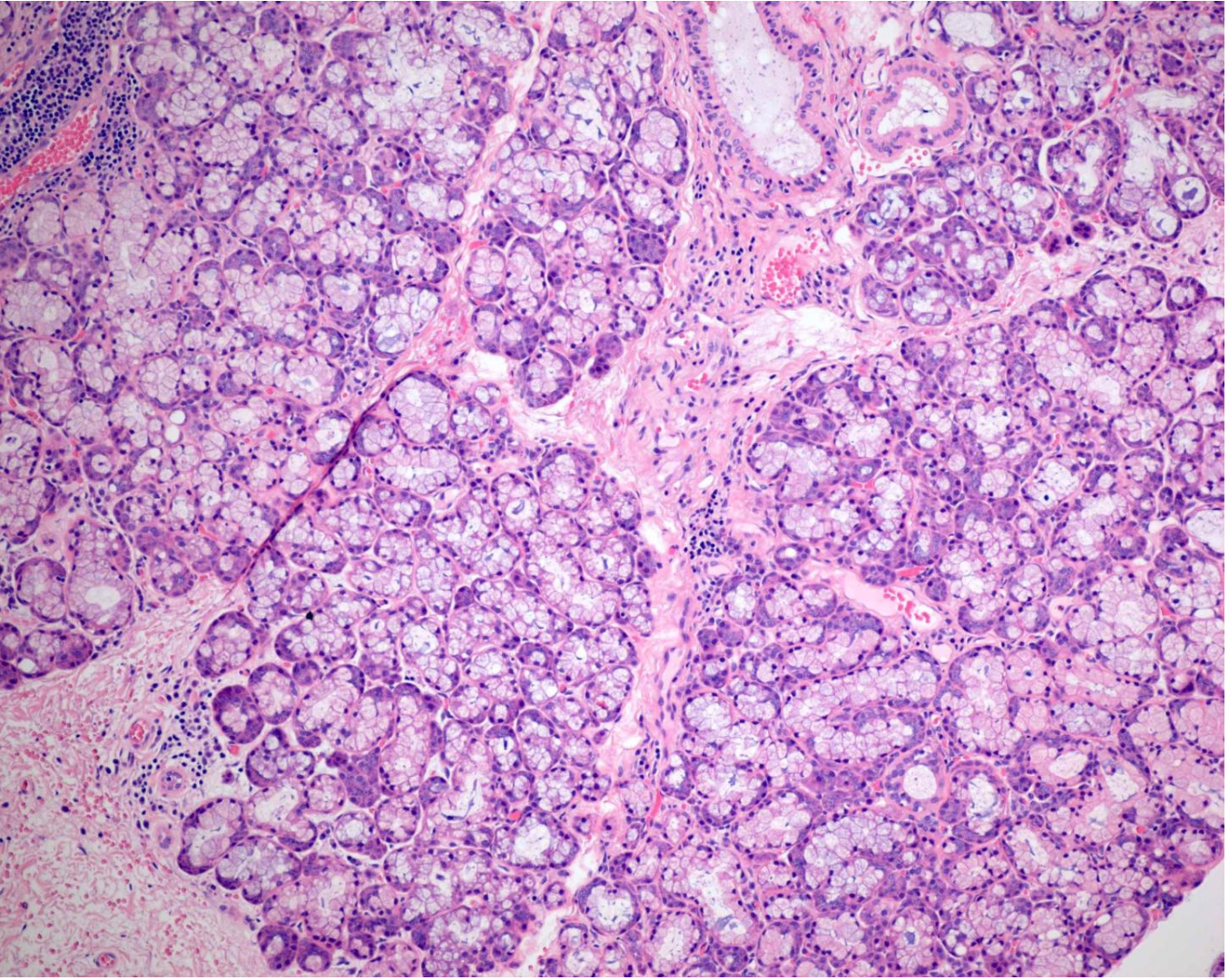
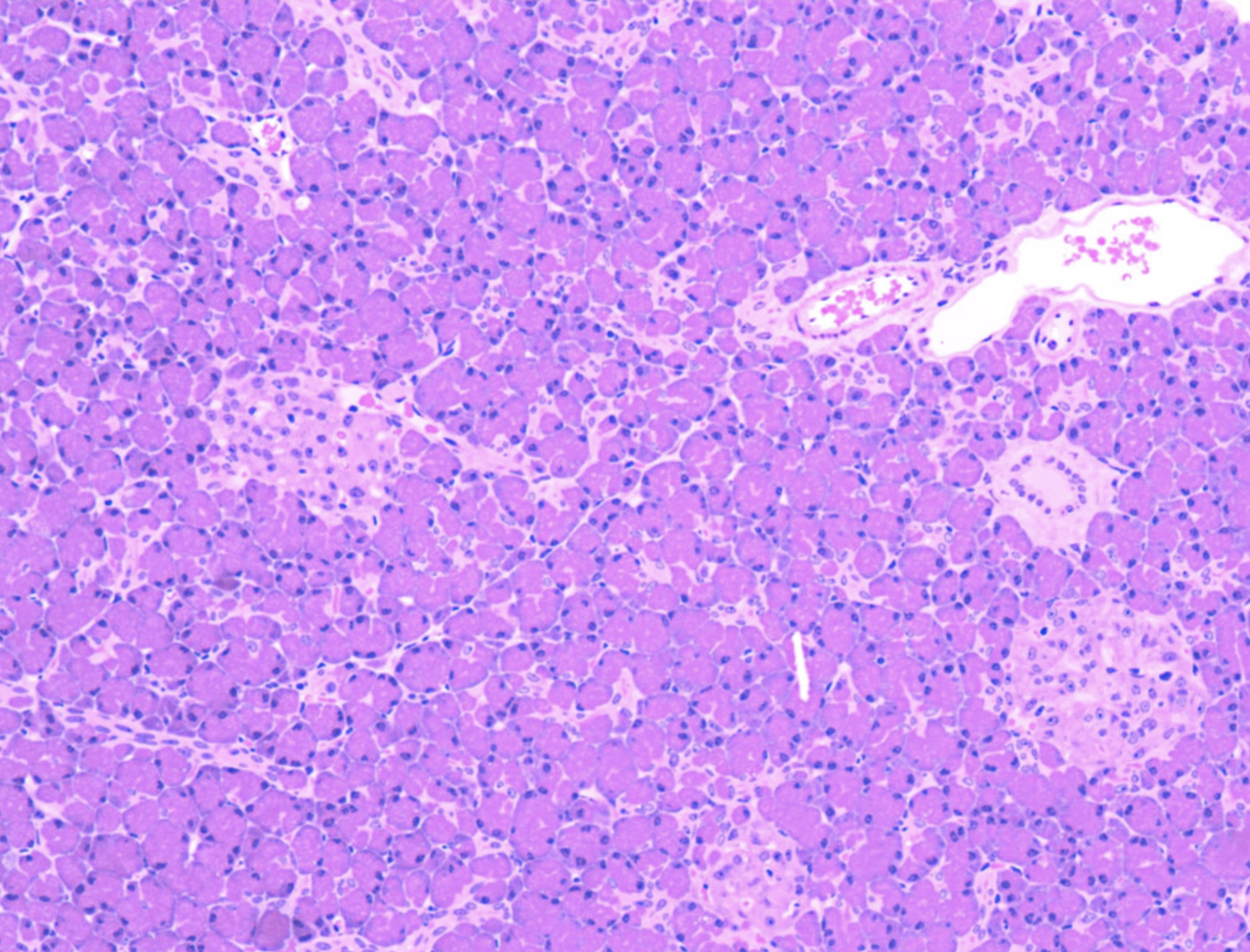
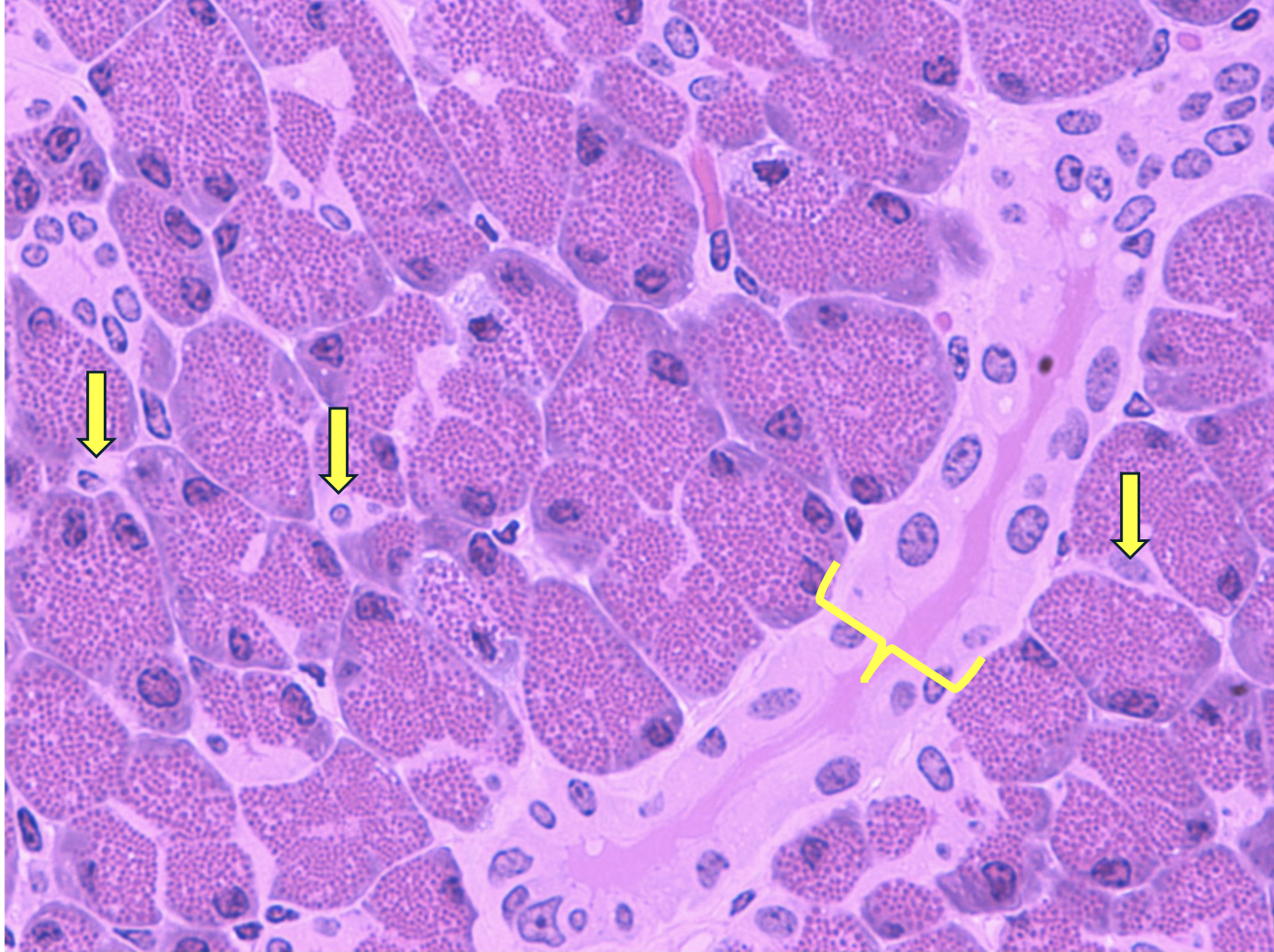
Pancreas
ID the structure
Pancreatic acinar cells
ID the structure. BE SPECIFIC
Parotid gland
ID the structure
Portal triad
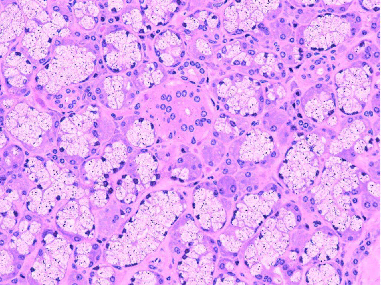
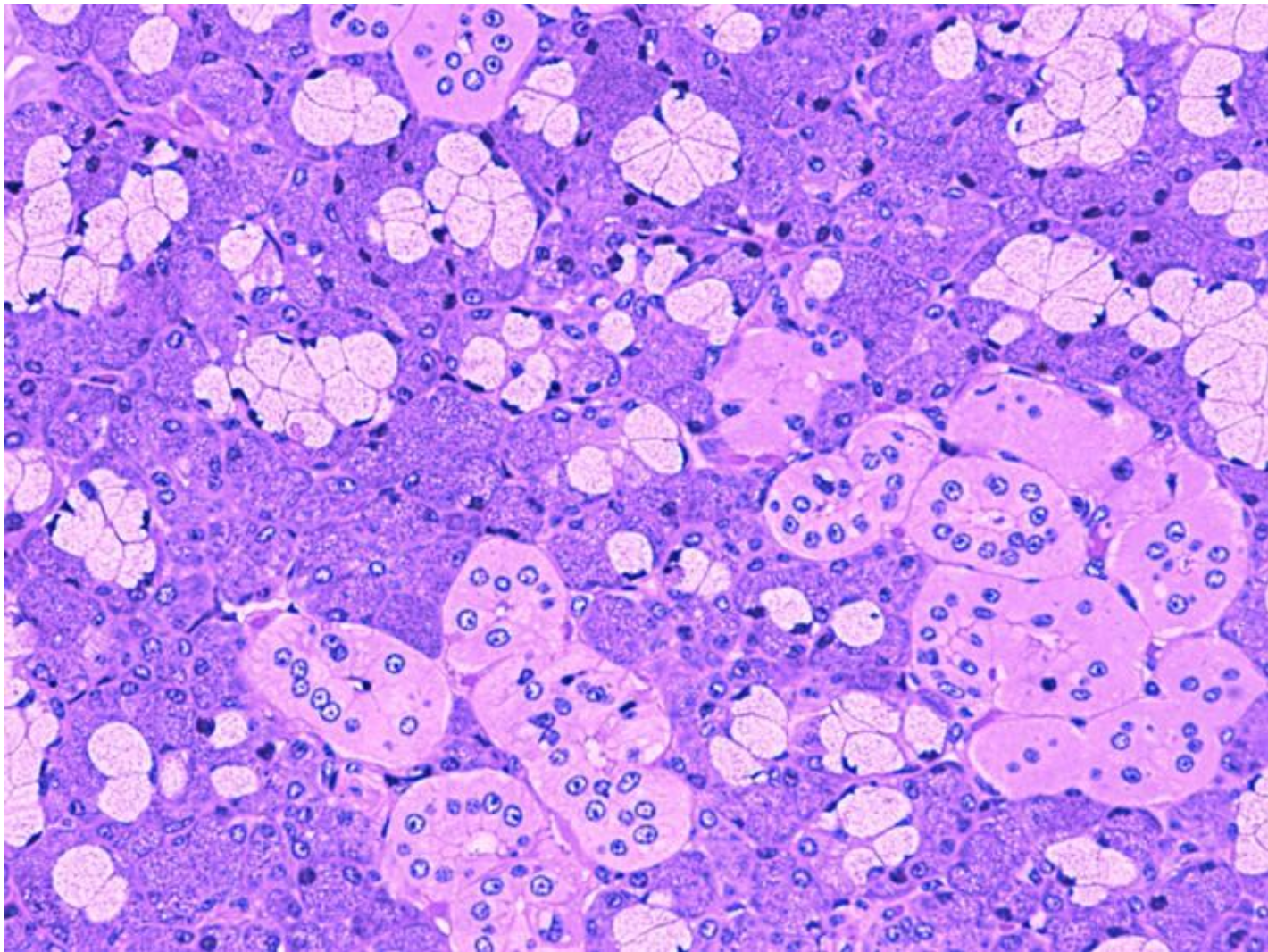
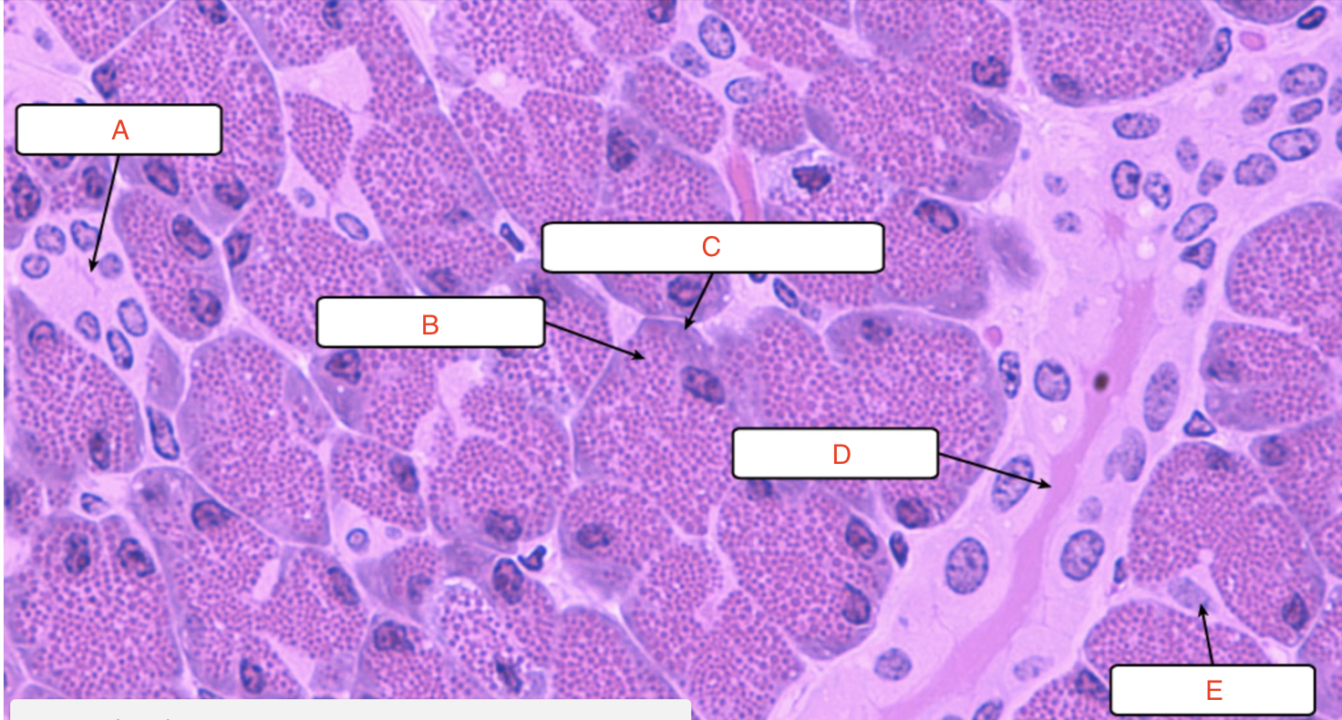
a) acini
b) Islets of Langerhans
c) duct
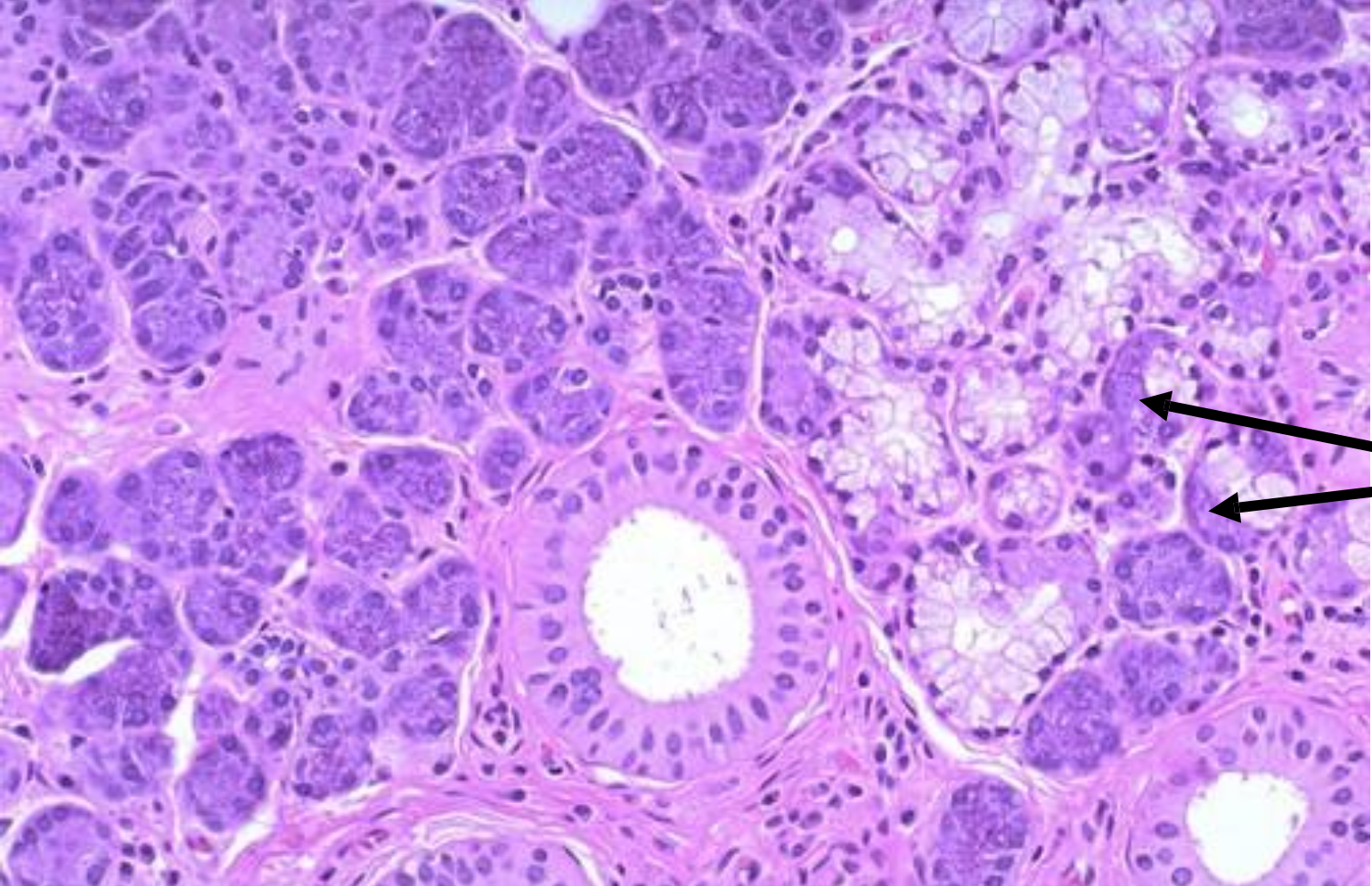
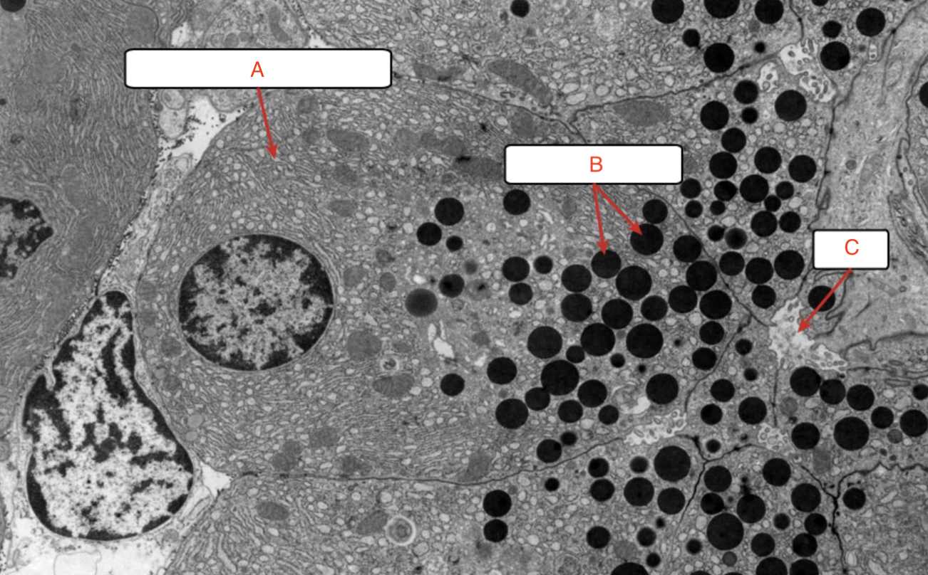
a) intercalated duct
b) zymogen granules
c) rough ER
d) intercalated duct
e) centroacinar cell
a) rough ER
b) zymogen granules
c) lumen

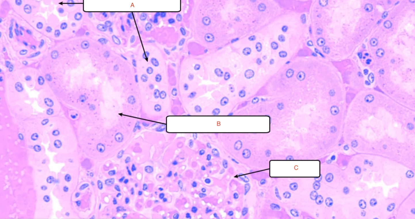
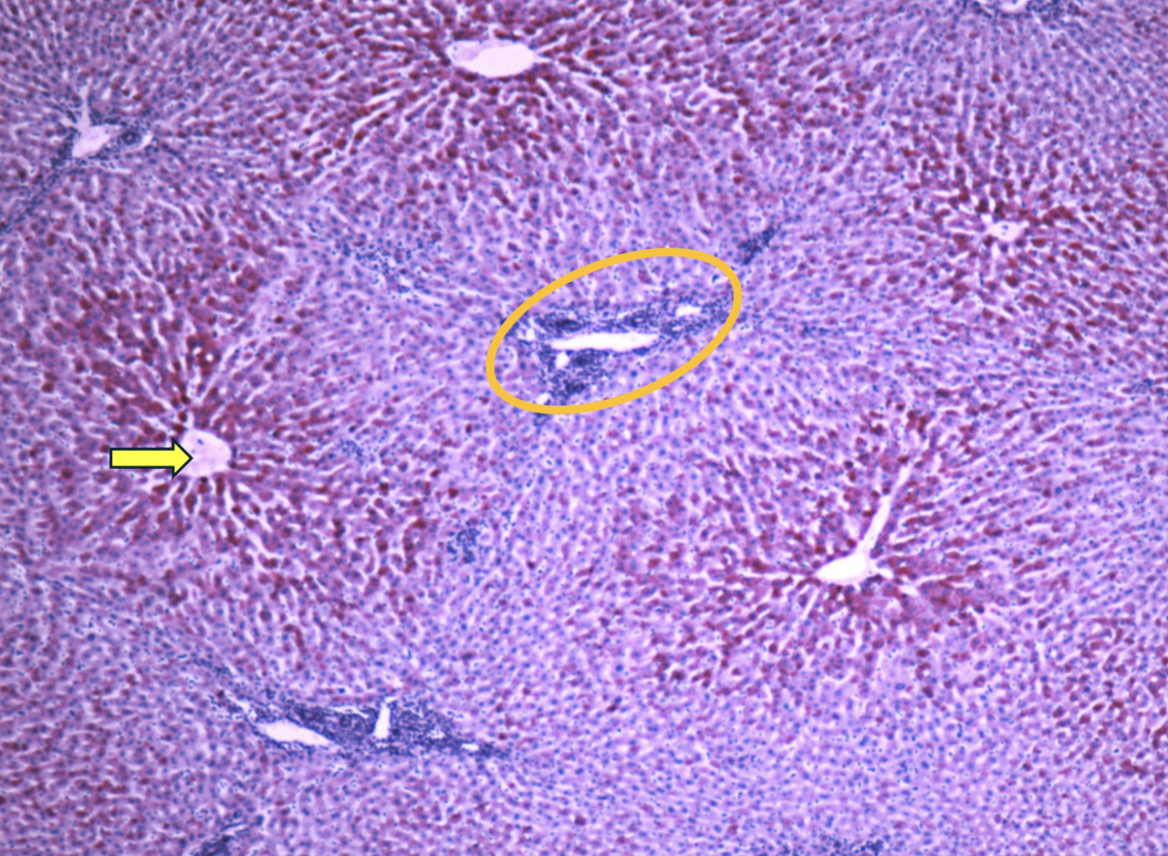
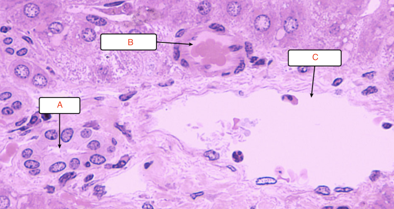
a) hepatic venules
b) zone 3
c) zone 2
d) zone 1
e) portal triad
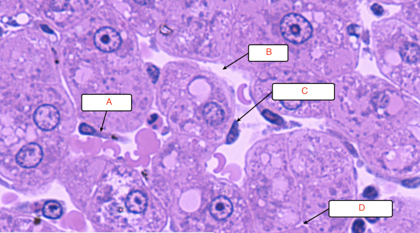
a) bile duct
b) hepatic artery
c) portal venule
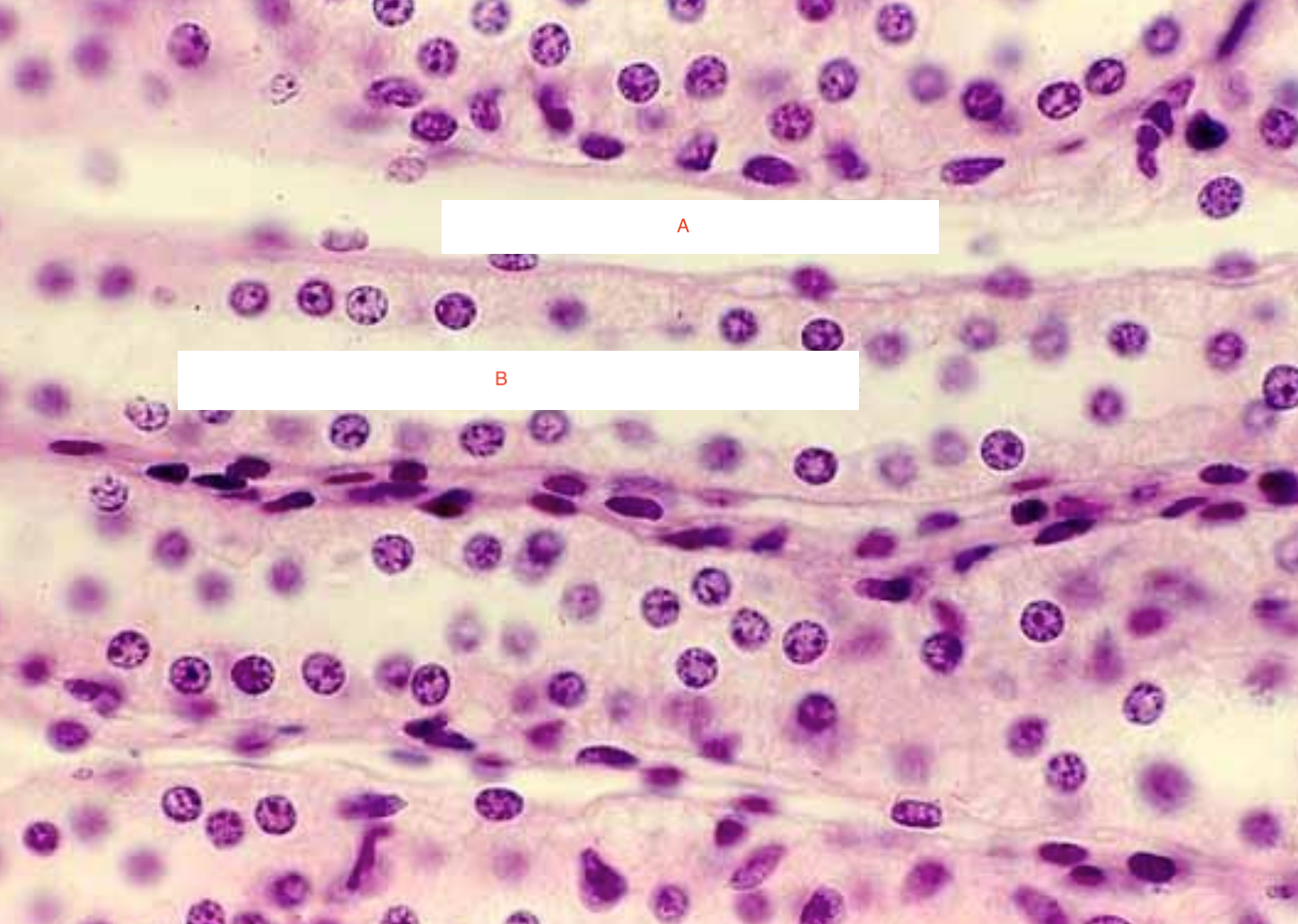
a) Kupffer cell
b) sinusoid
c) endothelial cell
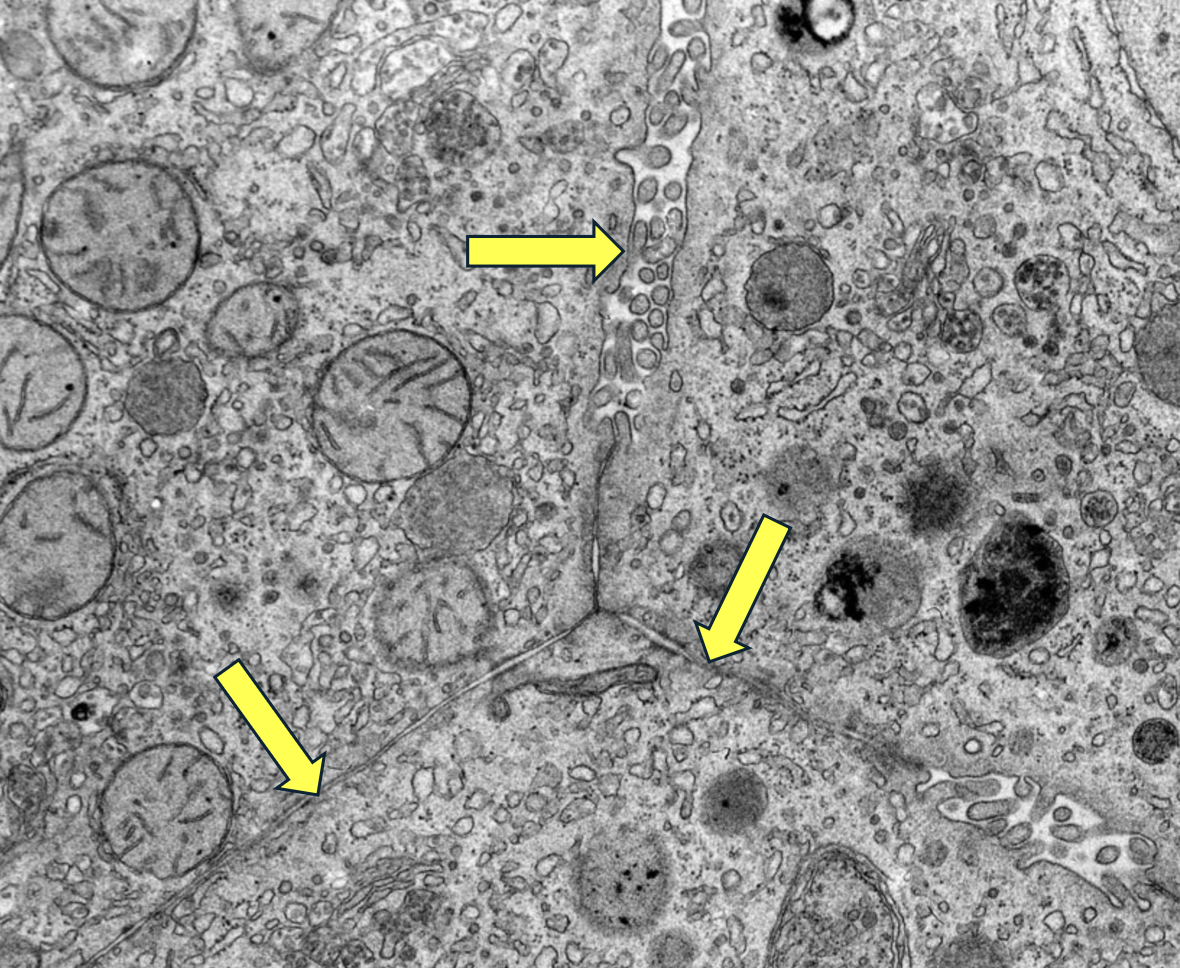
d) bile canaliculus
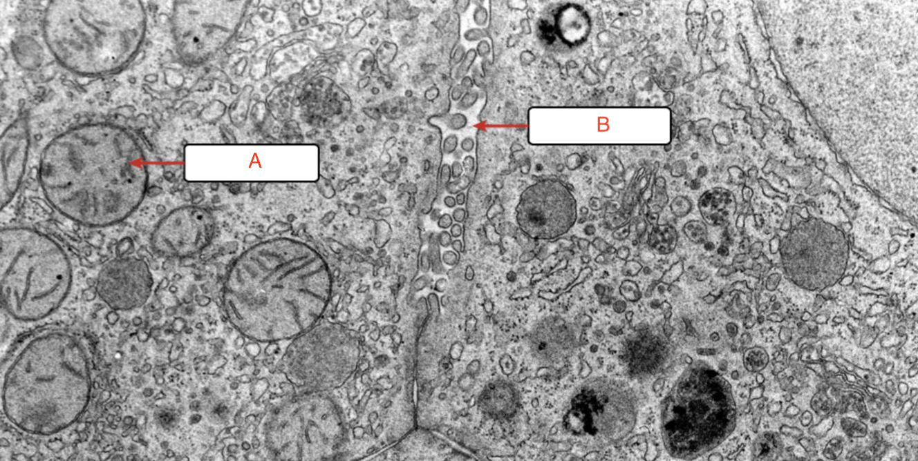
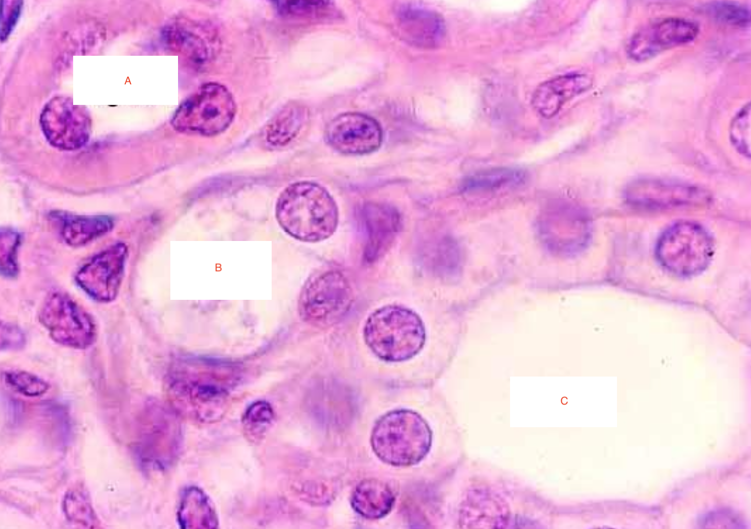
a) mitochondrion
b) bile canaliculus
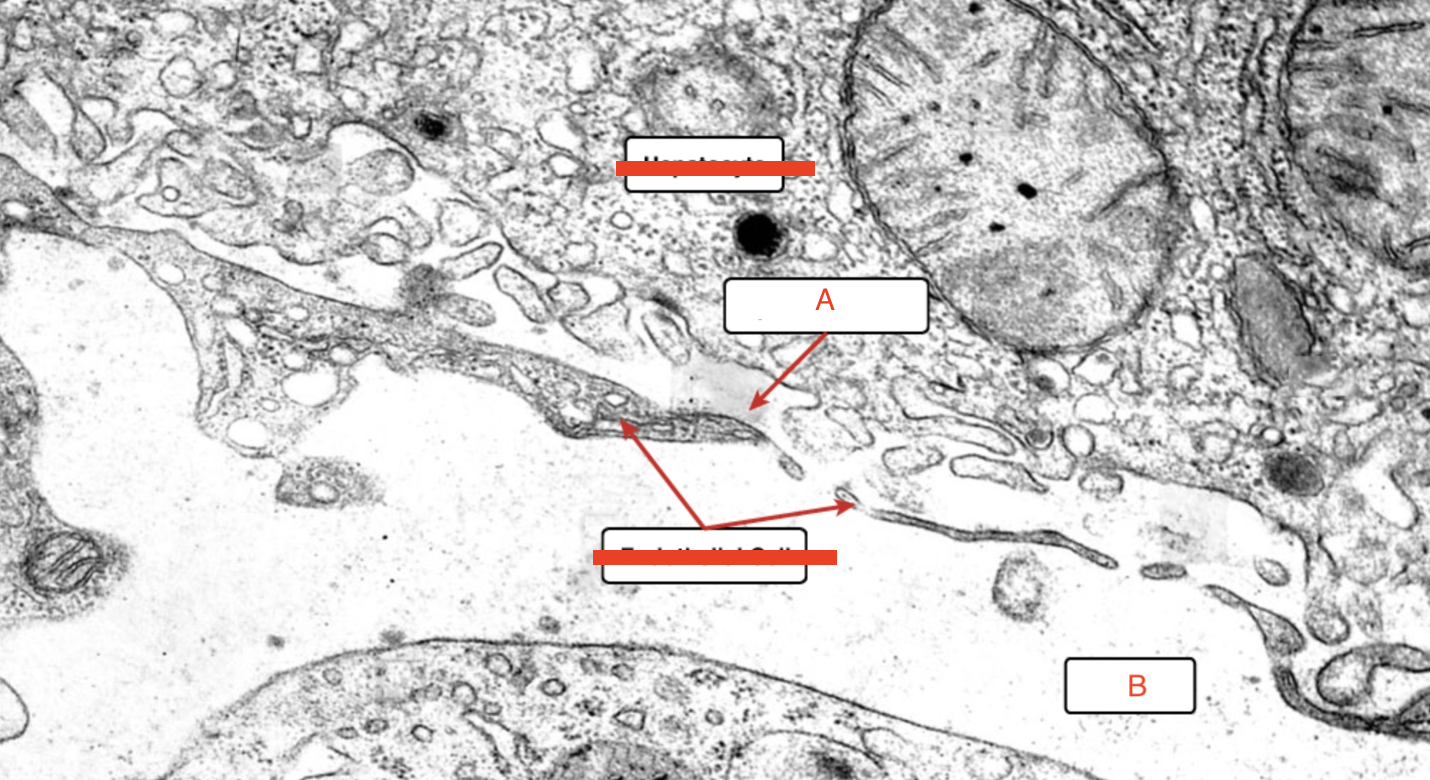
a) Space of Disse
b) sinusoid
a) smooth muscle
b) epithelial cells
c) villus
a) lumen
b) mucosa
c) muscularis
d) adventitia
a) intercalated duct
b) acini
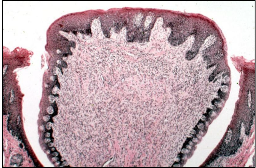
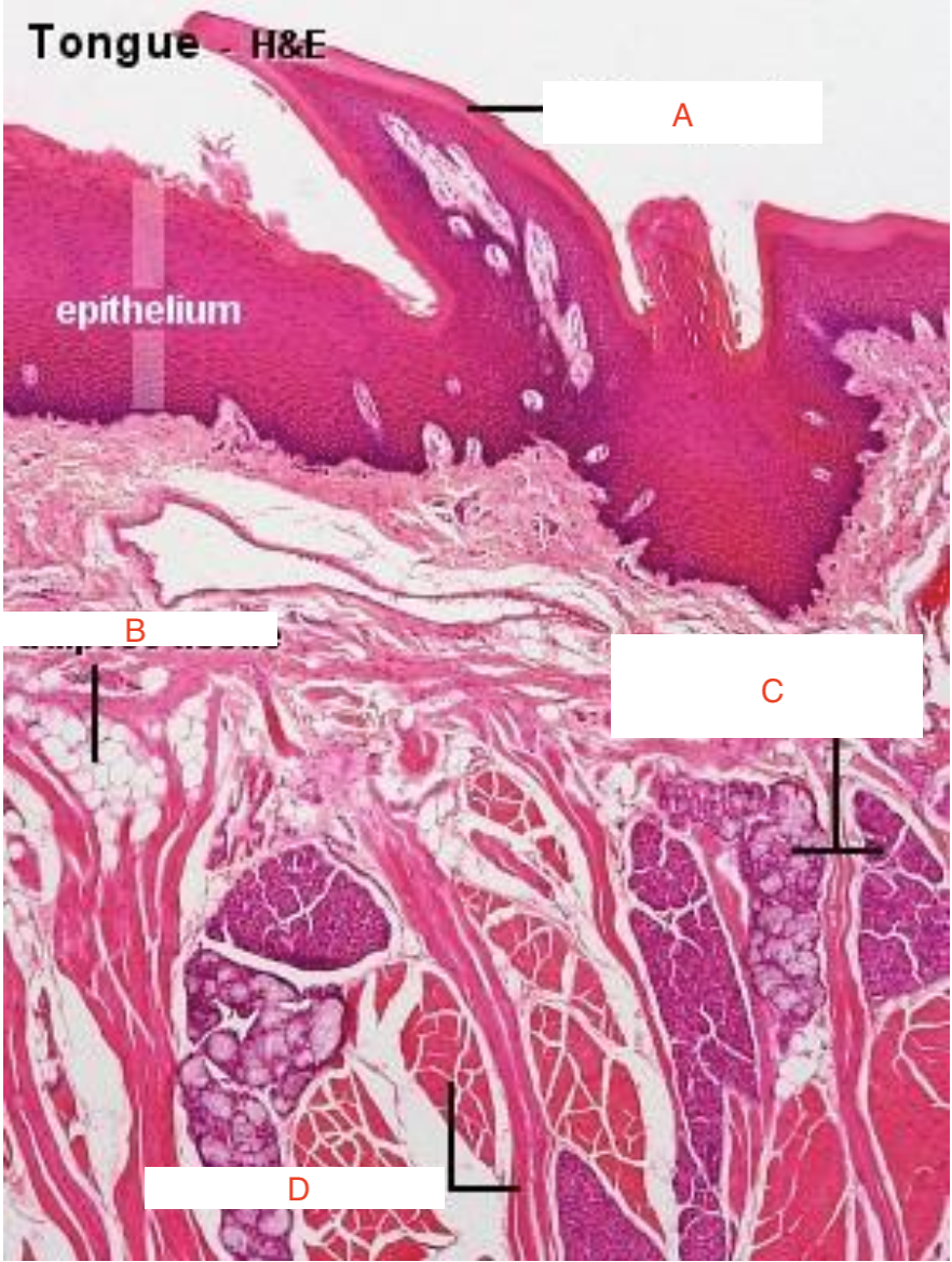
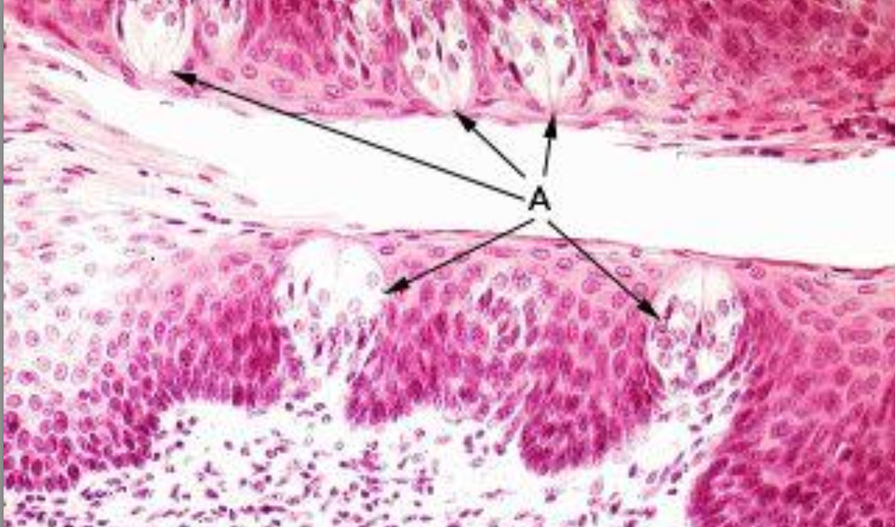
ID A
Circumvallate papillae
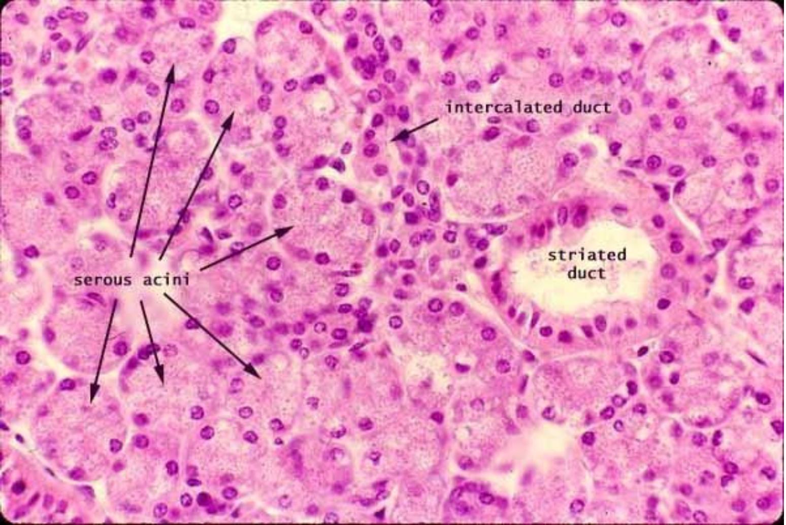
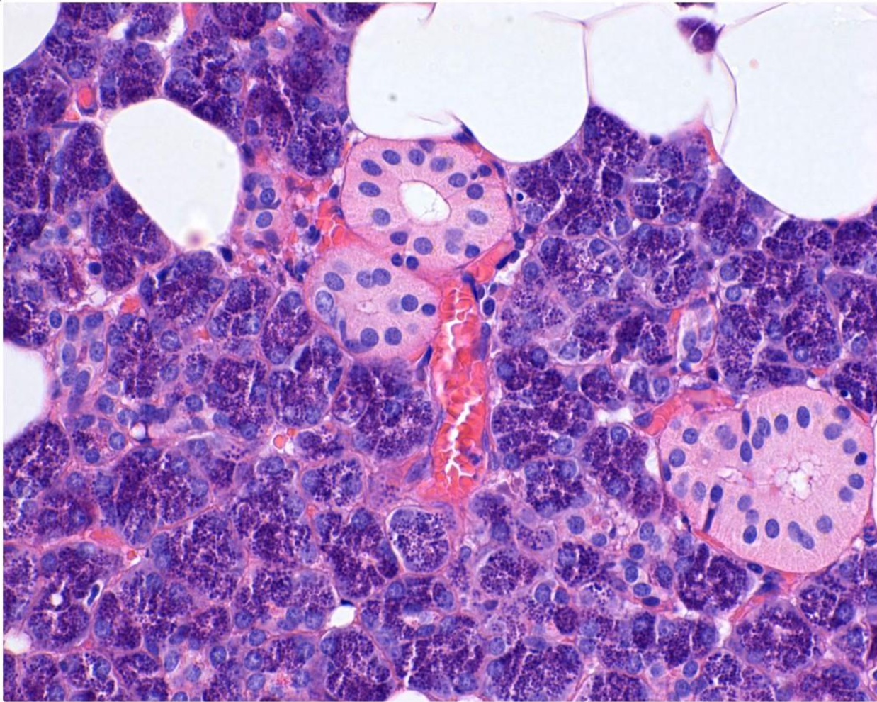
a) intercalated duct
b) striated duct
What are the arrows pointing to?
Parietal cells
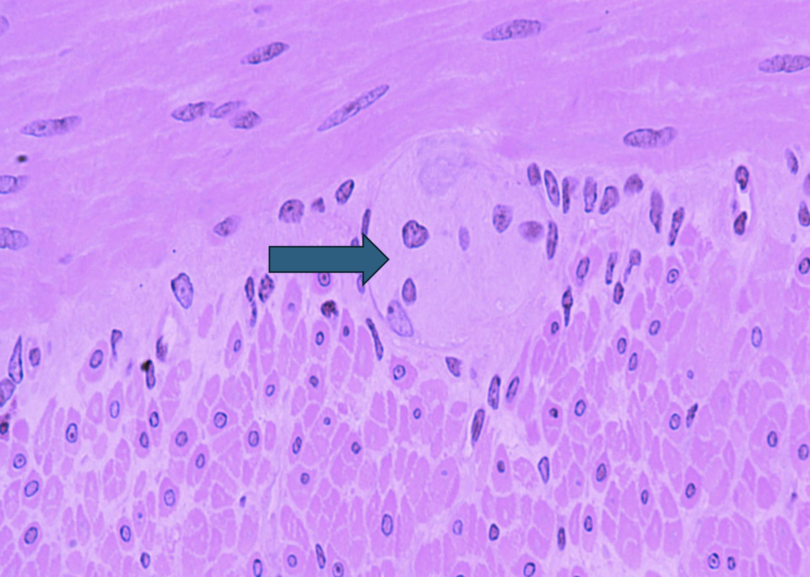
What is the arrow pointing to?
Auerbach plexus
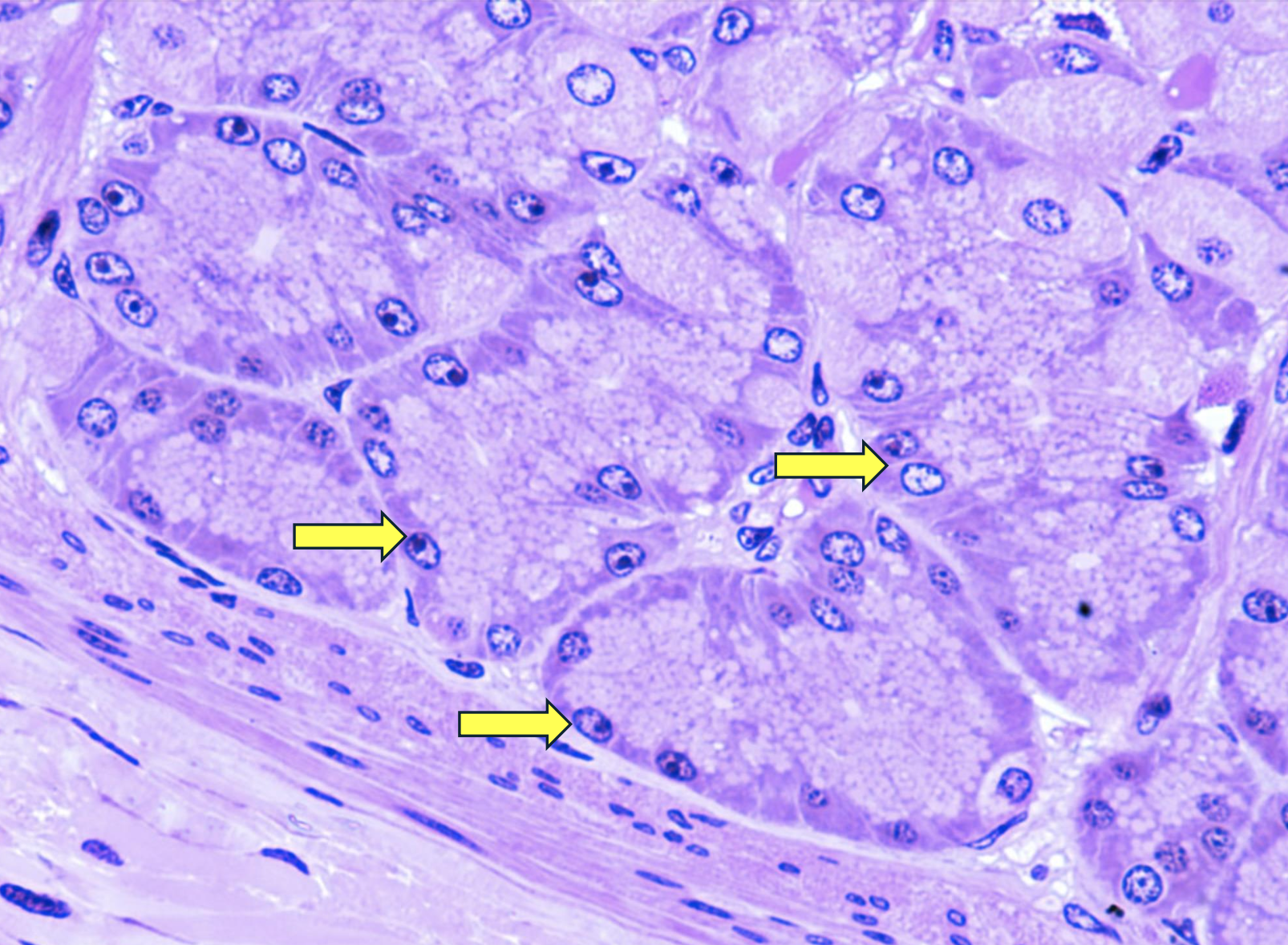
What are the arrows pointing to?
Chief cell
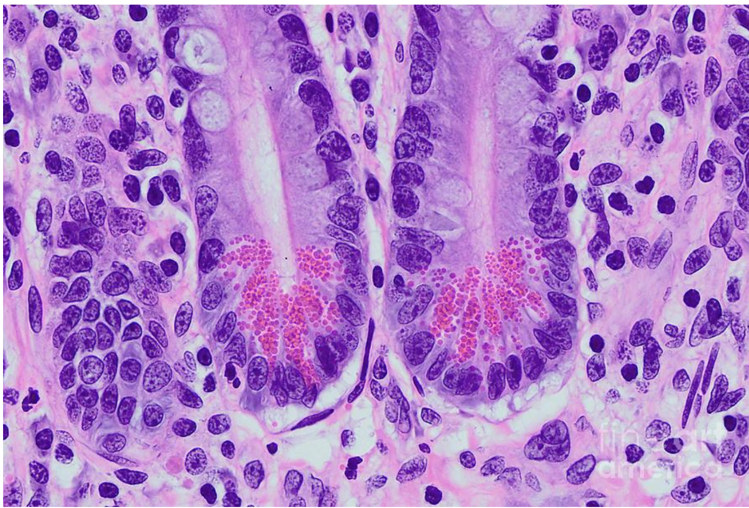
What are the arrows pointing to?
Taste buds
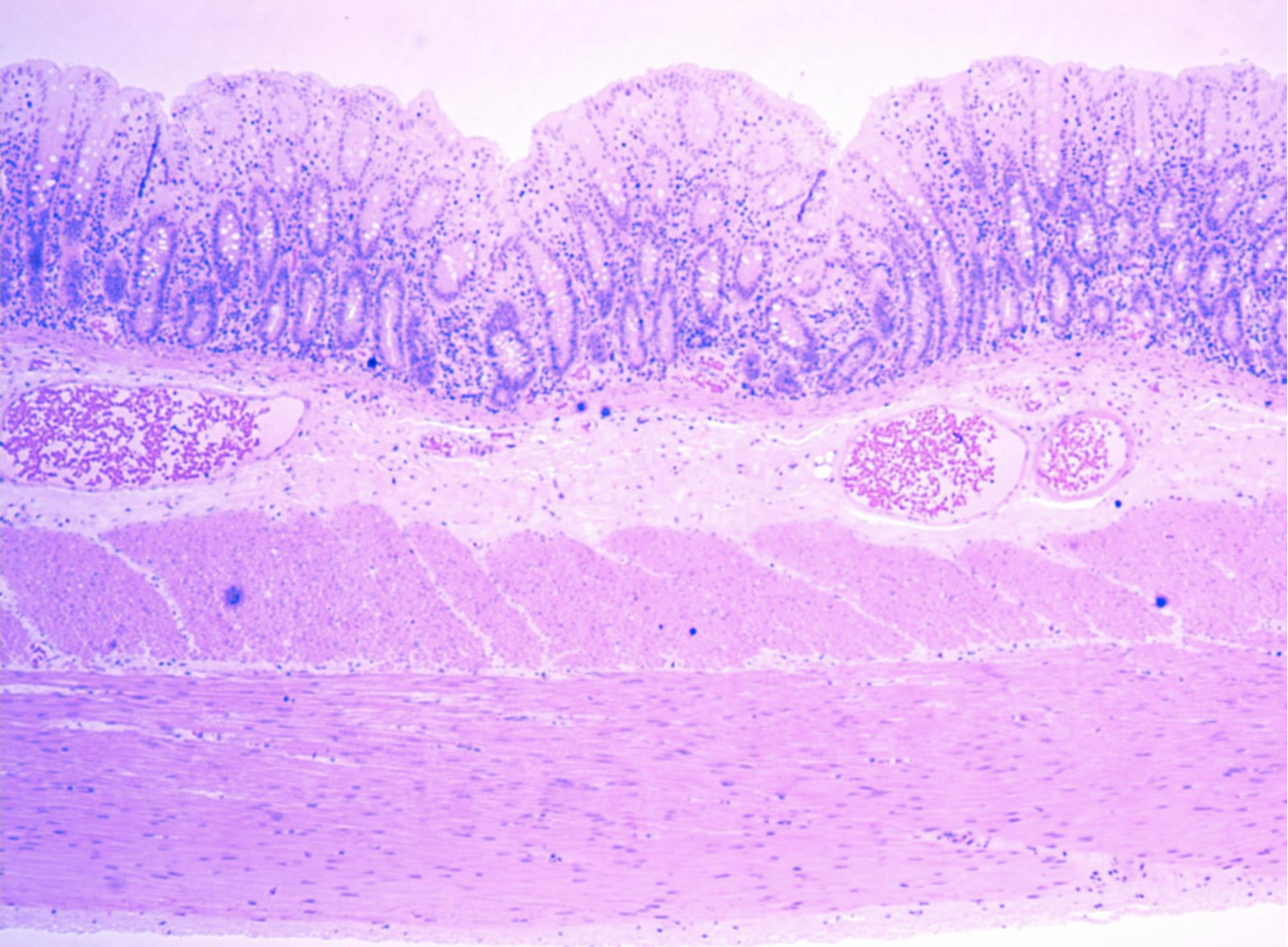
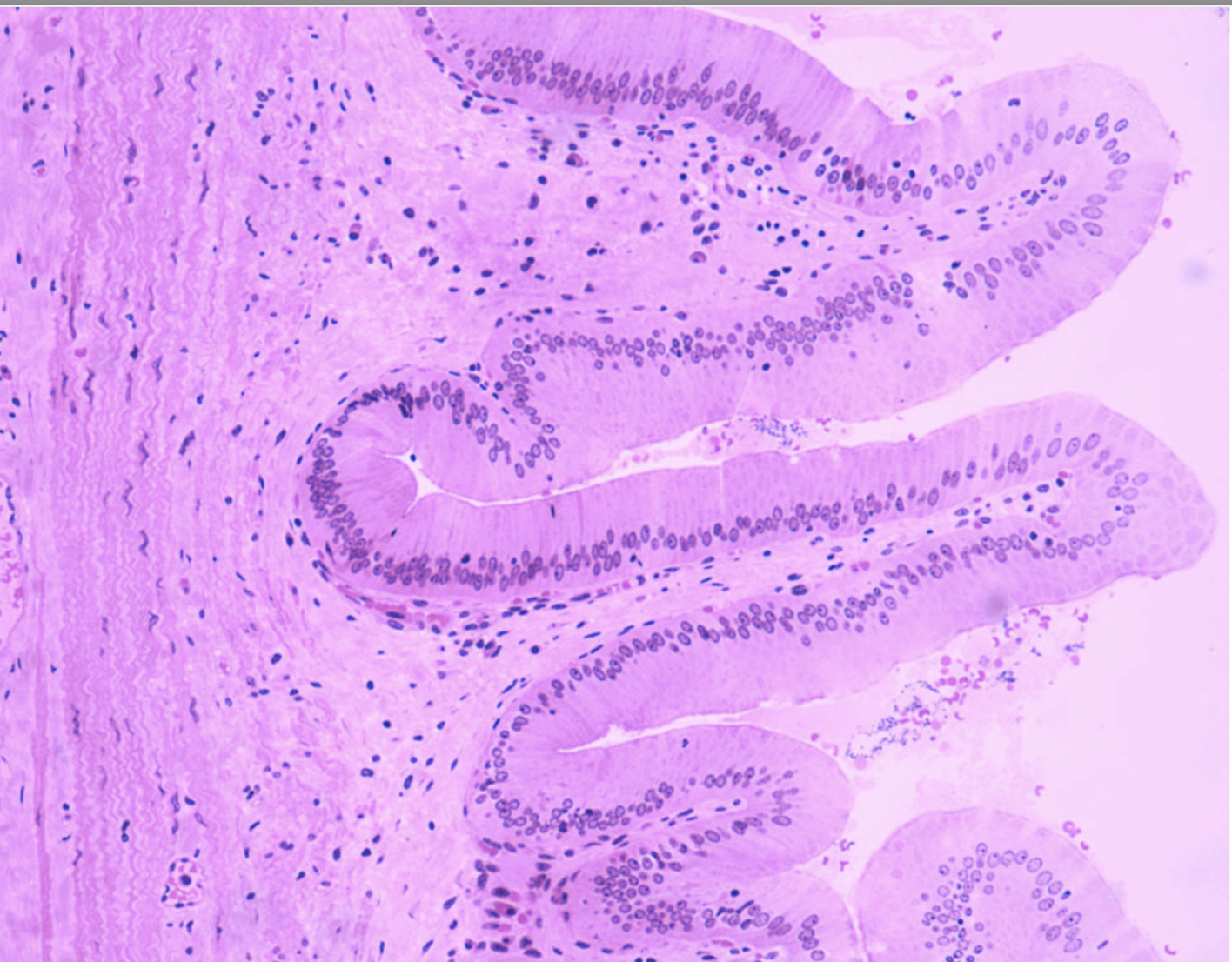
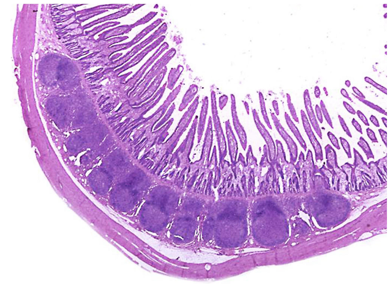
ID the structure
Colon
ID the structure
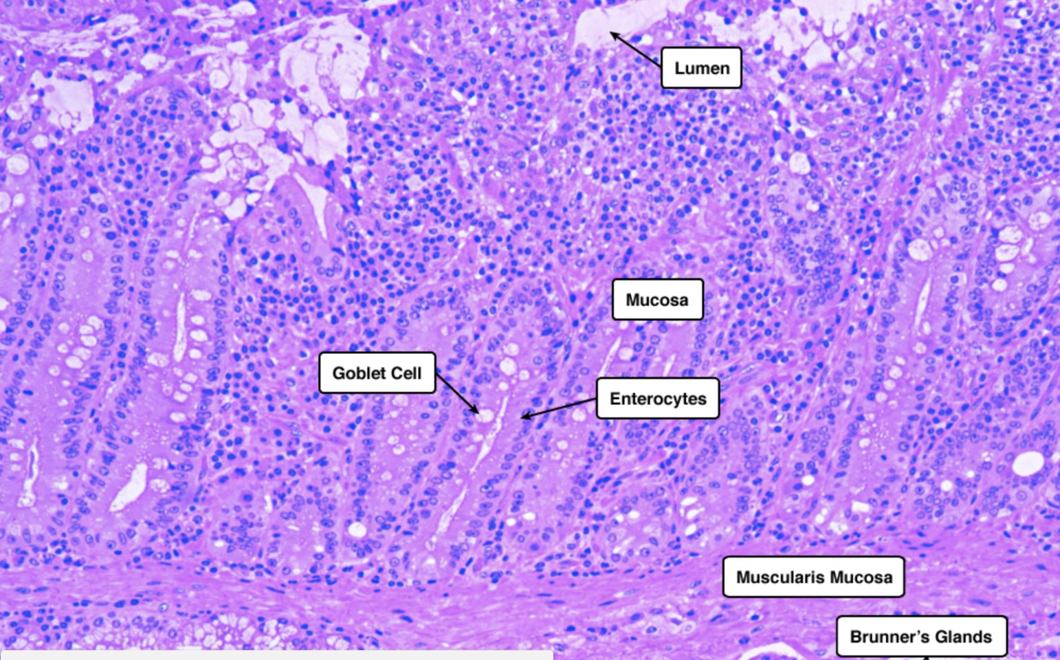
Duodenum
ID the structure
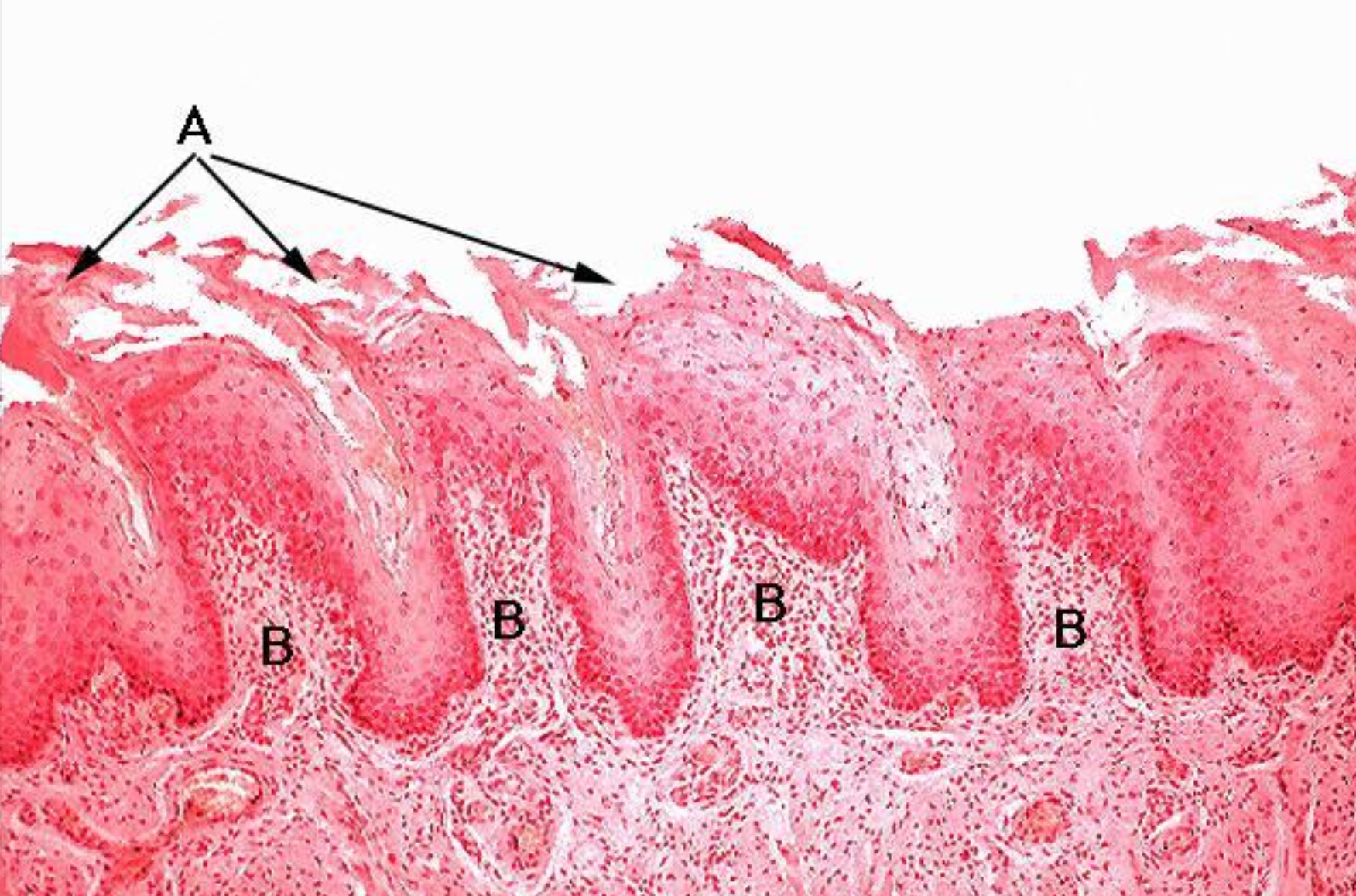
Esophagus
ID the structure. BE SPECIFIC
Filiform papillae
ID the structure. BE SPECIFIC
Foliate papillae
ID the structure. BE SPECIFIC
Gastric gland
ID the structure
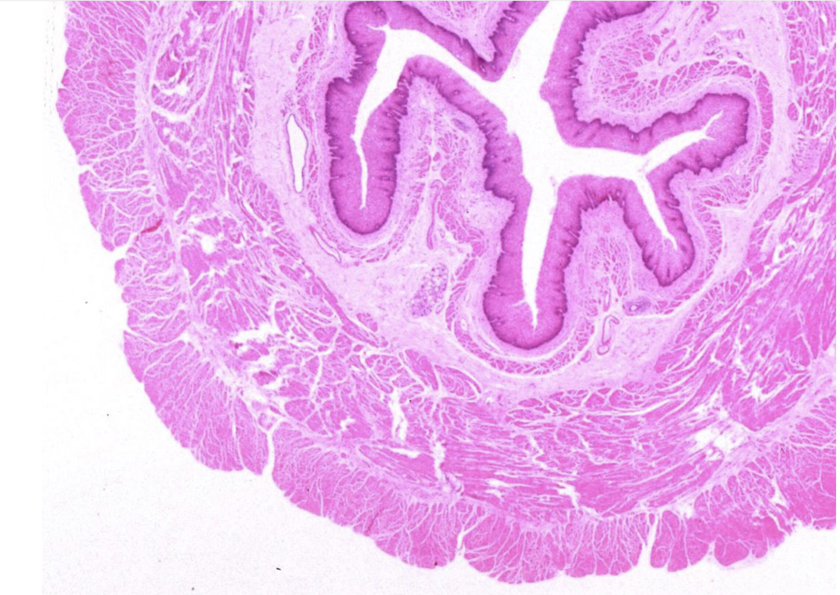
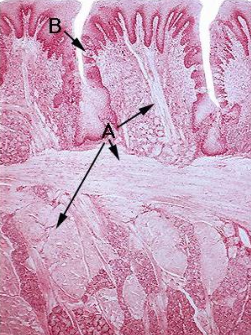
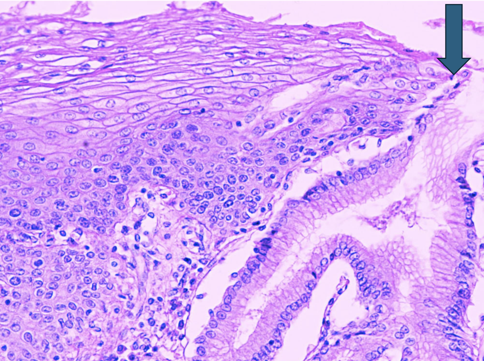
Gasto-esophageal junction
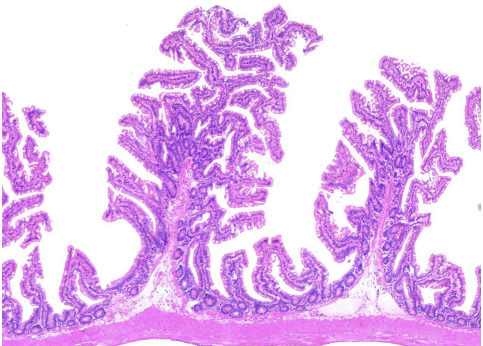
ID the structure
Ileum
ID the structure
Jejunum
ID the structure
Paneth cells
ID the structure
Salivary glands
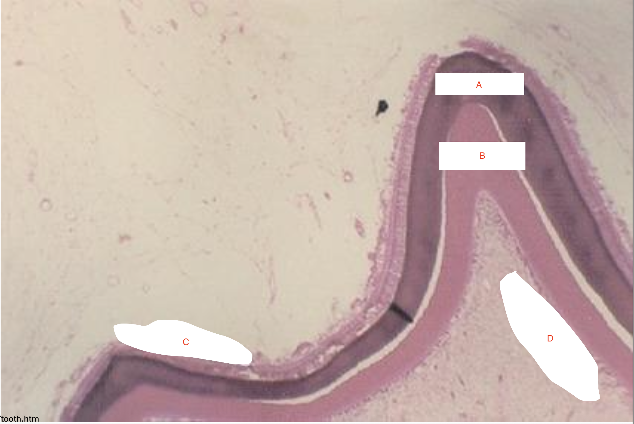
a) Enamel
b) dentin
c) ameloblasts
d) odontoblasts