1b- functional anatomy and biomechanics
1/20
There's no tags or description
Looks like no tags are added yet.
Name | Mastery | Learn | Test | Matching | Spaced | Call with Kai |
|---|
No analytics yet
Send a link to your students to track their progress
21 Terms
What is the TMJ?
Area connecting jawbone to skull
Mandibular condyle articulates in base of skull through temporal bone
Fibrocartilage surface- greater repair capacity than hyaline cartilage
Function of ginglymoid?
Hinge movement- rotation in inferior compartment
Function of arthrodial?
Gliding movement (translation) in superior compartment
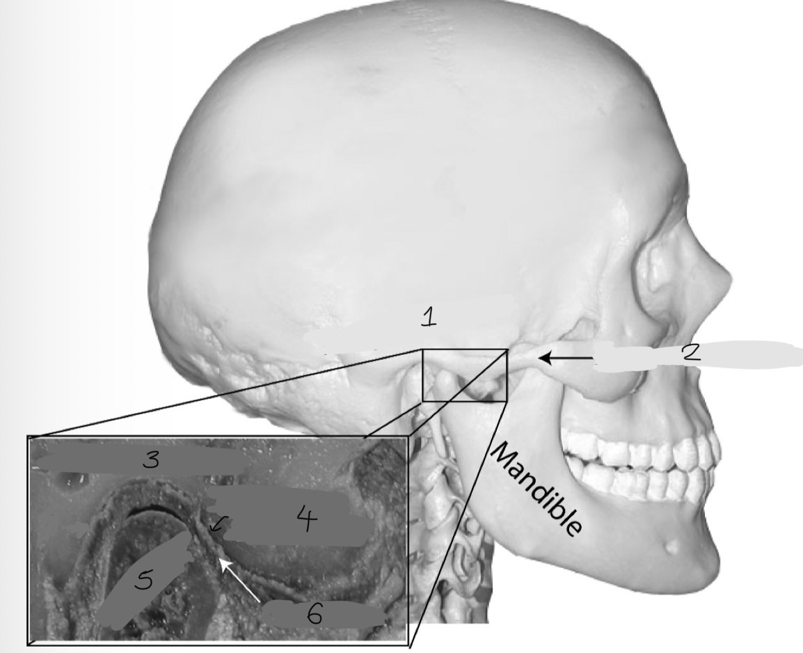
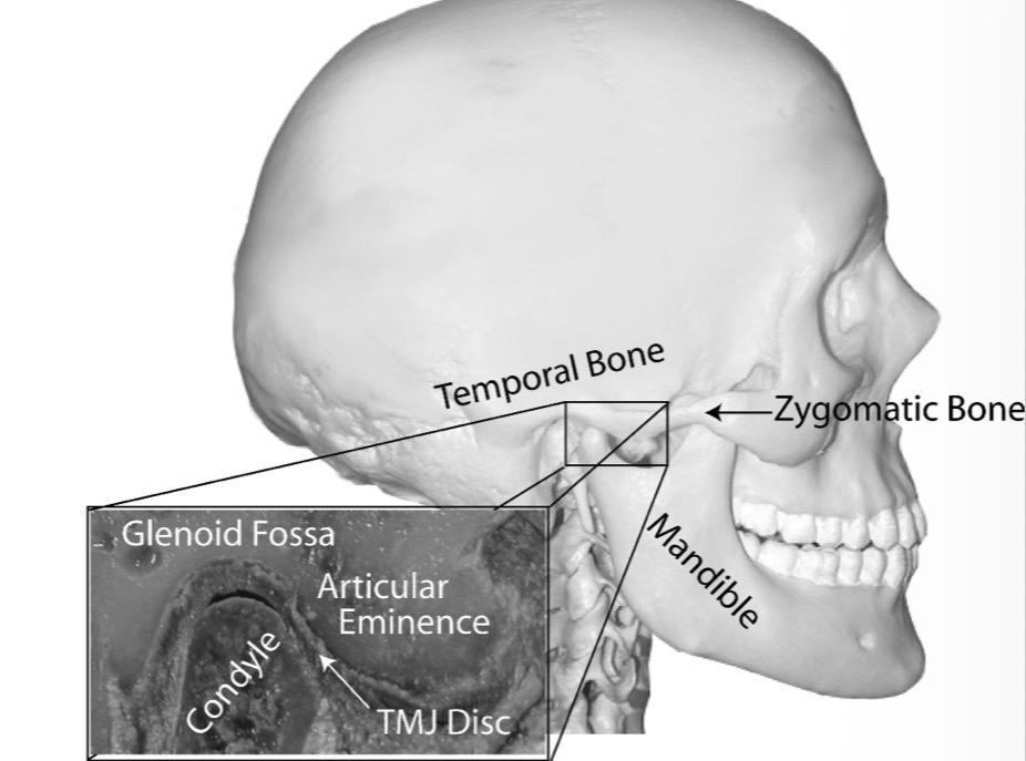
Label this
Temporal bone
Zygomatic bone
Glenoid/Mandibular fossa
Articular eminence
Condyle
Disc
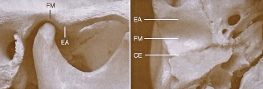
What is the glenoid fossa?
Area formed by concave mandibular fossa where condyle is situated
Thin roof
What is the articular eminence?(4)
Protruding dense convex bone in front of the fossa
Varying level of convexity
Guides movement of condyle, primary load bearing area
Ginglymoarthrodial joint- Ginglymoid (hinging) AND arthrodial (sliding joint)
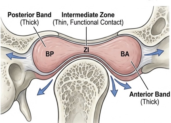
What is the articular disc made of? its structure and mechanism?
Dense fibrous ct
Intermediate zone is its thinnest points, functional contact between condyle and eminence
Has self centring mechanism- inter articular pressure aligns the thin zone with condyle
Thick posterior band prevents displacement
What are the dynamics of synovial lubrication?
Weeping lubrication- Fluid expelled from tissues under load- metabolic exchange
Fluid moves to surface during movement- reduces friction
1.2ml vol- only nutrient source for avascular disc
Clenching (static loading) exhausts weeping lubrication- leads to hypoxia
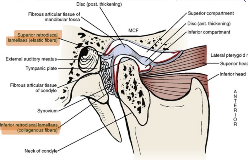
Describe the parts of the bilaminar zone/retrodiscal tissue
Superior retrodiscal lamina- elastic fibres, opposes forward translation
Inferior- collagenous (nonelastic), limits rotation
Venous plexus- rapid volumetric filling during condylar translation
Rich in nociceptors
If compressed here- causes intracapsular pain- retrodiscitis
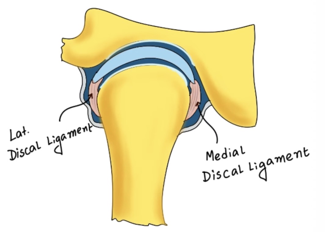
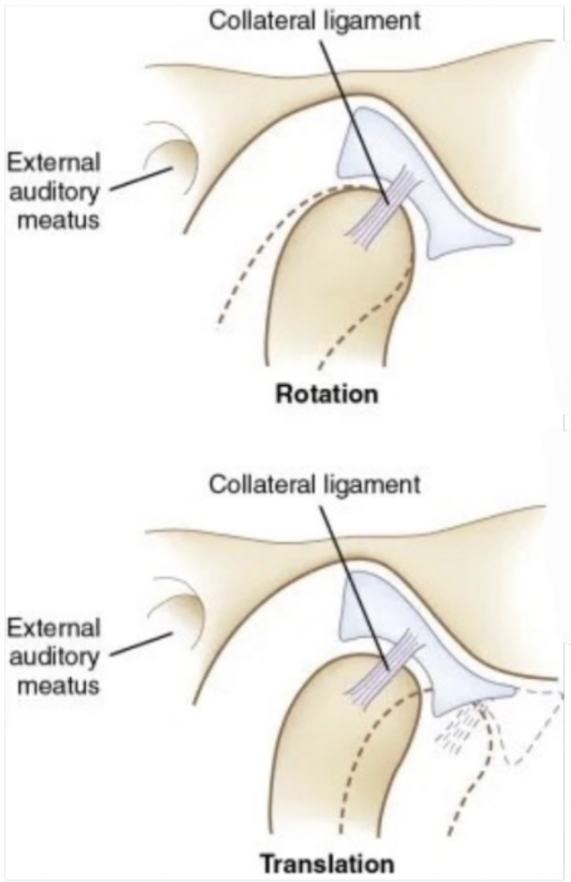
Function of collateral (discal) ligaments?
Passive restriction- they elongate or tear but don’t stretch
Anchor disc to condyle poles
Allows disc to rotate with condyle
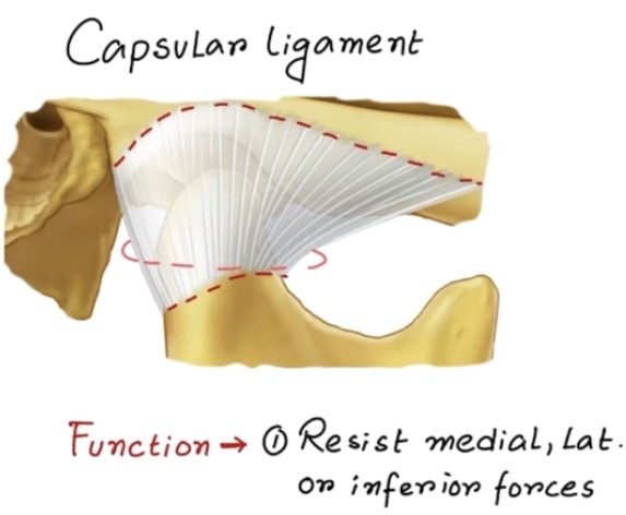
Function of capsular ligament
Retains synovial fluid and resist separation forces
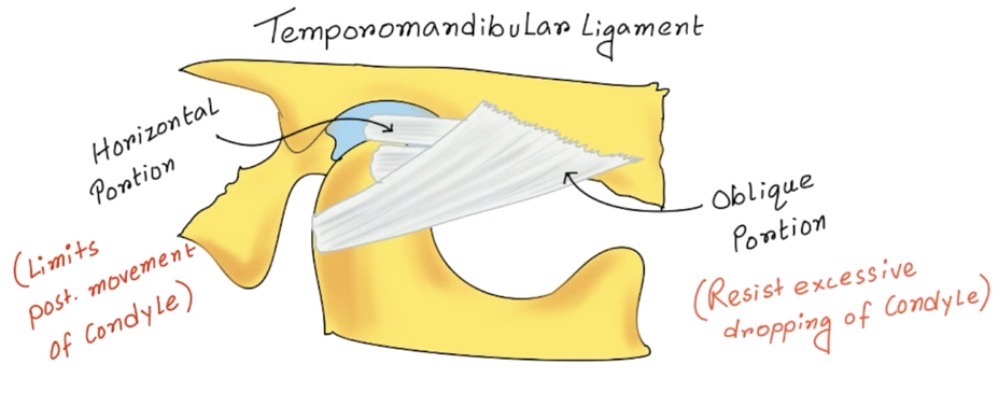
What is the temporomandibular ligament made of and its effect?
Outer oblique portion- limits pure rotational opening to 20-25mm
This forces condyle to translate after initial rotation (move down eminence)
Inner horizontal portion- limits posterior movement- protects retrodiscal tissue
This prevents compression of vital neck structures during opening
What are the jaw elevator muscles?
Temporal
Masseter
Pterygoid
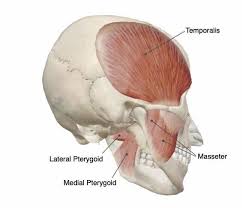
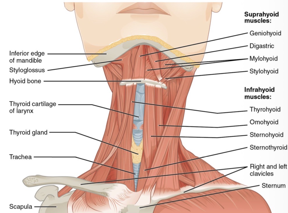
What are the jaw depressor (opening) muscles?
External or lateral inferior pterygoid muscle
Suprahyoid- digastric, stylohyoid, mylohyoid, geniohyoid
Infrahyoid
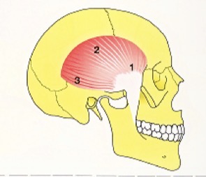
Structure and function of temporal muscle
Fan shaped
Anterior, medial, posterior portion
Contraction of anterior portion elevates jaw , medial and posterior elevate and retract
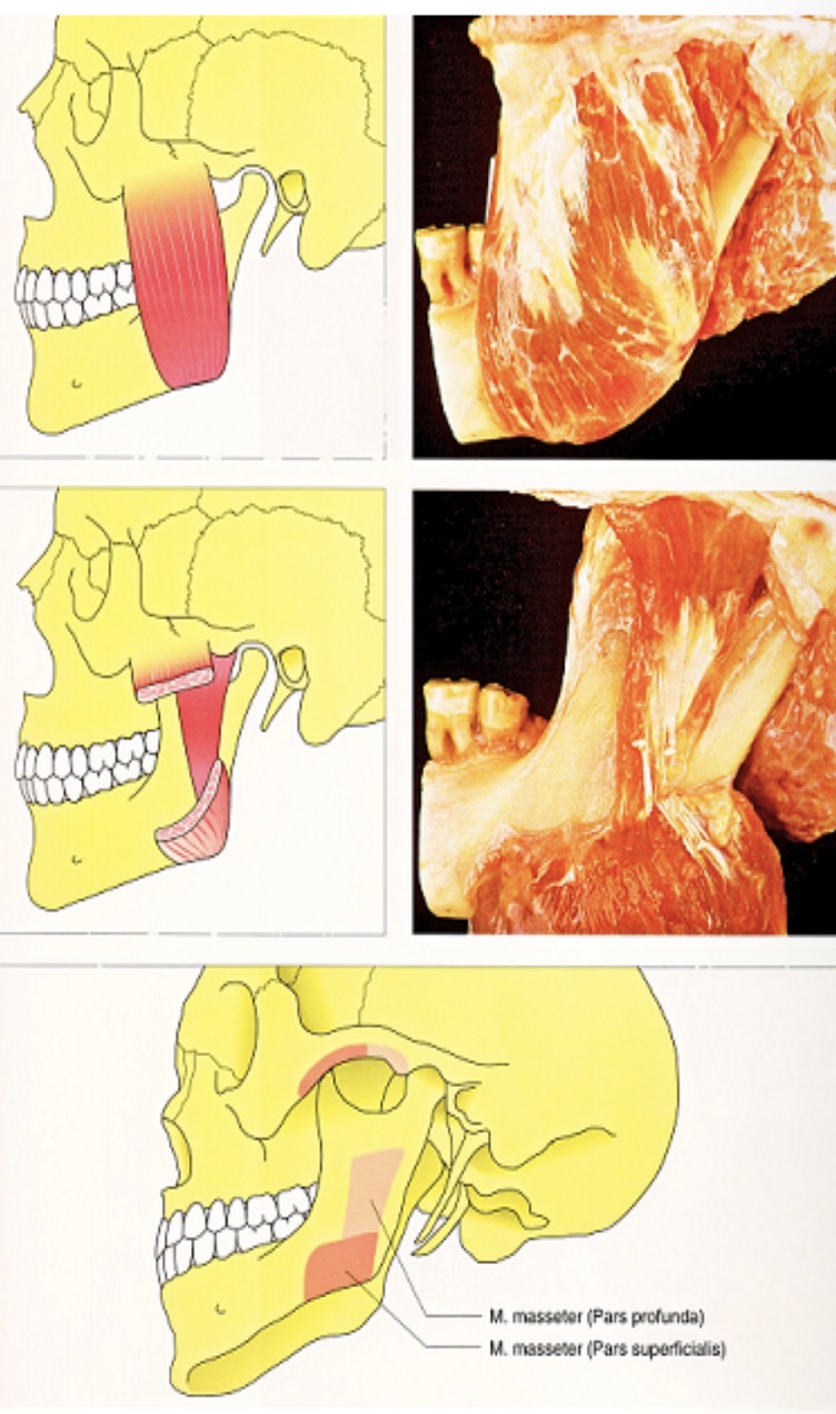
Structure of masseter muscle- where, insertion, portions
Rectangular muscle that goes from zygomatic arch to inferior edge of mandible
Insertion in jaw from 2nd molar at lower edge to angle
Superficial (for protrusion) and deep (retrusion) portions
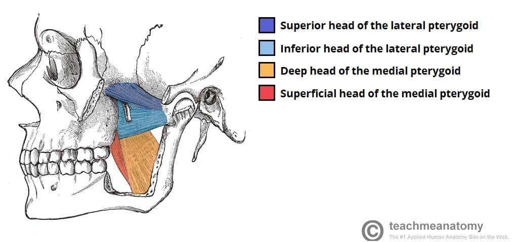
Explain the lateral pterygoid paradox
Inferior head is active during opening- pulls condyles forward for translation
Superior head active during closing- stabilises disc against eminence during biting
Incoordination causes disc displacement
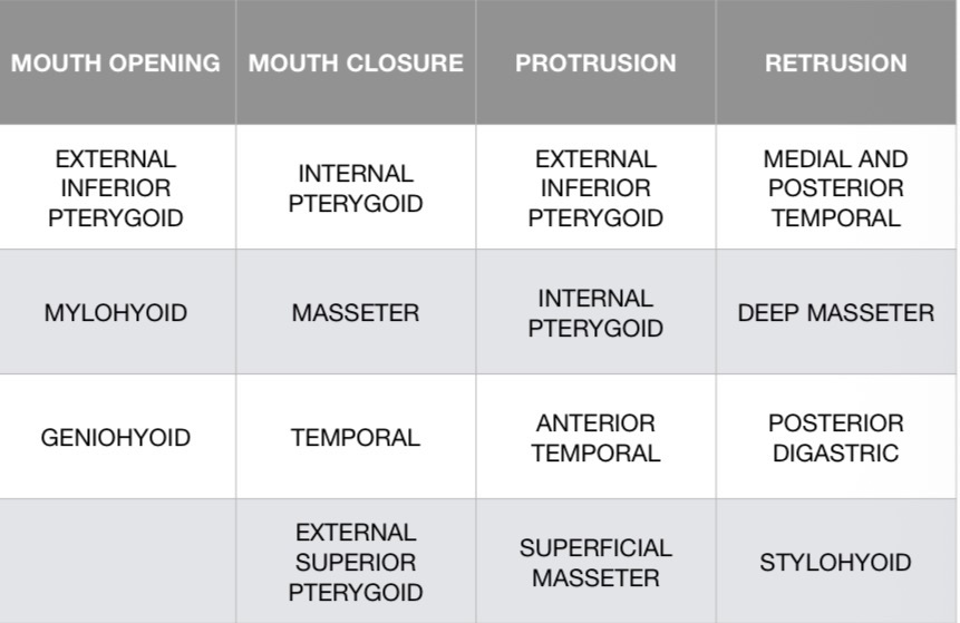
What muscles are responsible for- mouth opening, mouth closure, protrusion, retrusion?
What is the rotation phase constrained by? Its dynamic?
Limited by tightening of TML outer oblique portion
Condyle rotate, disc remains superiorly stable
What is translation phase triggered by and stopped by?
TML tightens forcing condyle to translate
Elastic superior retrodiscal lamina stretches to control movement and recoil
What is the criteria for CR (orthopedic stability)?
Most superior posterior position
Loading on intermediate zone
Elevator muscles released, minimal tonicity in resting position
MIC coincides with this joint position