AQA A Level Biology - Cell Structure
1/68
There's no tags or description
Looks like no tags are added yet.
Name | Mastery | Learn | Test | Matching | Spaced | Call with Kai |
|---|
No analytics yet
Send a link to your students to track their progress
69 Terms
List all the organelles found in all eukaryotic cells.
centrioles
rough endoplasmic reticulum
smooth endoplasmic reticulum
nucleus
ribosomes
cell-surface membrane
lysosome
golgi apparatus + vesicules
mitochondria
which organelles do only plant cells contain?
chloroplasts
cell wall
permanent vacuole

what is organelle 1?
chloroplast

what is organelle 2?
cell wall

what is organelle 3?
cell membrane

what is organelle 4?
cytoplasm

what is organelle 5?
permanent vacuole
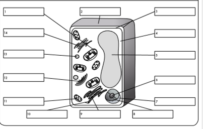
what is organelle 6?
nucleolus

what is organelle 7?
nuclear envelope

what is organelle 8?
nucleoplasm

what is organelle 9?
rough endoplasmic reticulum

what is organelle 10?
ribosome

what is organelle 11?
mitochondrion

what is organelle 12?
golgi body

what is organelle 13?
lysosome

what is organelle 14?
smooth endoplasmic reticulum
describe the structure of the Nucleus
it is a membrane bound organelle, containing a nucleolus and chromatin (DNA stored in the nucleus in an uncoiled form), which are contained in the nucleoplasm
it has a double membrane called the nuclear envelope which has small holes in called the nuclear pores
the outer nuclear membrane is continuous with RER membranes
largest organelle
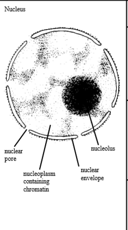
describe the function of the nucleus
envelope encloses and protects DNA
nuclear pores allow entry and exit of substances such as nucleotides and mRNA
chromatin condenses to form chromosomes for cell division, produces semi-complete ribosomes, coenzymes, nucleotides, proteins
describe the structure of the mitochondria
double membrane
inner membrane folded to form cristae
the space in between the cristae is called the matrix and is filled with fluid
the mitochondrion contains ribosomes (70S, smaller) , DNA and enzymes
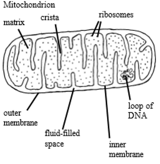
describe the function of the mitochondria
site of aerobic respiration, release energy and synthesise ATP
enzymes in matrix catalyse reactions
cristae hold these enzymes in place
produce some of the proteins they require (ribosomes)
cristae also increase surface area for aerobic respiration + metabolic reactions
describe the structure of the rough endoplasmic reticulum
system of hollow tubes and sacs, that are interconnected and flattened
covered with ribosomes
channels are called cisternae
describe the function of the rough endoplasmic reticulum
folds and processes proteins that have been made at the ribosomes
cavities of RER allow for transport of proteins
describe the structure of the smooth endoplasmic reticulum
system of hollow tubes and sacs, that are interconnected and flattened
describe the function of the smooth endoplasmic reticulum
synthesises and processes lipids
modifies substances such as steroid hormones
describe the structure of the golgi body
flattened cisternae - fluid filled membrane bound cavities/sacs - which are stacked on top of each other
connected to RER
has vesicles at edge
describe the function of the golgi body
processes and packages new lipids and proteins - modifies them before secretion (proteins)
makes lysosomes
cells that secrete a lot of enzymes contain a lot of RER and golgi apparatus
describe the structure of the golgi vesicles
small fluid filled sacs in the cytoplasm surrounded by a membrane
stores lipids and proteins made by the golgi apparatus and transports them out of the cell
describe the structure of the ribosomes
made up of a small subunit and a large subunit
made of rRNA and protein
float free in the cytoplasm or are attached to the RER
describe the function of ribosomes
site of protein synthesis and thus function relates to the steps of this
describe the structure of the lysosomes
vesicles that contain hydrolytic enzymes/digestive enzymes
type of golgi vesicle
membrane-bound
describe the function of the lysosomes
contains digestive enzymes called lysozymes - kept separate from cytoplasm by the membrane
can be used to digest cellular waste or invading cells
describe the structure of the chloroplasts
bound by a double membrane
contains membranes called thylakoid membranes
membranes are stacked up in some parts of the chloroplast to form grana
grana are linked by lamellae - thin, flat pieces of thylakoid membrane
contains thick fluid called stroma
contain some DNA
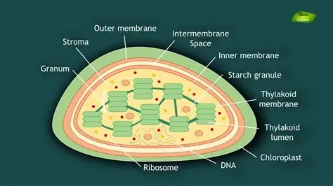
describe the function of the chloroplast
site of photosynthesis - some parts take place in grana, others in stroma
grana allow a large surface area for the assembly of chlorophyll and so photosynthesis
describe the structure of the cell membrane
found either on the surface of the cell or just inside the cell wall
made of a phospholipid bilayer and protein
describe the function of the cell membrane
regulates movement of substances into and out of the cell
has receptor molecules on out allowing it to respond to hormones
describe the structure of the cell wall
plants and algae - made of cellulose
microfibrils are embedded in a background material of pectin
describe the function of the cell wall
maintains the shape of the cell and allows it to remain turgid when water moves into it by osmosis, preventing it from bursting
describe the structure of the cell vacuole
membrane bound organelle
contains cell sap
membrane is called tonoplast
describe the function of the cell vacuole
maintains pressure inside the cell and keeps it rigid
what is the formula for magnification?
magnification = size of image/size of real object
define the term magnification
the number of times larger an image is compared with the real size of an object
define the term resolution
the ability to distinguish between two separate points
what is the limit of resolution of a light microscope?
½ the wavelength of the radiation used to view the specimen
the shortest wavelength of light is 400nm so the max resolution is 200nmwha
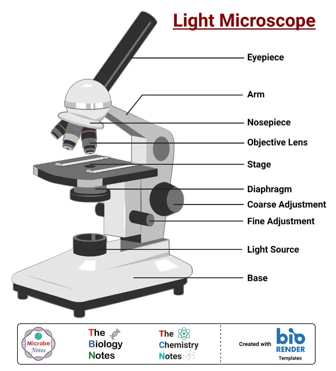
what does a light microscope look like?
what is the maximum useful magnification of an optical microscope?
x1500
which organelles can you not see using a light microscope?
ribosomes
lysosomes
mitochondria
endoplasmic reticulum
what is the other kind of microscope?
electron microscopes
what are the two types of electron microscope?
transmission electron microscope
scanning electron microscope
what is the maximum resolution of a transmission electron microscope?
0.1nm
what is the maximum resolution of a scanning electron microscope?
20nm
describe how a TEM works
use electromagnets to focus a beam of electrons, which are transmitted through the specimen
denser parts of the specimen absorb more electrons, making them look darker on the resulting image
describe how an SEM works
scan a beam of electrons across a specimen
this knocks off electrons from the specimen
these are gathered in a cathode ray tube to form an image
the resultant images show the surface of the specimen and the 3-D
what are the advantages of TEMs?
gives the highest resolution images so shows small objects
what are the advantages of SEMs?
can be used on thick specimens
produce 3D images
have a relatively high resolution
what are the disadvantages of TEMs?
can only be used on very thin specimens
can only be used on non-living specimens
the specimen must be viewed in a vacuum
images may contain artefacts which can make it difficult to identify organelles
what are the disadvantages of SEMS?
give lower resolution images than TEMs
can only be used on non-living specimens
need to be used in a vacuum
images may contain organelles
describe how to prepare a temporary microscope slide
place a small drop of water on the centre of the slide
use tweezers to place a thin section of your specimen on top of the water drop
add a drop of iodine on top
place a coverslip on top
blot off any excess iodine using filter paper
which organelles do prokaryotic cells (mainly bacteria) contain?
cell wall
slime capsule
plasmid
circular DNA
flagellum
cytoplasm
cell membrane
small ribosomes (70S vs 80S)
which organelles do prokaryotic cells not contain?
any membrane-bound organelles
mitochondria
nucleus
ERs
golgi apparatus
lysosomes
describe the structure of a prokaryotic cell wall
made of murein, which is a glycoprotein (protein with carbohydrate attached) - peptidoglycan
describe the function of the cell wall
physical barrier that protects against mechanical damage
excludes certain substances from the cell
describe the structure and function of the slime capsule
coats the cell wall, made up of secreted slime
protects the bacteria from attack by cells of the immune system
helps bacteria stick together
describe the structure and function of the flagellum
has a rigid, corkscrew shape and a rotating base
this helps the cell spin through fluids
describe the structure and function of prokaryotic enzymes
smaller than eukaryotic - 70S type
site of protein synthesis - can be inhibited by certain antibiotics
the difference between prokaryotic and eukaryotic ribosomes allows antibiotics to selectively target prokaryotes
describe the function of the circular DNA
possesses the genetic information for the replication of bacterial cells
describe the function of the plasmids
can reproduce independently, so are used as vectors in genetic engineering
possess genes that aid the survival of bacteria by producing enzymes that can break down antibodies
describe the structure of viruses
they have a core of genetic material (DNA or RNA)
this is covered by a protein coat called a capsid
attachment proteins stick out from the capsid
describe how viruses reproduce
virus attaches to the host cell receptor proteins
virus injects genetic material into the host cell
genetic material and proteins are replicated by the host cell machinery - ribosomes, RER
the viral components assemble
the replicated viruses are released from the host cell
the attachment proteins on the virus require different receptor proteins, affecting which types of host cell viruses can infect