Week 6: Neuroscience research methods- strengths and limitations
1/55
There's no tags or description
Looks like no tags are added yet.
Name | Mastery | Learn | Test | Matching | Spaced | Call with Kai |
|---|
No analytics yet
Send a link to your students to track their progress
56 Terms
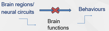
What are the aims of research methods in behavioural neuroscience?
To understand how the brain produces behaviour
Identify the functions that are required for performing a behaviour and determine what circuits of neurons are responsible for each of these functions
Name some methods and uses of neuroscience research methods
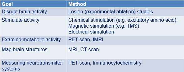
What does spatial resolution in imaging techniques refer to?
The ability to locate structures/activity in the brain ('where something happens').
What does temporal resolution in imaging techniques refer to?
The ability to detect changes in brain activity over a given time period ('when something happens').
What significant event happened to Phineas Gage? (1848)
An iron rod was driven through his head, destroying much of his left frontal lobe.
Despite the traumatic brain injury, it didn't affect his cognitive functioning - yet he experienced a personality change
Outline the case of 'Tan' (1861)
Louis Victor Leborgne could understand language but only utter the syllable 'tan'
French surgeon Paul Broca connected the clinical symptoms (loss of speech) to specific anatomical damage (left frontal lobe) → Broca’s area
What has been suggested about the behavioural effects of brain damage?
If removing structure X changes behaviour Y, then structure X contributes to behaviour X
What do lesion studies tend to involve?
Naturally occurring lesions (human research participants e.g. Phineas Cage)or induced lesions (animal studies)
Experimental ablation: The oldest method used in neuroscience, still in common use in animal studies.
Brain tissue is deliberately destroyed, removed or inactivated and alterations in behaviour observed.
What is experimental ablation?
A method in neuroscience where brain tissue is deliberately destroyed or removed to observe changes in behavior.
Which 4 ways are lesions created?
Surgical lesions
Radiofrequency (RF) lesions
Excitotoxic lesions
Temporary inactivation
What are radiofrequency (RF) lesions?
Lesions created by passing electrical current through tissue to destroy it.
→ can damage both neurons and passing axons
What are excitotoxic lesions?
More precise lesions compared to RF lesions
How is temporary inactivation caused?
GABA receptors
Reversible effects
What are 2 historical methods to visualise lesions?
Histological staining
Immunocytochemical methods
What is histology?
The study of cells and tissues at the microscopic level.
What is histological staining?
Interactions between charged dyes and cells to visualise cell and tissue structure - not specific proteins/molecules
What is the purpose of immunocytochemical methods?
To visualise specific molecules, neurotransmitters, or receptors in the brain.
What is meant by tracing neural connections?
Injecting tracers into specific brain regions
What is the difference between anterograde and retrograde labelling within tracing neural connections?
Anterograde labeling traces efferent neurons from cell body to axon terminals, while retrograde labeling traces afferent neurons from axon terminals to cell body.
How does tract tracing work?
Use a stereotaxic apparatus to target a specific brain region
They would be injected into a living brain, transported along the axons,
Animal is euthanised
Tissue is sectioned & histological staining reveals the labelled pathways.
What is Diffusion Tensor Imaging (DTI)?
A non-invasive imaging method used to visualise brain structure and connectivity.
→ technique that maps the brain's white matter by measuring the direction and speed of water molecule diffusion
Name some strengths and limitations of lesion studies
STRENGTHS
moderate to high spatial resolution
CONS
other structures might be damaged
poor temporal resolution
does not account for compensation
Name some strengths and limitations of tract tracing
STRENGTHS
high/very high spatial resolution
CONS
poor temporal resolution
invasive, not used in humans
Name some strengths and limitations of Diffusion Tensor Imaging (DTI)
STRENGTHS
non-invasive, can be used in humans
CONS
moderate resolution
cannot show individual axons/connections
What are neuroimaging studies?
The use of methods to visualise the structure and function of the brain
What do structural brain scans tell us?
They tell us what the brain looks like and allows locating an area that has been affected by a condition like a stroke or a lesion
What do functional brain scans tell us?
They tell us which part of the brain is actively doing something (i.e. which area of the brain activates under particular conditions).
What does a Computerised Tomography (CT) scan do?
Uses X-ray measurements to generate horizontal images of the brain, revealing structural abnormalities.
contrast dye helps differentiate between normal and abnormal structures
What are the pros and cons of CT scans?
PROS
good spatial resolution
widely available, fast
cheap
CONS
radiation exposure
poor temporal resolution; cannot track brain activity in real time
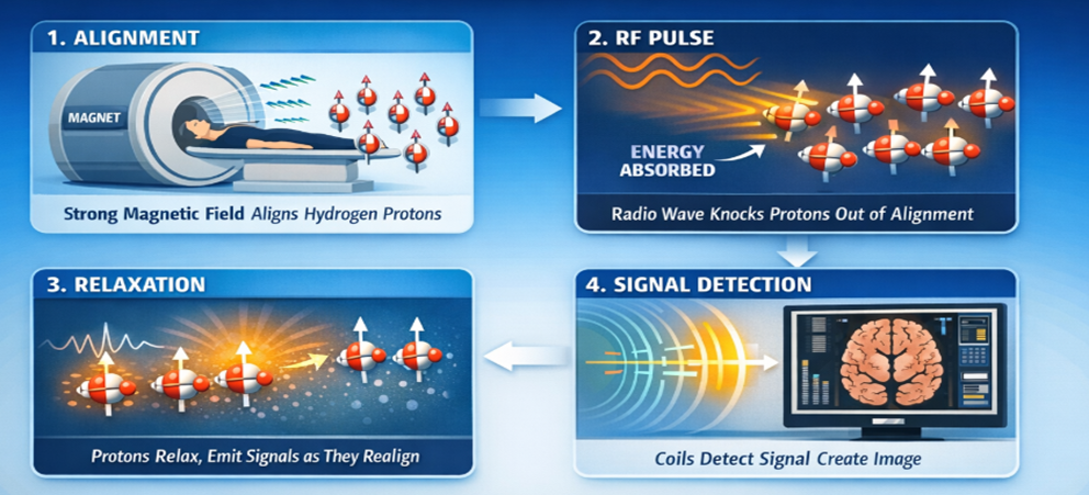
What is the function of Magnetic Resonance Imaging (MRI) and what does it measure?
To create detailed images of the brain using strong magnetic fields.
Changes in brain activity by detecting blood flow variations.
How do MRI’s work?
What are the pros and cons of MRIs?
PROS
good spatial resolution
relatively accessible
non-invasive
CONS
poor temporal resolution
expensive
noisy
participant lies perfectly still
cannot be used with metal implants
What did the Maguire et al (2000) study show?
Found that London taxi drivers had more grey matter in the hippocampus compared to non-taxi drivers
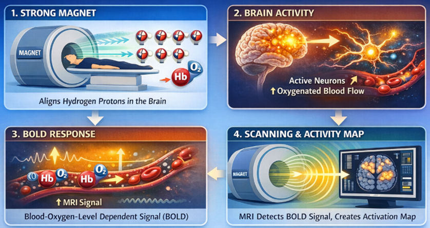
How do functional magnetic resonance (fMRI) work? What do they show?
Works: When neurons are active, they use oxygen, blood flow increases fMRI measures changes in blood flow
Shows: visualisation of brain activity associated with performing a cognitive task and/or behaviour
What are the pros and cons of fMRIs?
PROS
good spatial resolution
non-invasive
relatively accessible
CONS
poor to moderate temporal resolution (seconds)
loud environment
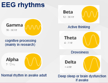
How do Electroencephalograms (EEGs) work?
When many neurons fire together they produce tiny electrical fields; EEG records this electrical activity.
During an EEG procedure, small disc-type electrodes are placed on the scalp surface.
Electrodes pick up brain’s electrical signals and send them to electroencephalogram.
EEG records neuronal impulses as wavy lines (brain waves) onto a computer screen.
Outline the differences in EEG rhythms
Frequency band - how fast the waves are
Morphology - shape
Topography - where on scalp
Amplitude - how tall the waves are
Reactivity - response to simulation
Symmetry
What are some applications of EEGs?
Used in clinical settings to diagnose conditions such as epilepsy or sleeping disorders
EEG-based research on functional networks in cognitive and effective processing
What are some pros and cons of EEGs?
PROS
excellent temporal resolution (milliseconds)
tolerant of subject movement
non-invasive/salient
CONS
low spatial resolution compared to fMRI
analysis of acquired data can be very complex
poorly measures neural activity that occurs below the upper layers of the brain (cortex)
What is Transcranial Magnetic Stimulation (TMS)? Outline the steps involved
A method that uses magnetic pulses to influence neuronal activity in the brain.
A TMS machine sends a strong electric current to a coil.
This gives rise to a fluctuating magnetic pulse which goes through the skull into the brain.
The pulses trigger electrical charges changing the activity of nearby neurons.
How is TMS used to treat aphasia? (Fernandez-Romero, 2025) - excitatory TMS
-TMS over left dorsolateral cortex in people with progressive aphasia
-TMS Vs controls
-Slower decline in brain metabolism and improvements in language with active TMS
Reconnect trial
How is TMS used for motor perception? (Walsh and Cowey, 2000) - inhibitory
TMS is used to investigate motor perception by acting as a tool for producing temporary, localised "virtual lesions"
-TMS over MT/V5 area in visual cortex
- “If MT/V5 is active and necessary for motion perception then disrupting it with TMS during motion viewing should impair motion perception”
-Hypothesis confirmed- Region functionally necessary
What are the pros and cons of TMS?
PROS
good temporal resolution (milliseconds)
can be combined with other methods to record response to simulation
non-invasive
CONS
stimulates superficial cortical areas - cannot reach deep structures
moderation spatial resolution - compared to fMRI
interindividual variability due to anatomy/neurotransmitter levels
How does Positron Emission Tomography (PET) work?
Radiotracer is injected into blood stream
Crosses blood brain barriers
Binds to specific receptors
PET scanner detects positron emissions
Signal reflects binding potential (BP)
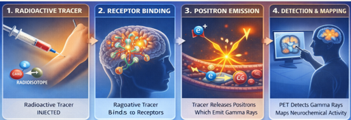
What do PET scans show?
Metabolic and neurotransmitter activity
What are the pros and cons of PET scans?
PROS
can measure specific molecules or neurotransmitter systems, e.g. dopamine uptake
silent
great diagnostic value
CONS
comparatively poor spatial and temporal resolution (minutes) compared t0fMRI
invasive - required use of radioactive tracers
Name 3 future directions in neuroimaging
Use of AI
Human Connectome Project
UK Biobank Imaging Study
How can AI be used as a future direction of neuroimaging?
AI allows to:
enhance image resolution;
reconstruct 3D brain images from MRI, CT or PET scans,
identify and labels brain structures
predict disease progression
What is the Human Connectome Project?
Aims to connect brain structure, function, and behaviour, providing open-access data to study brain connectivity.
What is the UK Biobank Imaging Study?
A study using MRI/fMRI/DTI to understand risk factors for conditions like dementia and depression.
What is the significance of the Reconnect trial in TMS research?
It showed that TMS over the left dorsolateral cortex can slow decline in brain metabolism and improve language in people with progressive aphasia.
What does the term 'resolution' refer to in neuroimaging?
The clarity and detail of the images produced by imaging techniques.
What is the role of contrast dye in CT scans?
To help differentiate between normal and abnormal brain structures.
What are the applications of EEG in clinical settings?
To diagnose conditions such as epilepsy or sleep disorders.
What is the significance of the frequency band in EEG?
It indicates how fast the brain waves are oscillating.
What does the term 'morphology' refer to in EEG analysis?
The shape of the brain waves recorded.