A&H lab quiz
1/49
There's no tags or description
Looks like no tags are added yet.
Name | Mastery | Learn | Test | Matching | Spaced | Call with Kai |
|---|
No analytics yet
Send a link to your students to track their progress
50 Terms
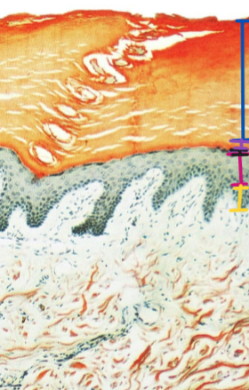
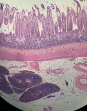
Thick skin
-Stratum corneum
-Stratum lucidum
-Stratum granulosum
-Stratum spinosum
-Stratum basale
Thin Skin
-Stratum corneum
-Stratum granulosum
-Stratum spinosum
-Stratum basale
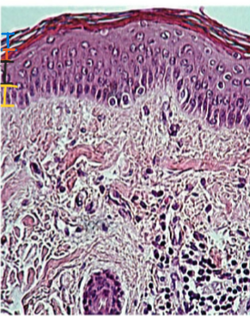
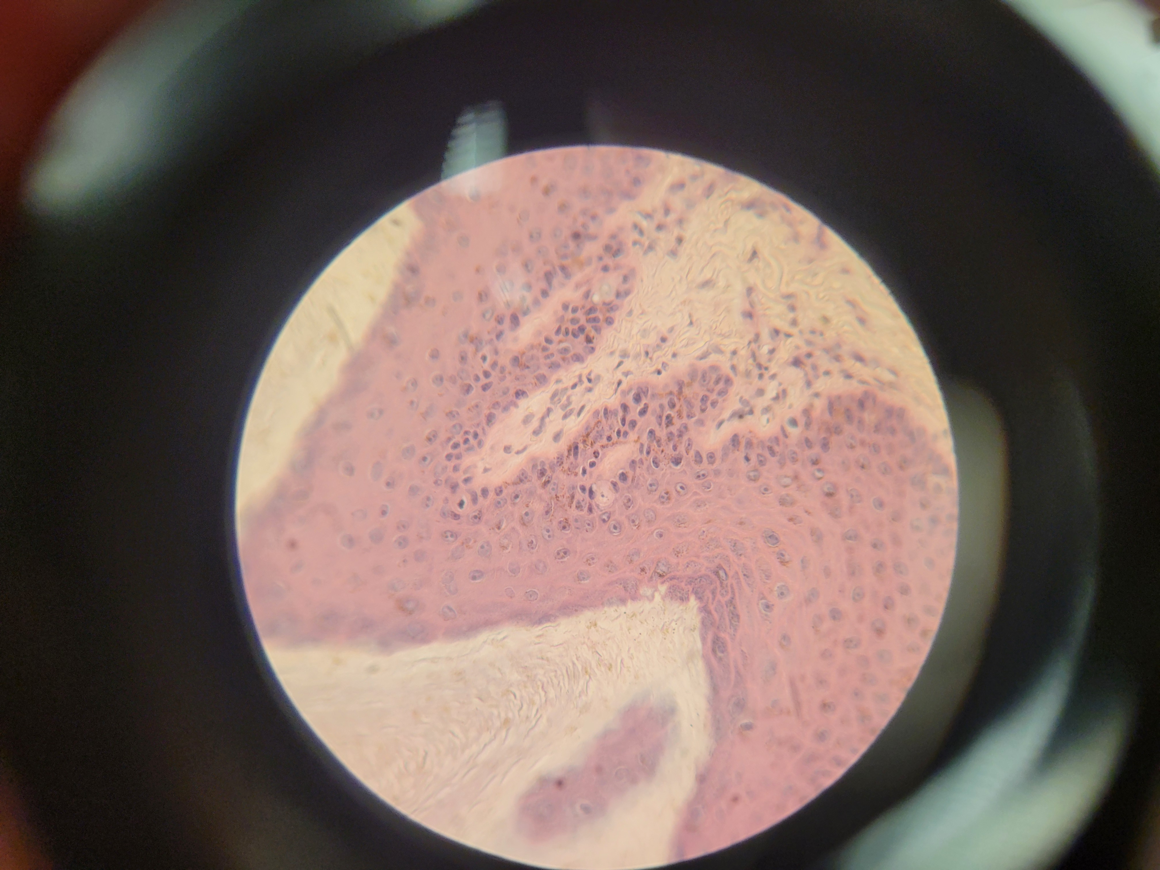
What is this a picture of?
Thick skin
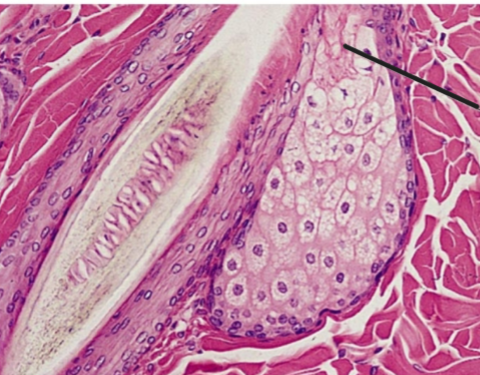
What is the black line pointing to? What is the overall picture?
-sebacous gland duct
-sectioned sebacious gland
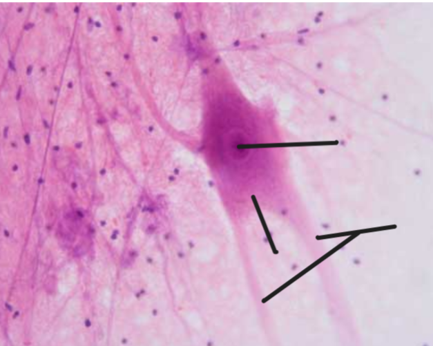
What are each of the black lines pointing to? What is the overall image?
-Overall image is a neuron
-First line is cell body
-Second line is axon hillock
-Third lines are dendrites
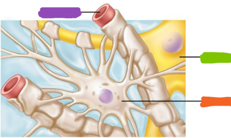
Name the following highlighted items on the image
-Capillary
-Neuron
-Astrocyte
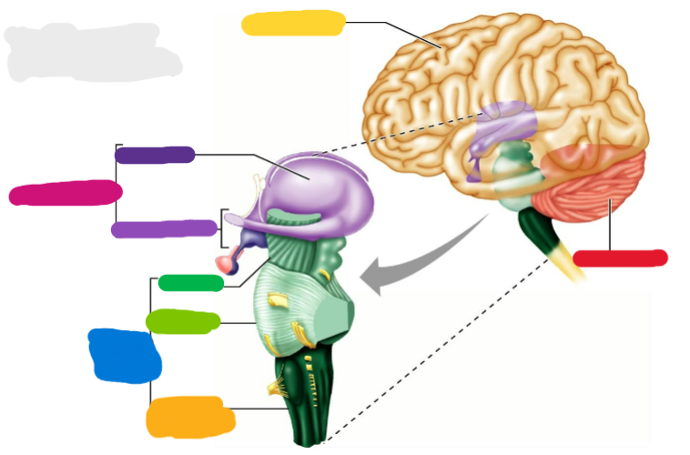
-Cerebrum
-Cerebellum
-Diencephalon
-Brain stem
-Thalamus
-Hypothalamus
-Mid-brain
-Pons
-Medulla oblongata
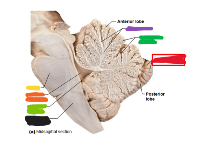
What is this an image of? Name all of the highlighted parts
-Cerebellum
-Arbor vitae
-Cerebral cortex
-Follia (gyrus)
-Pons
-4th ventricle
-Choroid plexus
-Medulla oblongata
What is this? What type of skin is it located in?
-Meissner’s corpuscle
-Located in thick skin
-It is a receptor
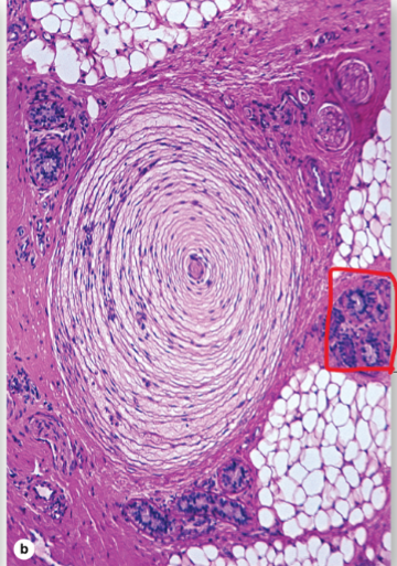
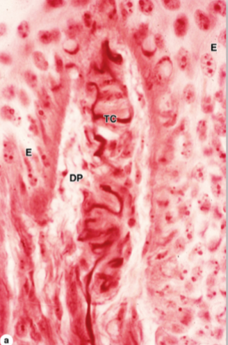
What is this? Where is it found? What is circled?
-Pacinian corpuscle
-A receptor in the thick skin
-The eccrine sweat gland is circled
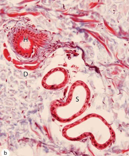
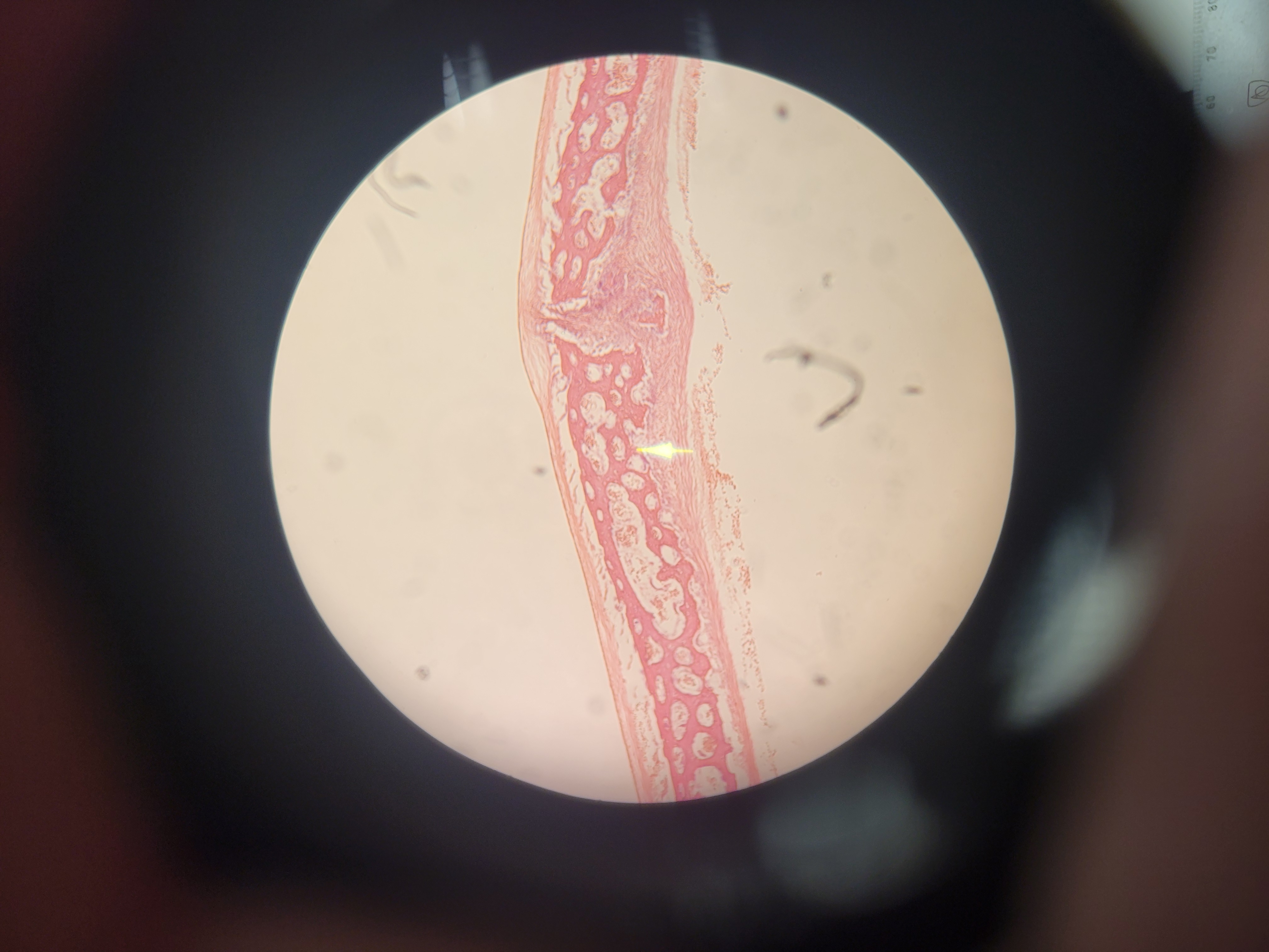
Name the image and the shape of the lumen
-Eccrine sweat gland
-Narrow lumen
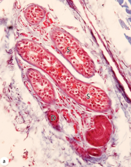
What is this and what is the shape of the lumen
-Apocrine sweat gland
-Wide lumen
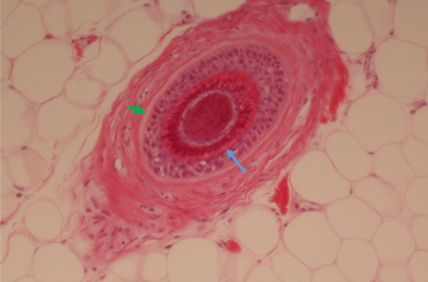
What is this and what are the arrows pointing to?
-Cross section of a hair follicle
-Internal epithelial root sheath
-External epithelial root sheath
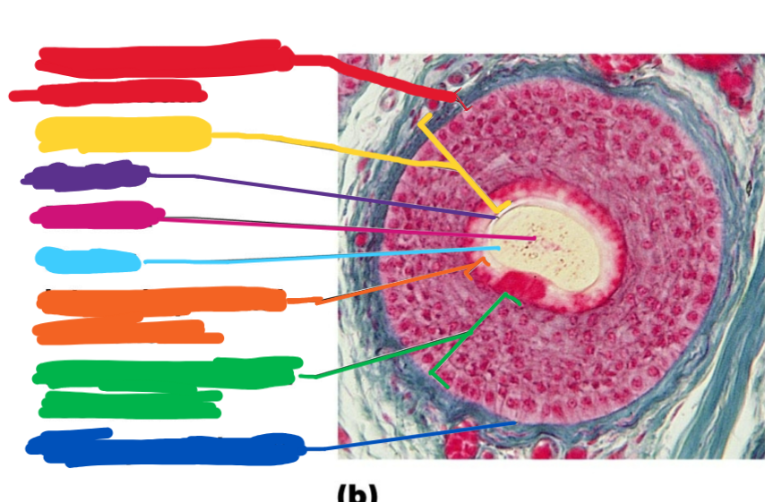
This is another hair follicle section. Name all of the items highlighted
-Connective tissue root sheath
-Follicle wall
-cuticle
-Medulla
-Cortex
-Internal epithelial root sheath
-External epithelial root sheath
-Glassy membrane
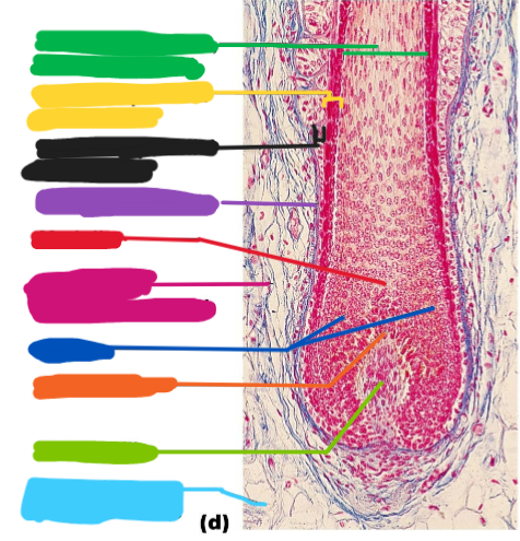
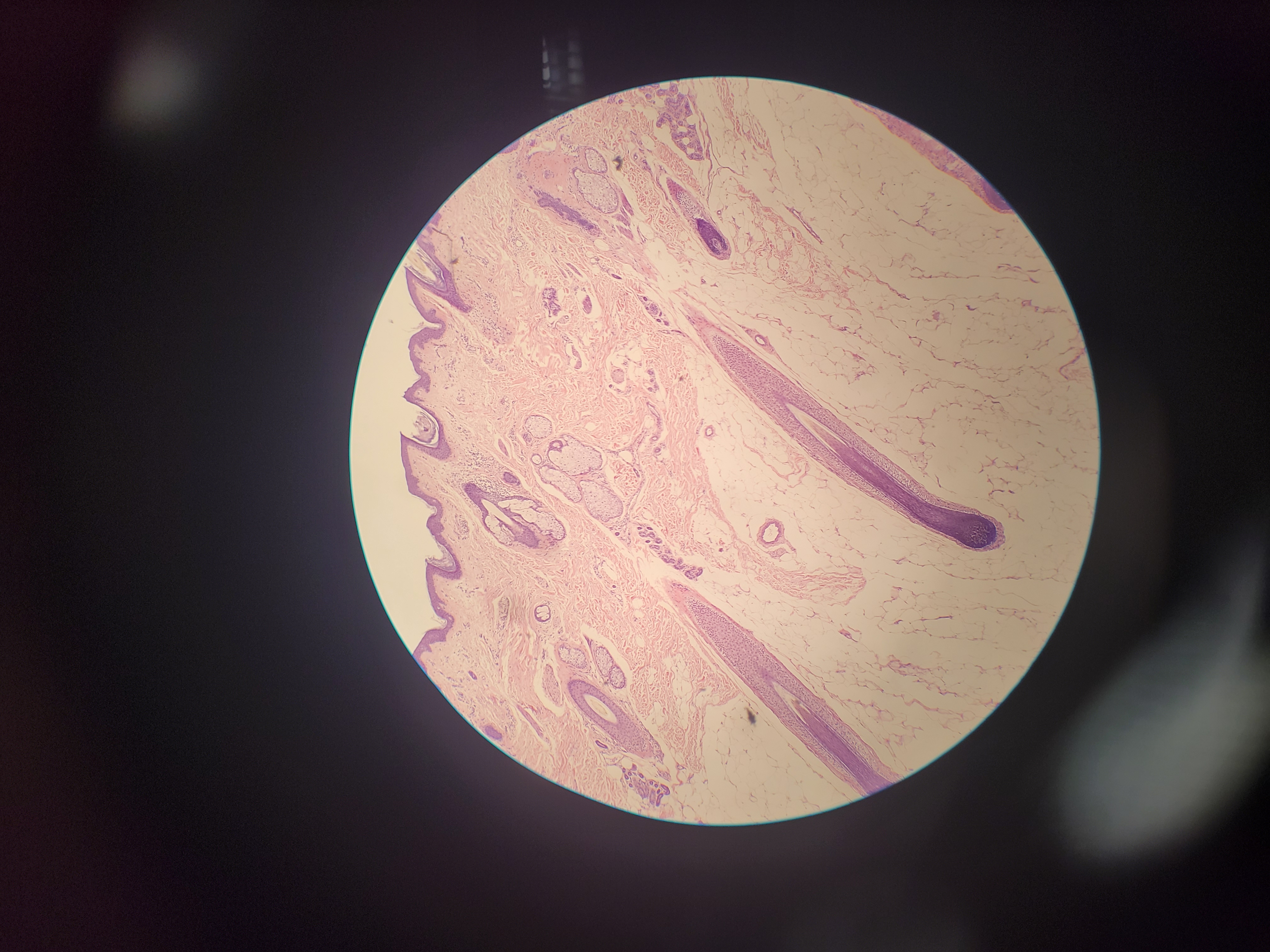
This is a hair follicle. Name the parts
-Hair shaft
-Internal epithelial root sheath
-External epithelial root sheath
-Glassy membrane
-Medulla
-Connective tissue root sheath
-Cortex
-Hair matrix
-Hair papilla
-Subcutaneous adipose tissue
What is shown? What are the pink and black fibers?
-Areolar Loose Connective Tissue
-Collagen fibers are pink
-Elastic fibers and black
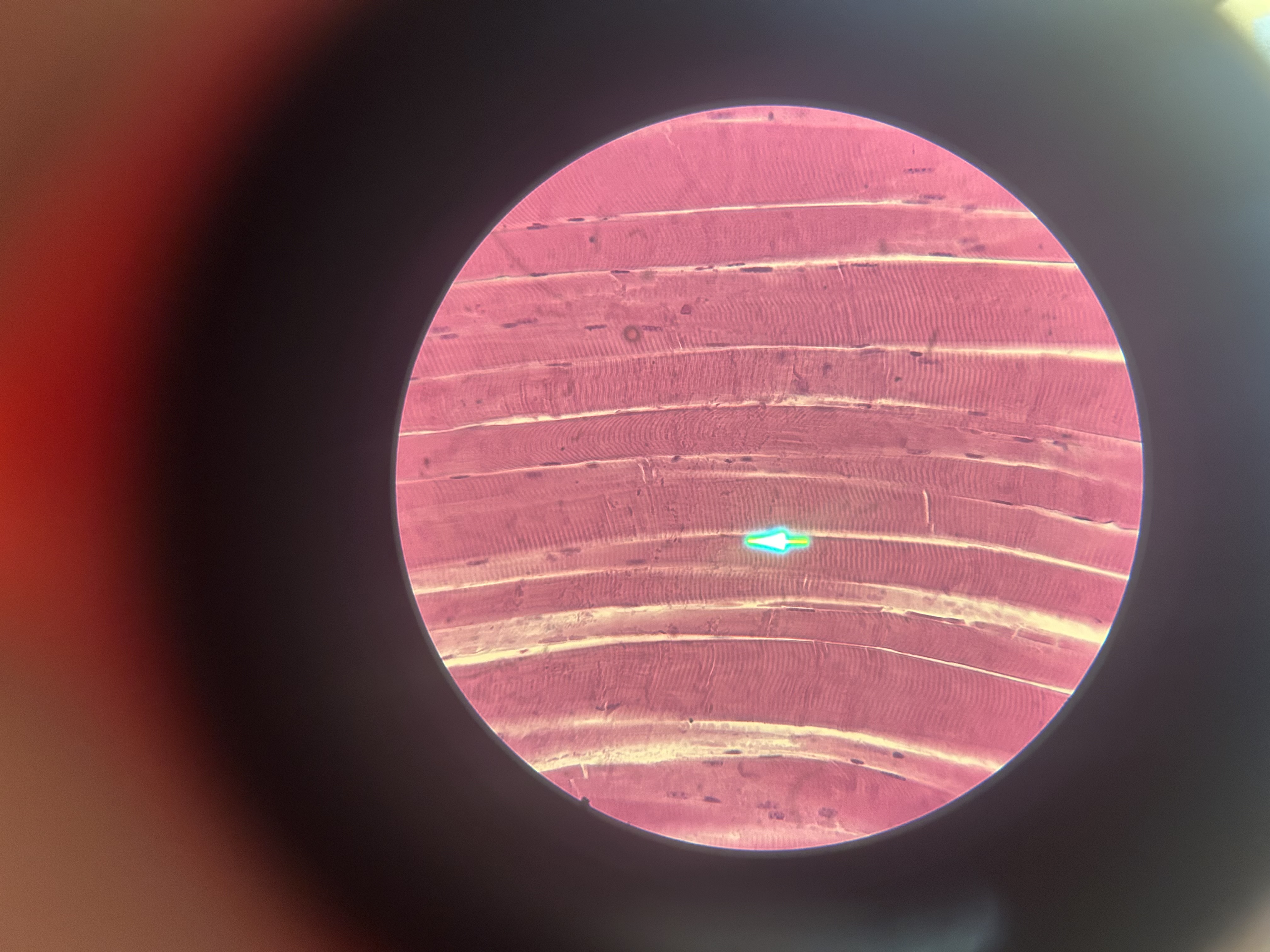
What is shown? What are some features displayed?
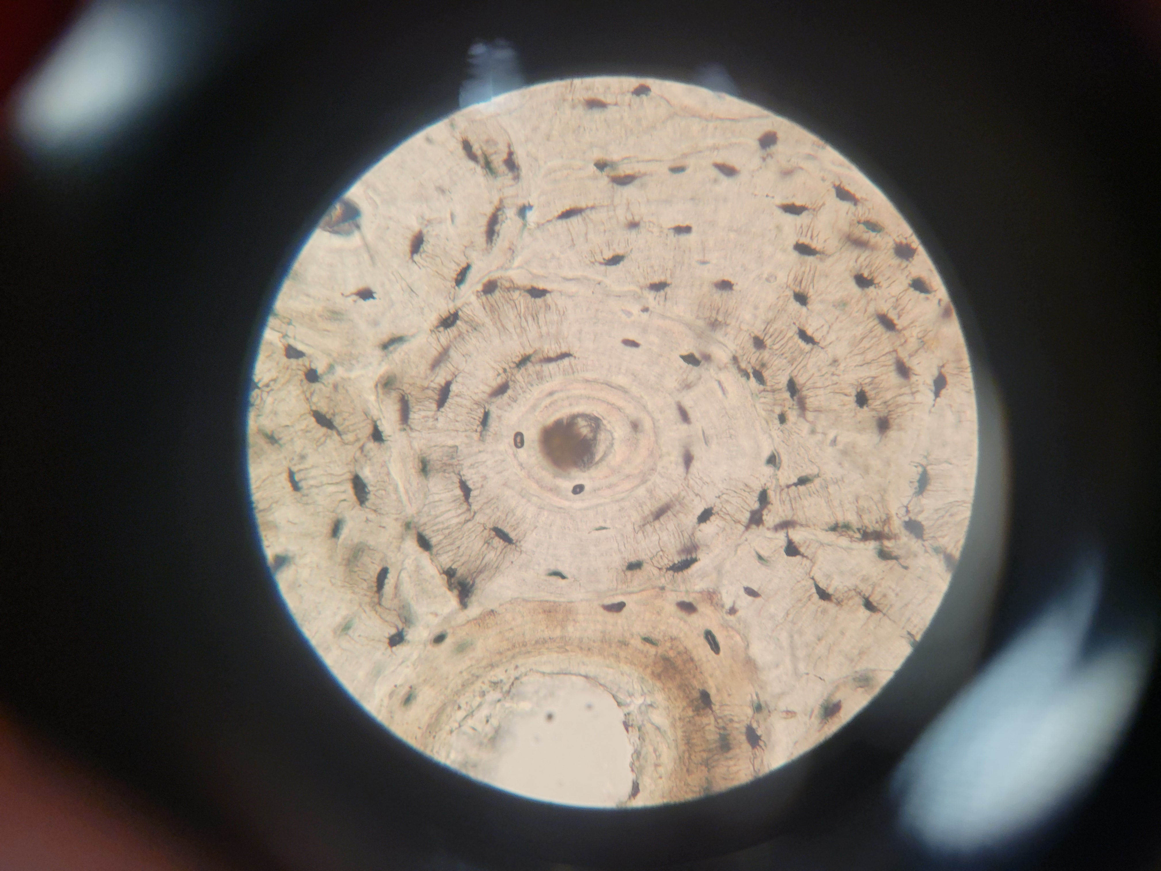
-Ground bone
-Osteon is the circle
-Volkmans canal are clear pathways
-Haversian’s canal is the center of the osteon
-Lacuna are the flat black dots that form a circle in the osteon
-Canaliculi are the striation marks between the lacuna
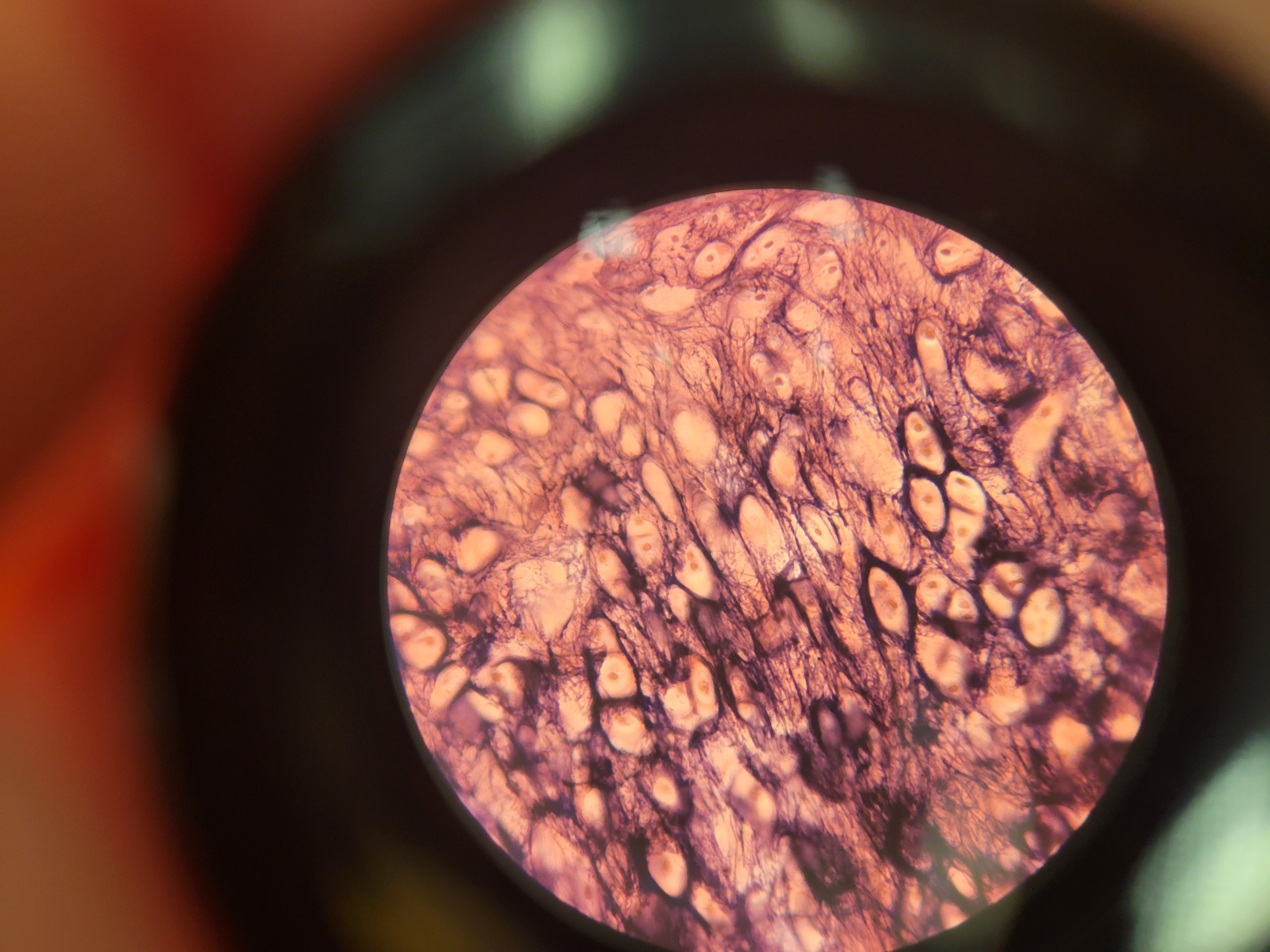
What is shown?
-Elastic cartilage
-Surrounded by perichondrium
-Predominant cell is a chondrocyte
What image is shown? What are the features?
-Another image is provided
-Growth plates
-Diaphysis and Epiphysis
-The zones
-Osteoblast and osteoclasts
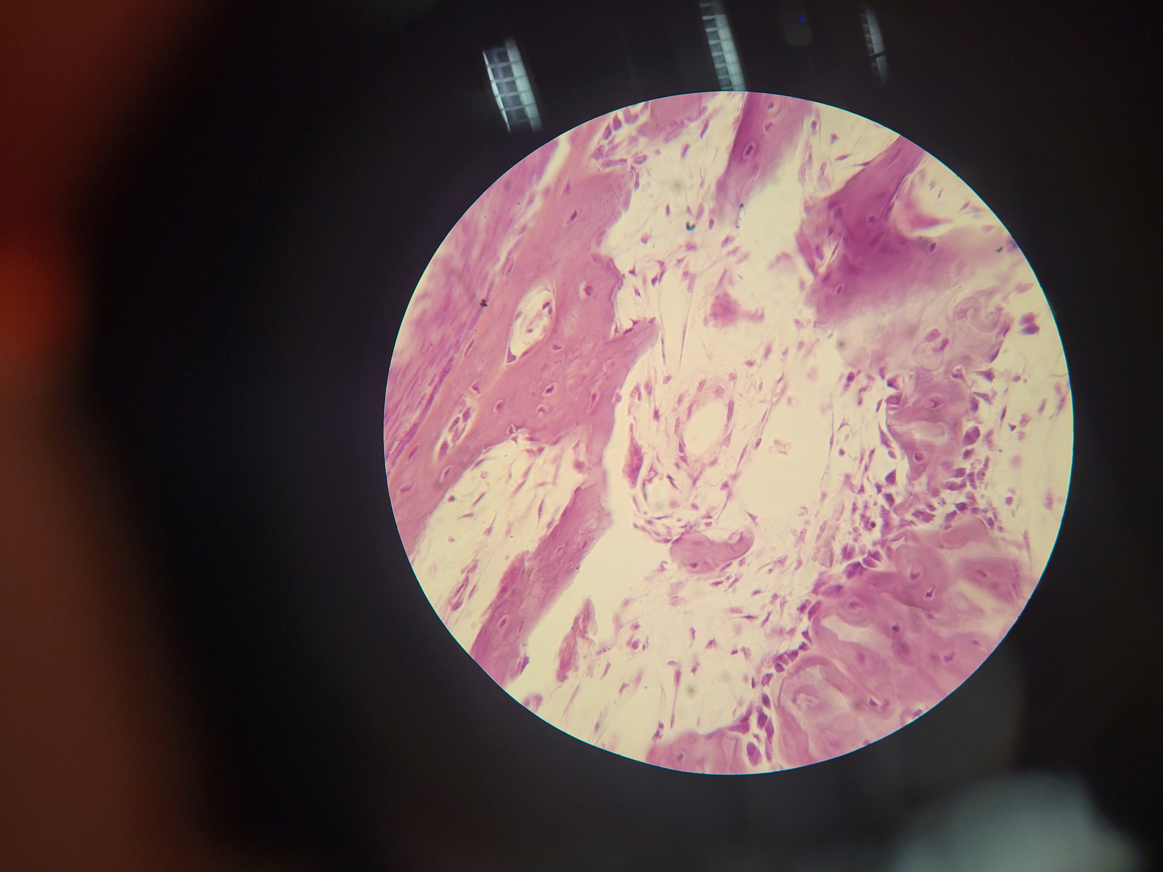
What is this?
Decalcified bone
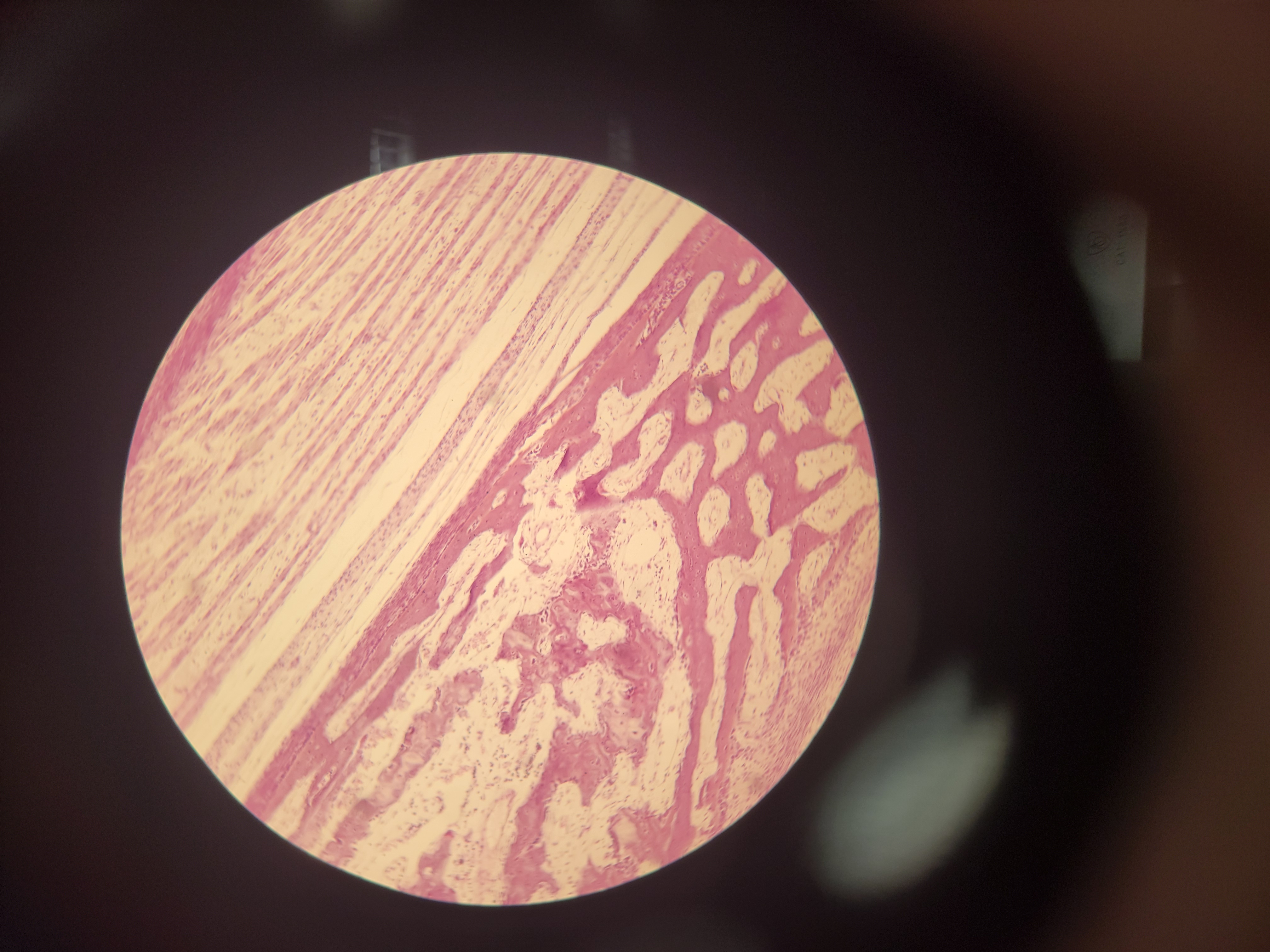
What is shown?
-Developing bone (intramembraneous)
-NO hylaine cartilage
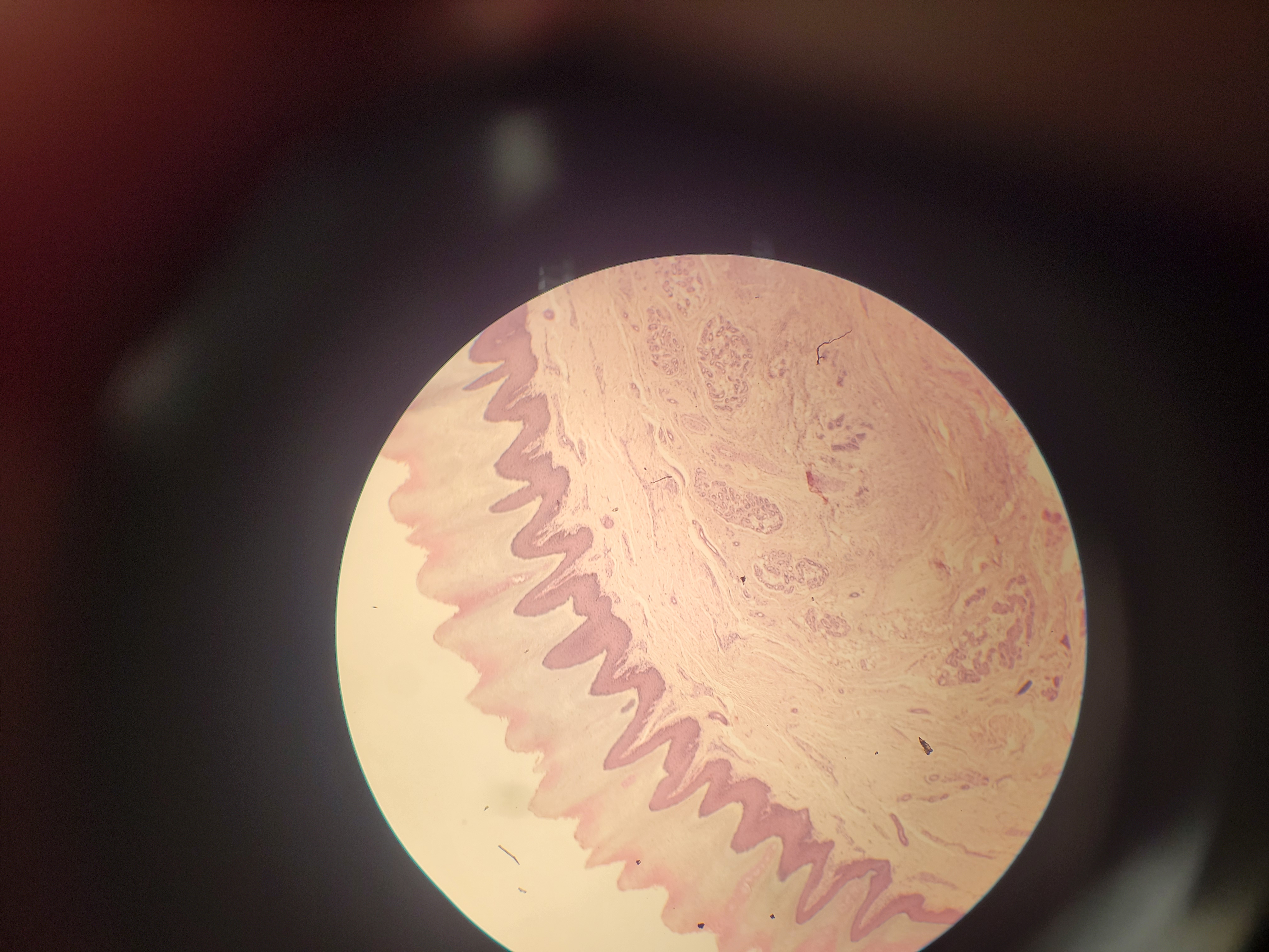
What is this and what features are shown?
-Human scalp
-The epidermis is shown (stratified squamous nonkeratinized)
-The reticular layer is the mostly white region
-Hypodermis contains adipose
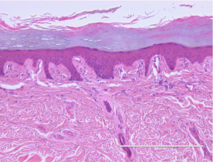
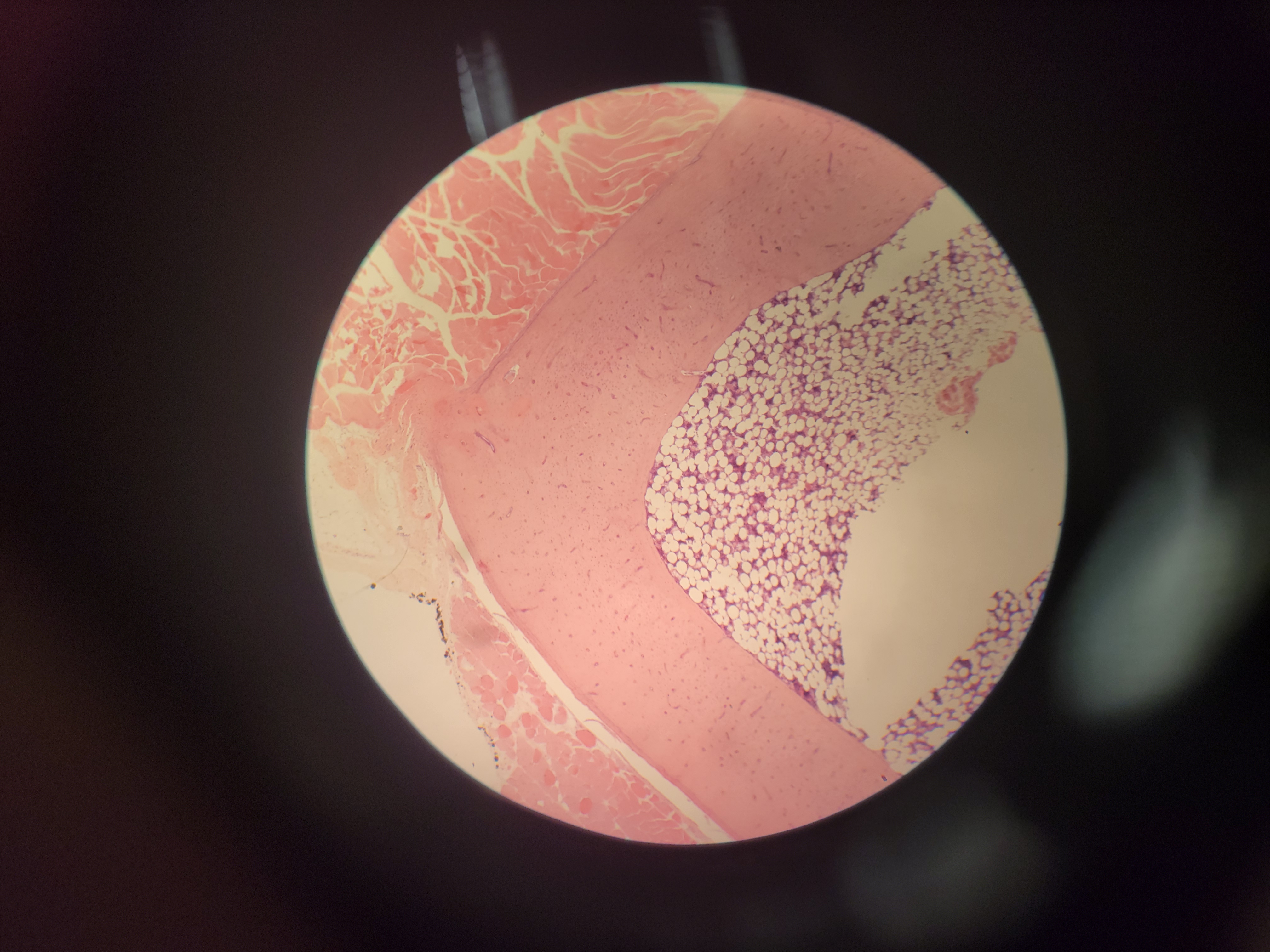
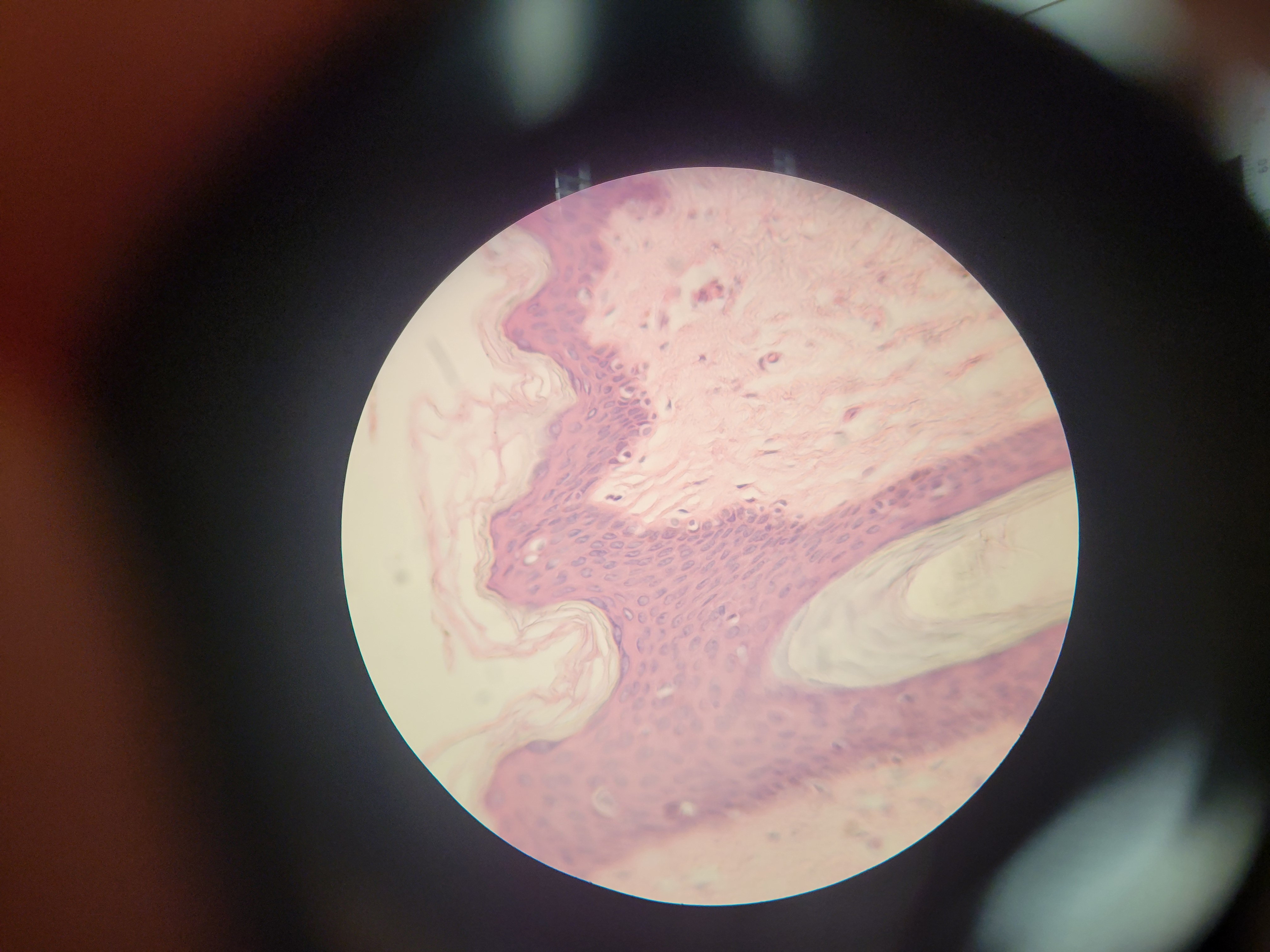
What is this?
-Skin cornified
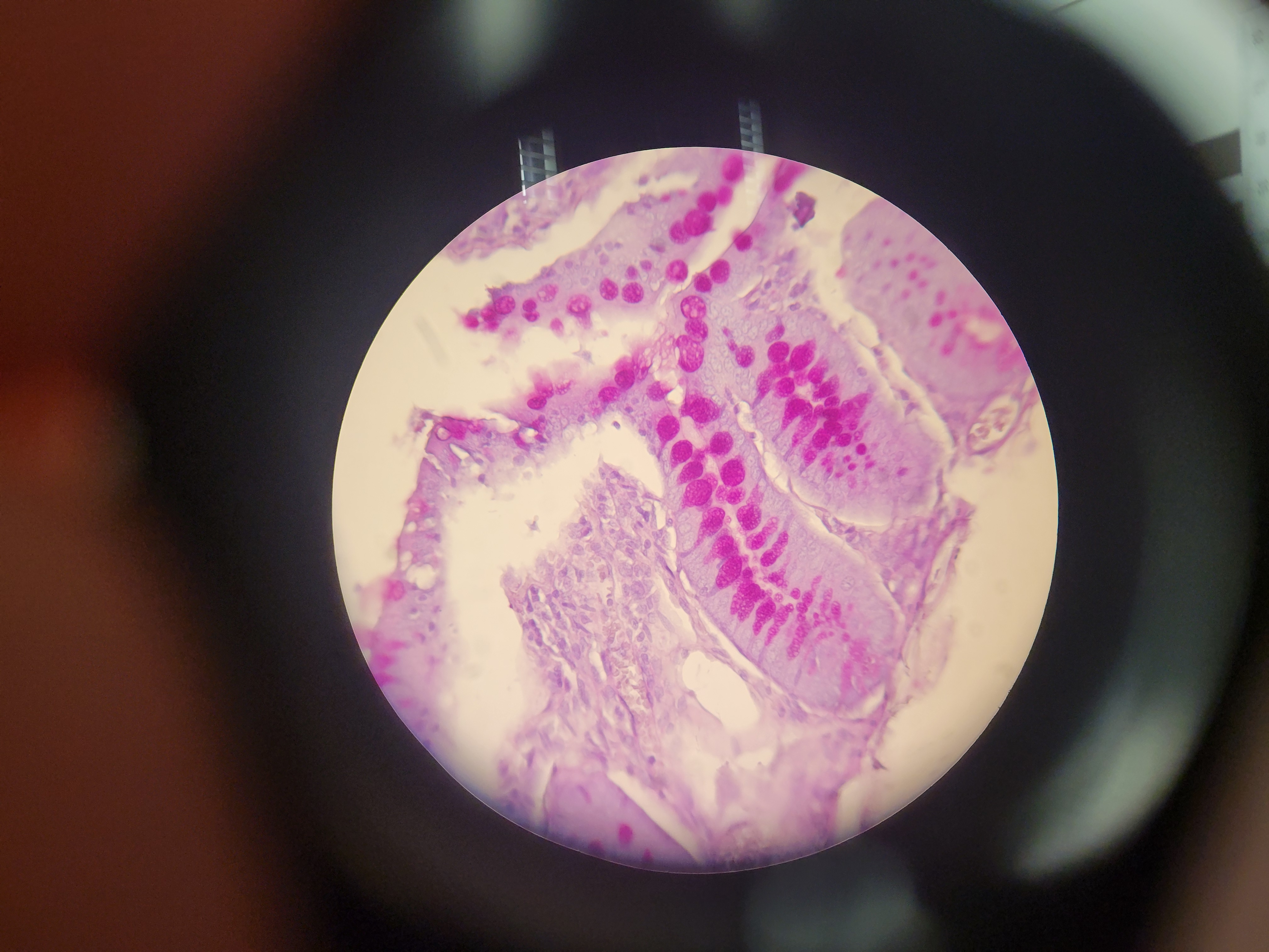
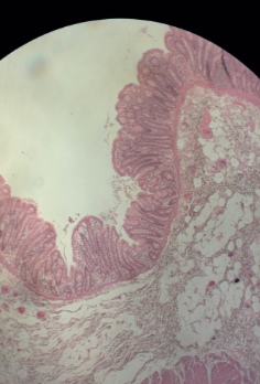
What organ is this? What type of cells are down
-Small intestine
-Goblet cells
What is shown?
Axillary skin
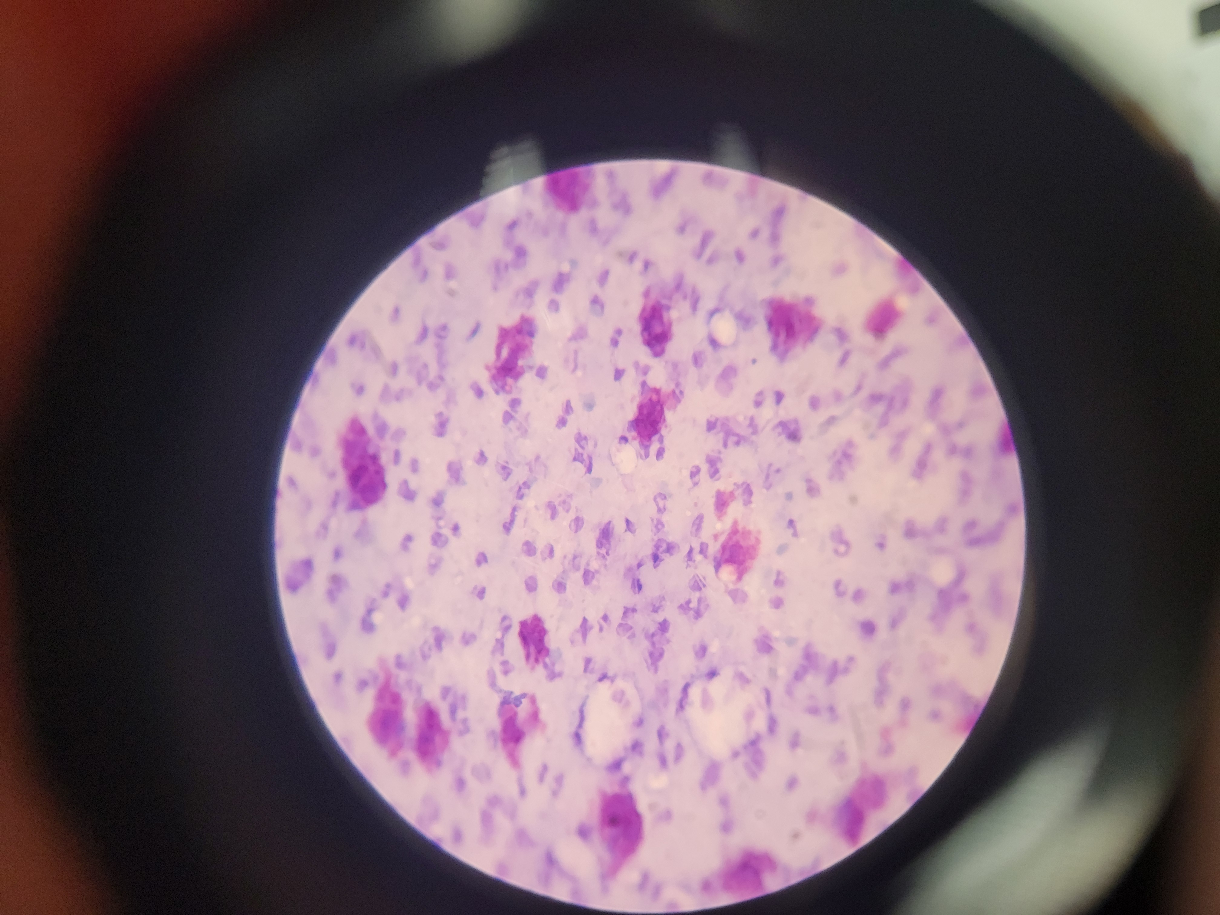
What is shown? What 2 things are featured in this image?
-Mesentery
-Mast cells (blue tiny guys)
-Adipocyte (red/purple bigger guys)
What is shown? What tissue makes it up?
-White fibrous tissue
-Tendon
-DRCT
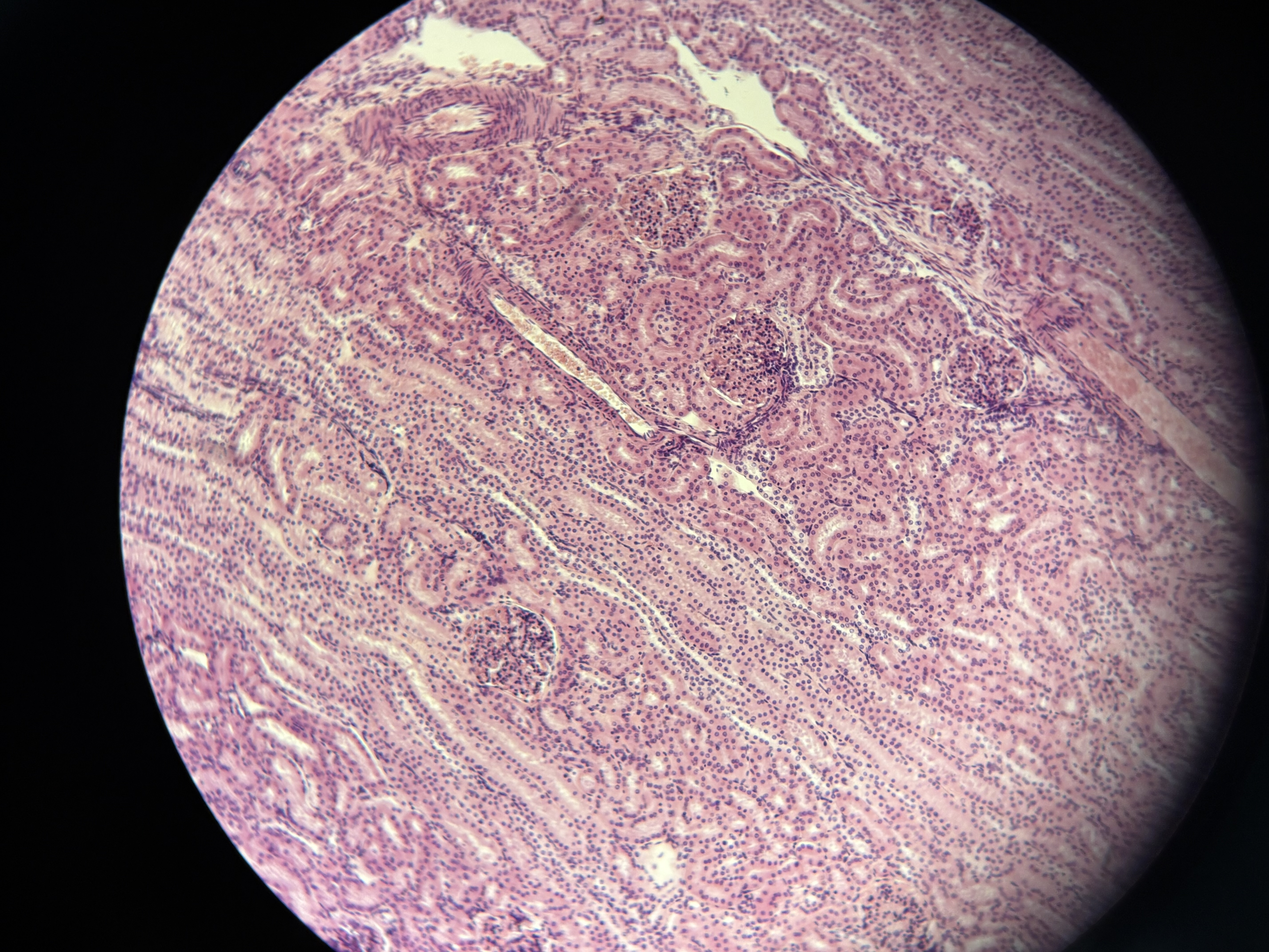
What is the tissue shown as well as the epithelium?
-Kidney
-Simple cubodial epi
What is the tissue shown and its epithelium
-Urinary Bladder
-Transitional epi
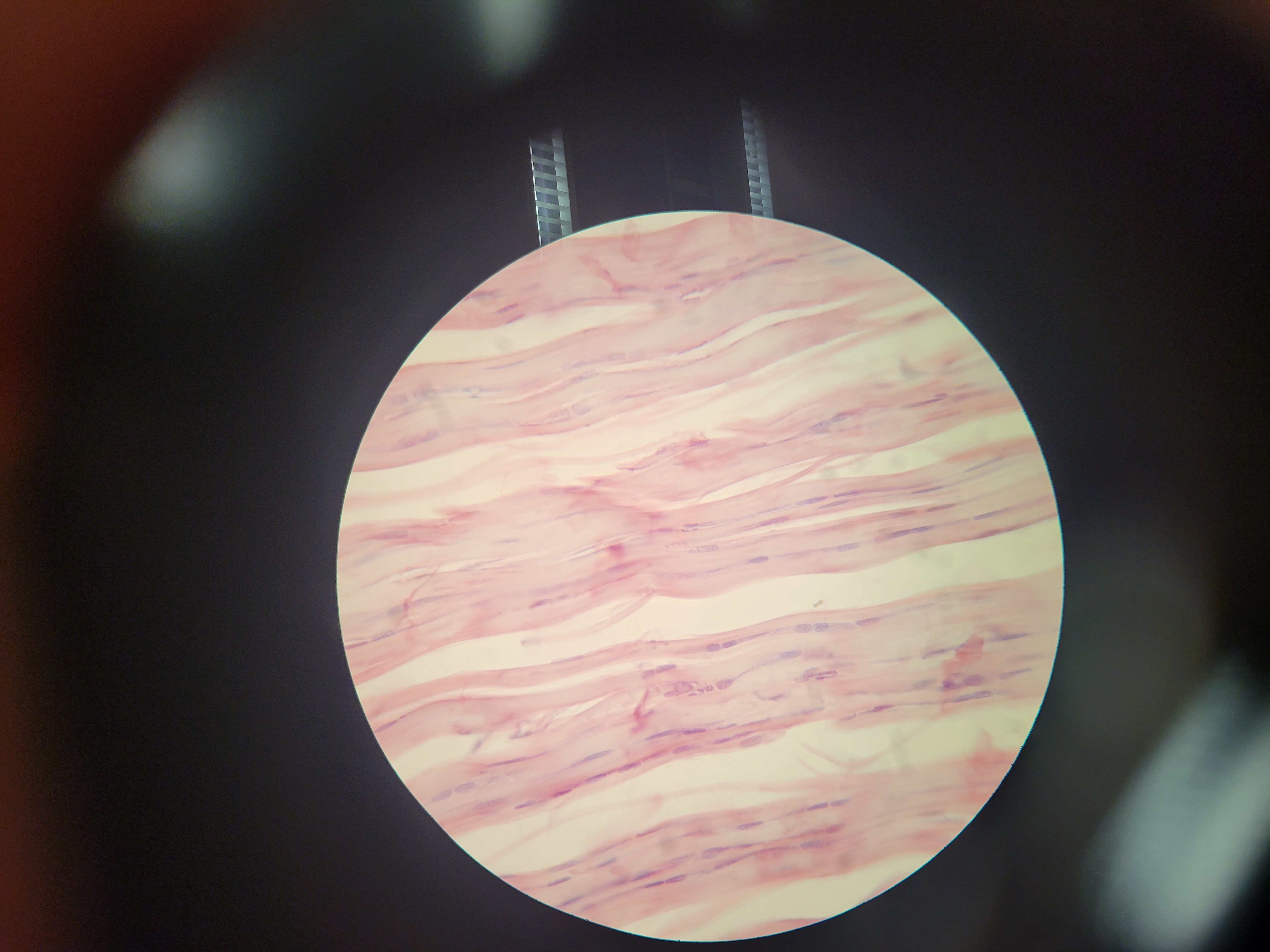
What is shown?
-Smooth muscle
-Lacks striations
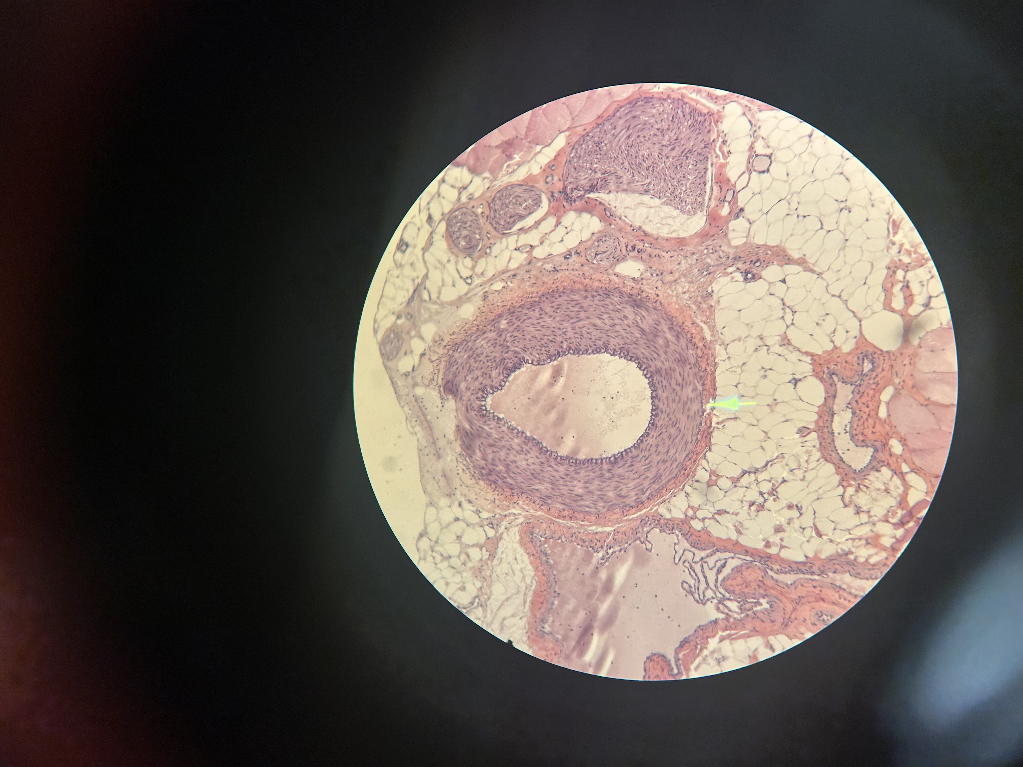
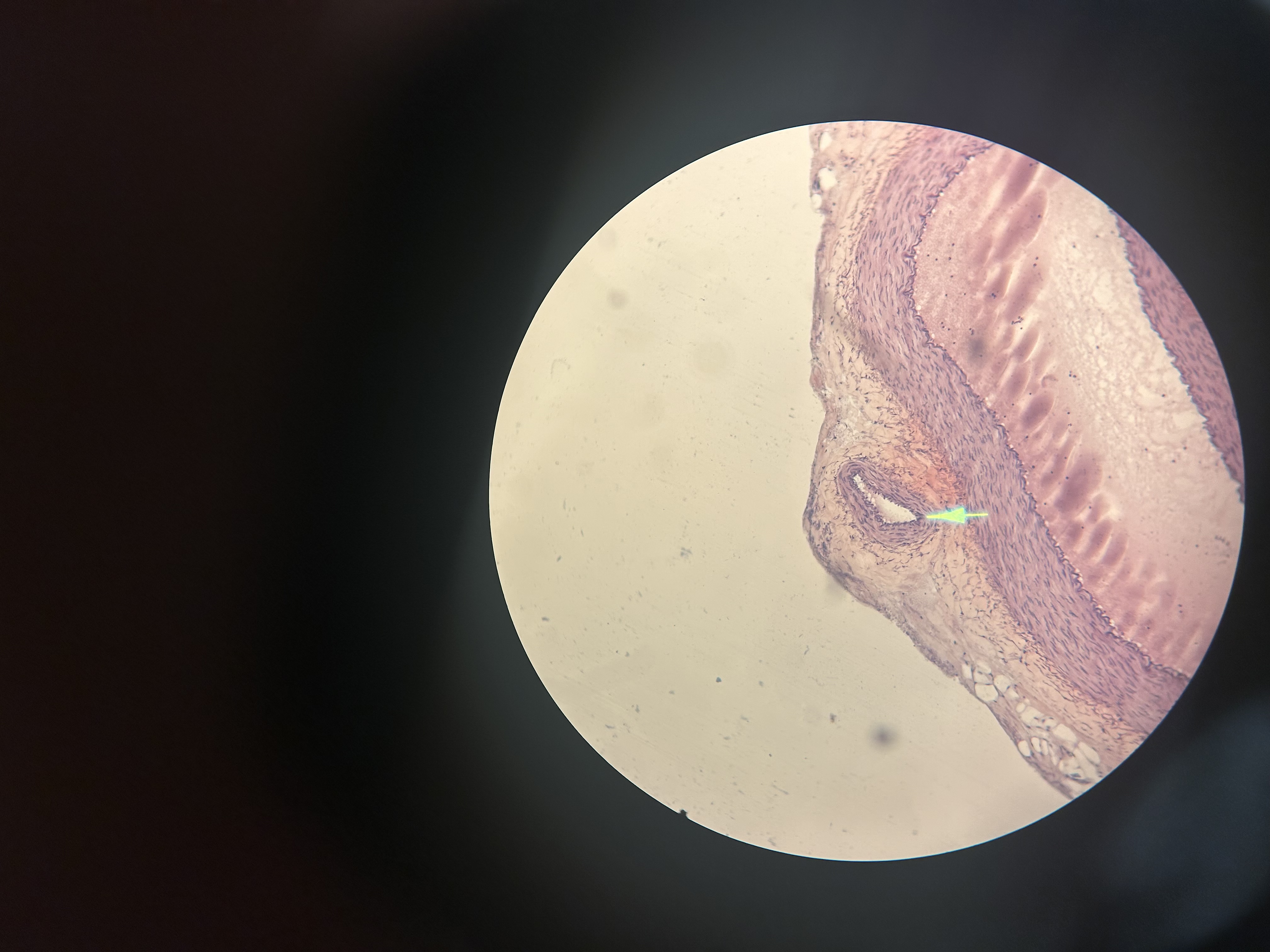
Name the image shown. What feature is the arrow pointing to? (on the back)
-Artery
-Arrow points to a vein
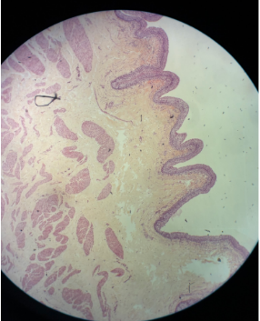
Name the tissue and its epithelium
-Esophagas
-Stratified squmous nonkeratinized
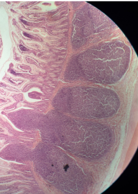
What is shown?
-Appendix
-very short with few glands in the mucosa
-abundant lymphocytes
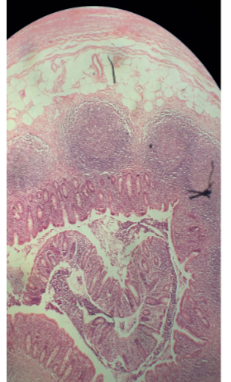
What is this? where is it located?
-Illeum
-Part of the small intestine
-MALT in mucosa
-Peyers patches are the blue staining collections of lymphocytes
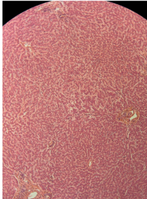
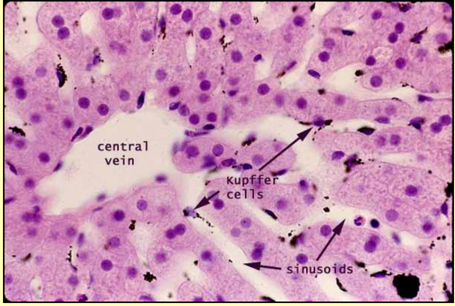
What is this and what are some noteable features?
-Liver
-Hepatocyte is the predominant cell
-Portal triads with central vein (the big white circles)
-Sinusoids are the spaces between the rows of hepatocytes
-Kupffer cells in sinusoids
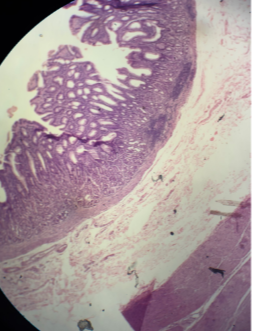
-What is this?
-Colon
-Has a lack of vili
-Abundant goblet cells

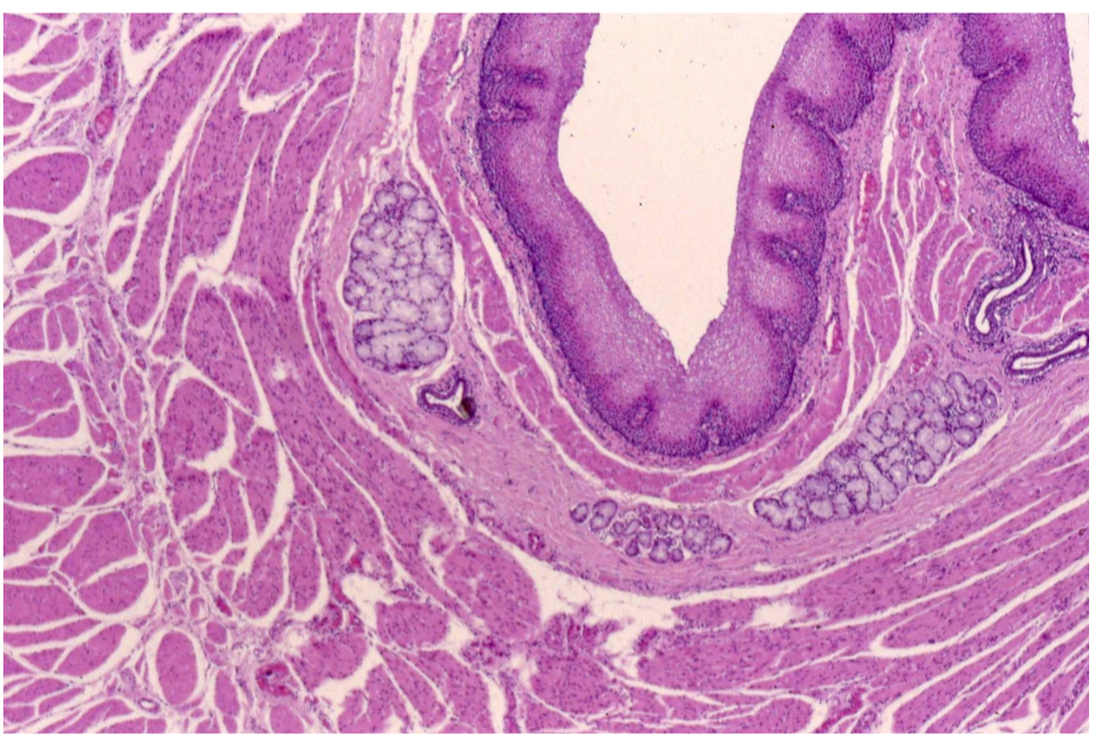
What is this? How do you know?
-Duodenum
-Villi of the mucosa
-Duct of pancreas is the circle guy surrounded in pink on the right
-Brunners glands are the pink lines in/under the villi
-Myentric nerve plexus
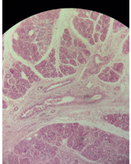
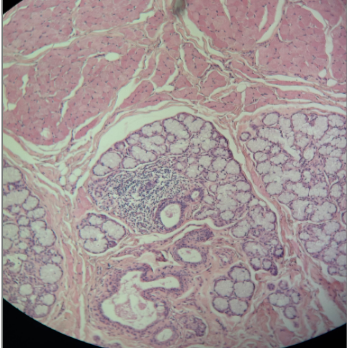
What is pictured?
-submaxillary gland
-mucos and serous secreting cells
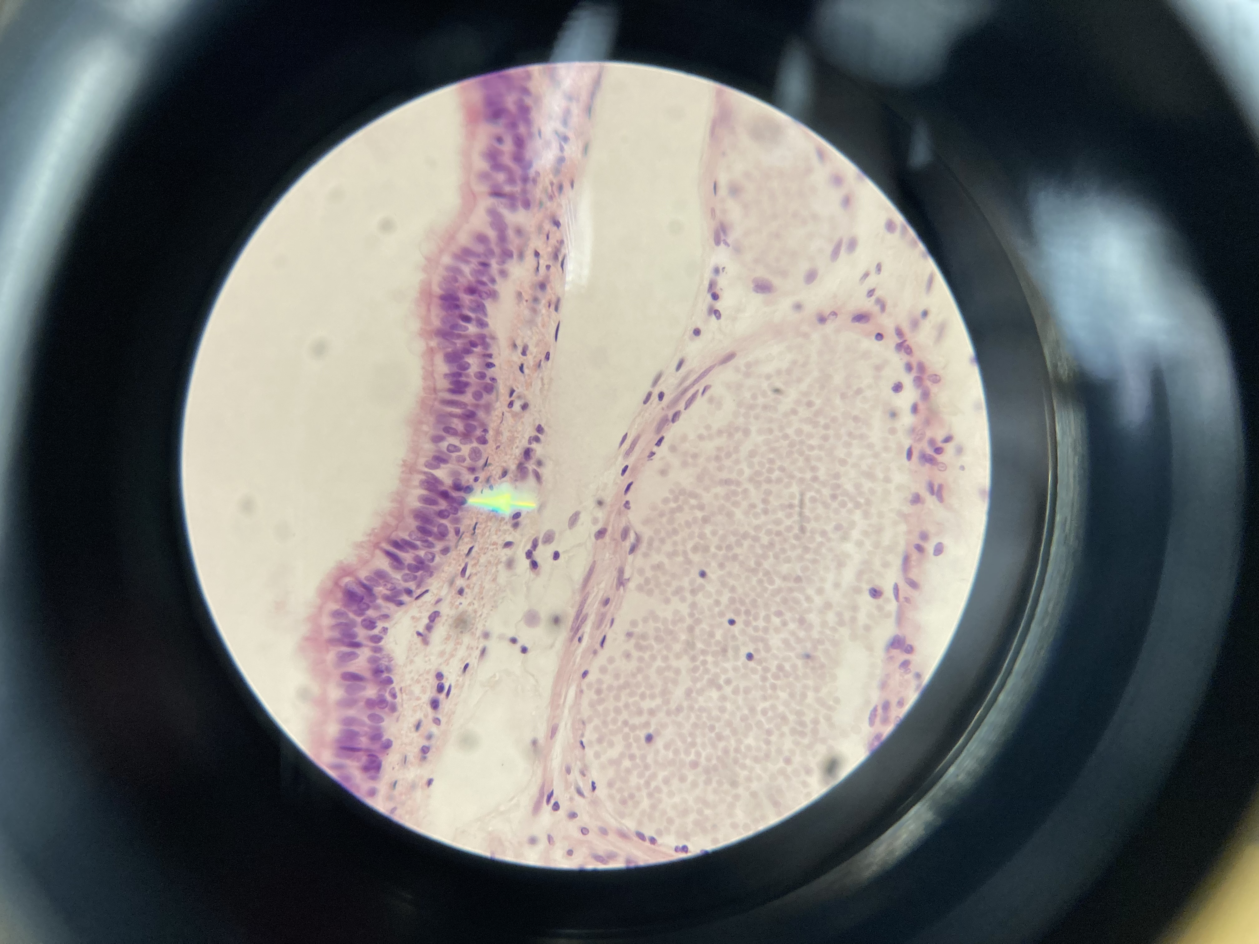
-What is pictured? What two cells are involved?
-Human Stomach (fundis)
-The image on the back is a mammal stomach
-Parietal cells (make HCl, eosinophilic cytoplasm)
-Chief cells (make pepsinogen, basoohilic cytoplasm)
What is this?
-Lip (specifically of a primate)
-Thin skin and mucous membrane
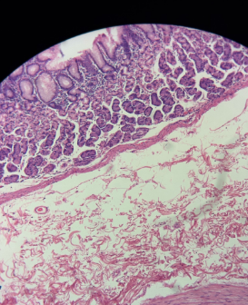
What is shown?
-Trachea
-stratified squamous nonkeratinized epi
-Mucosa with pseudo stratified ciliated columnar epi. with goblet cells
-Submucosa- seromucous glands
-‘C’ shaped hyaline cartilage
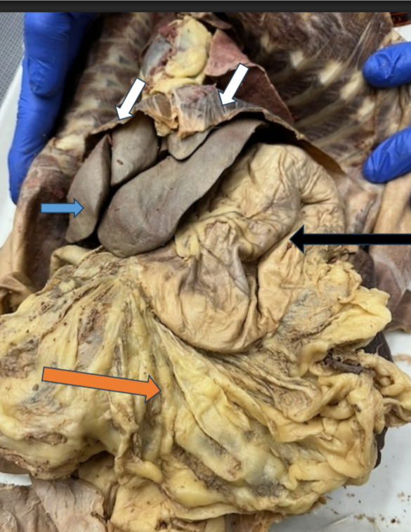
What are each of the arrows pointing to?
-Diaphragm (white arrows)
-Liver
-Stomach
-Greater omentum
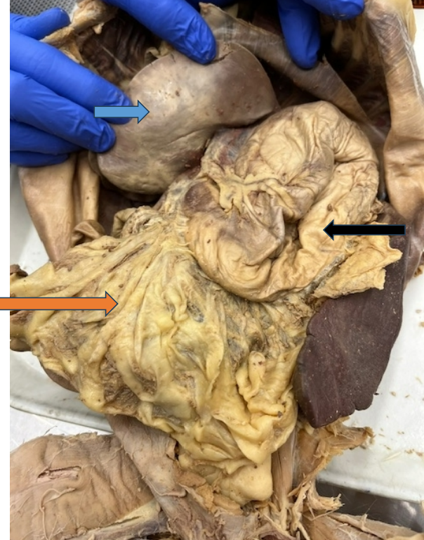
What are the arrows pointing to?
-liver reflected
-stomach
-greater omentum
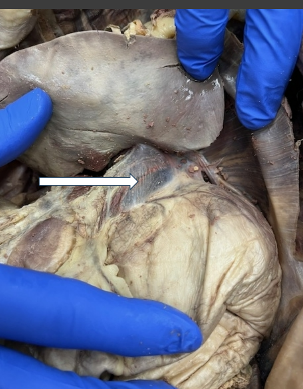
What is the arrow pointing to?
Lesser omentum
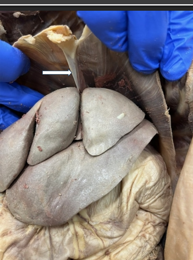
What is the arrow pointing to? What two organs is it between?
-The falciform ligament
-Between the diaphragm and liver
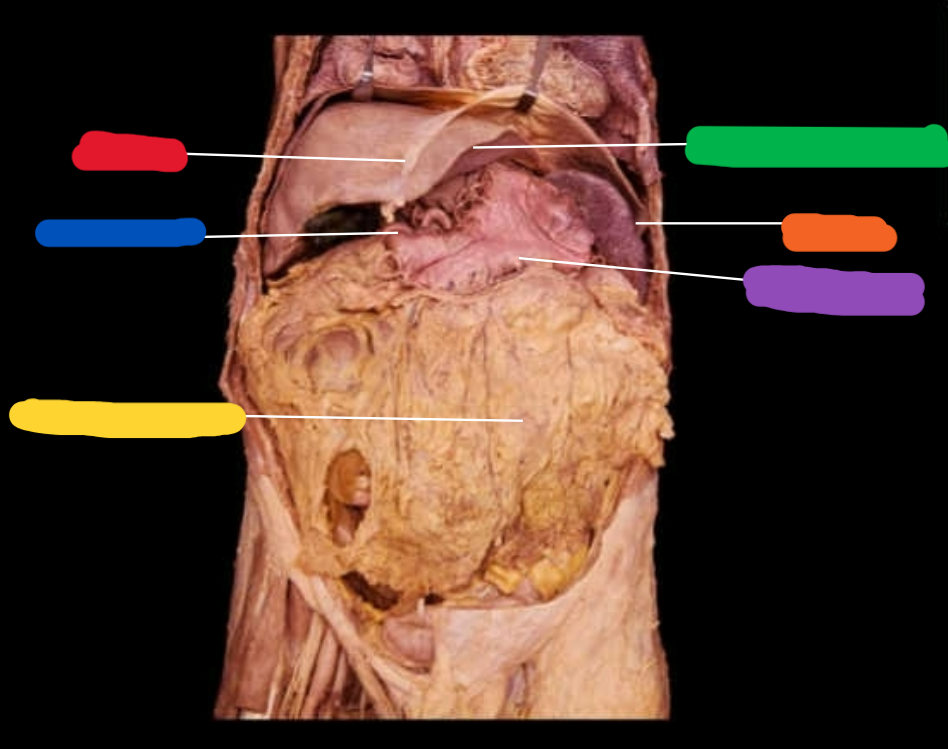
-What are the highlighted regions
-Falciform ligament
-Spleen
-Stomach
-Liver
-Gallbladder
-Greater omentum
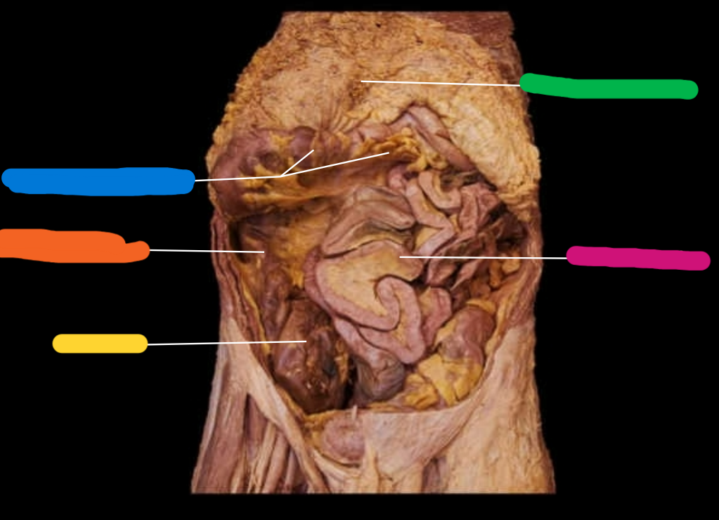
Name the following features highlighted
-Greater omentum
-Small intestine
-Epiploic appendages
-large intestine
-cecum
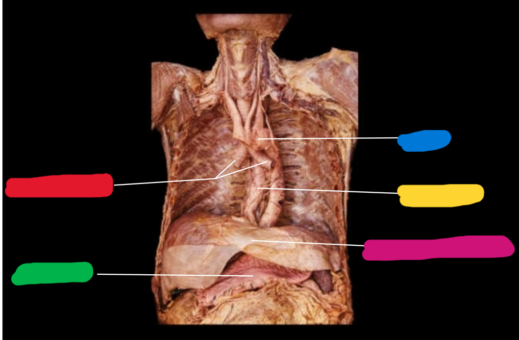
What features are highlighted?
-Aorta
-Esophagus
-Respiratory diaphragm
-Primary bronchi
-Stomach
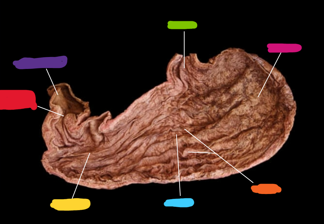
What organ is this? What are the features?
-Cardia
-Fundus
-Rugae
-Body
-Pylorus
-Pyloric sphincter
-Duodenum