BIO 202 (MCC) - The Cardiovascular System - Heart
1/18
There's no tags or description
Looks like no tags are added yet.
Name | Mastery | Learn | Test | Matching | Spaced | Call with Kai |
|---|
No analytics yet
Send a link to your students to track their progress
19 Terms
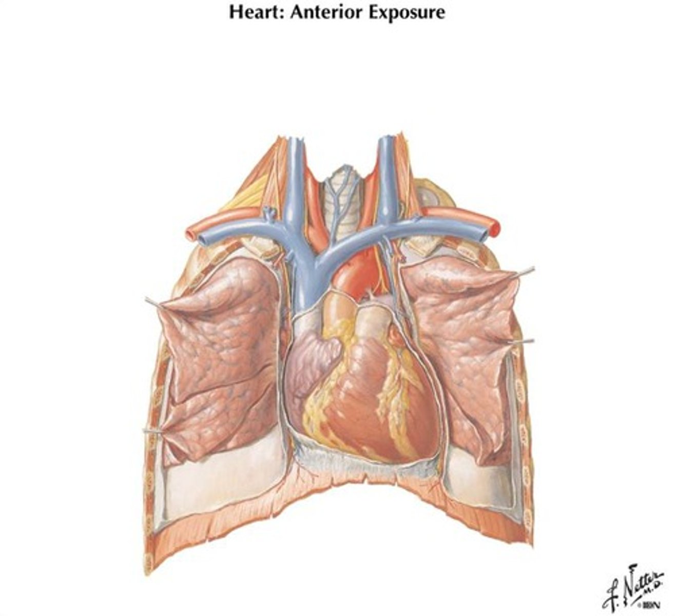
Where is the heart located?
Middle mediastinum.
2/3 of the heart's mass is just barely to the left of the midline.
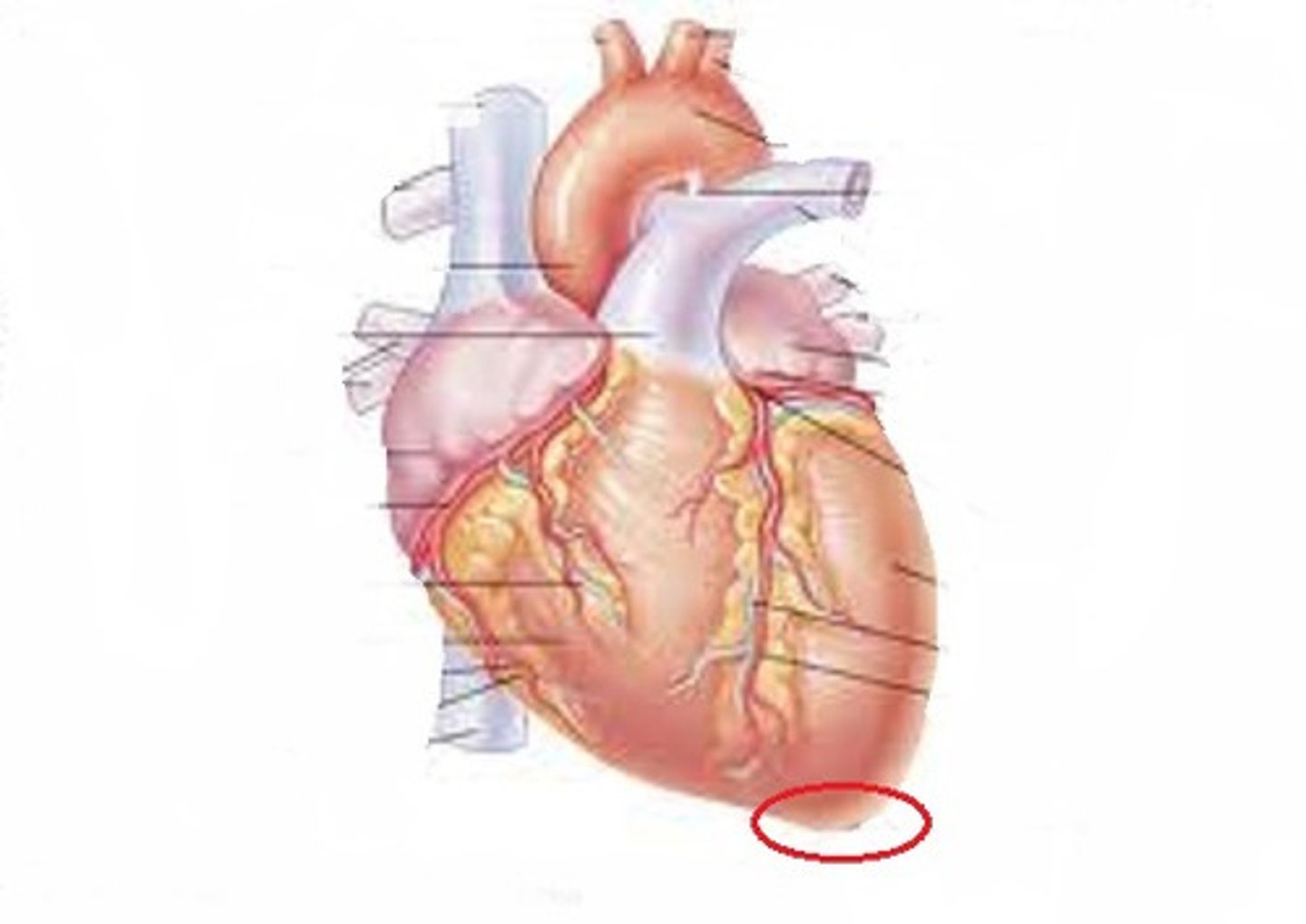
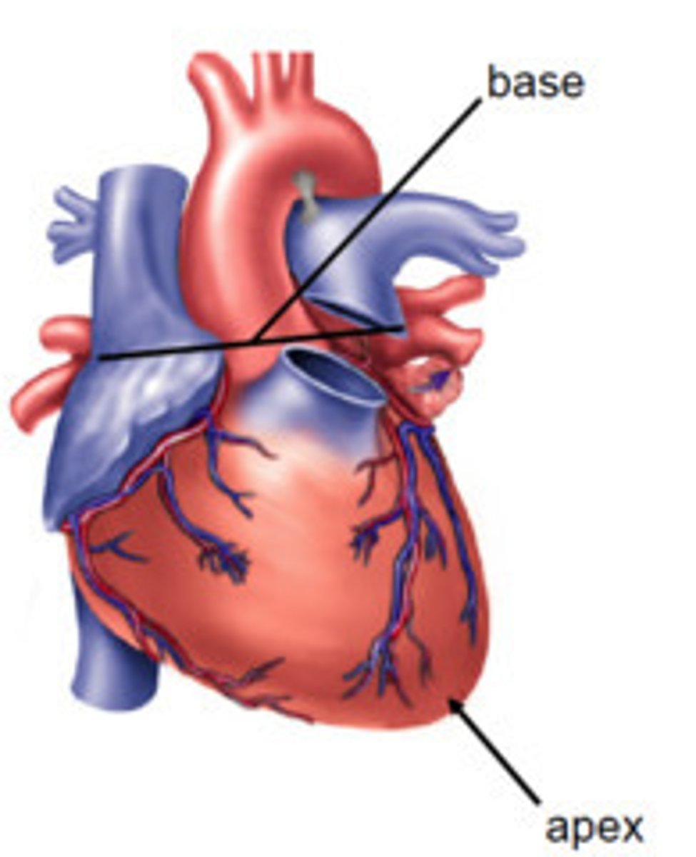
Apex of the heart
Projects inferiorly and laterally
Base of the Heart
Tipped up medially and posteriorly
Pericardium
Membrane that surrounds and protects the heart and retains its position in the mediastinum
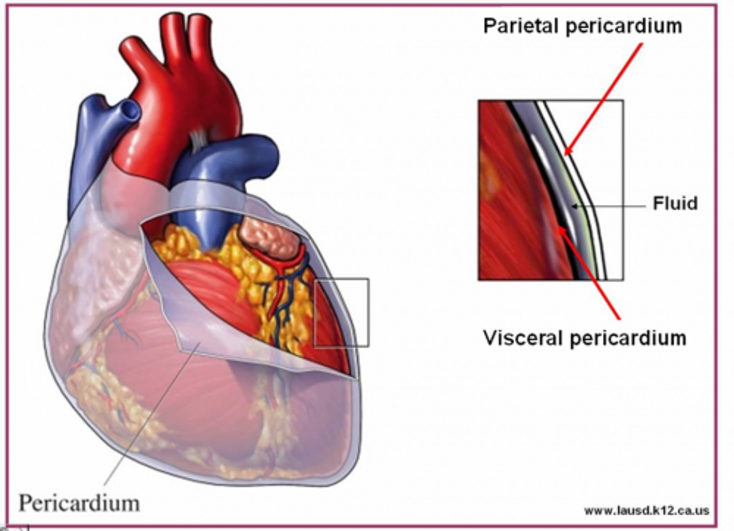
Fibrous Pericardium
A very dense and non-flexible connective tissue that helps protect and anchor the heart.
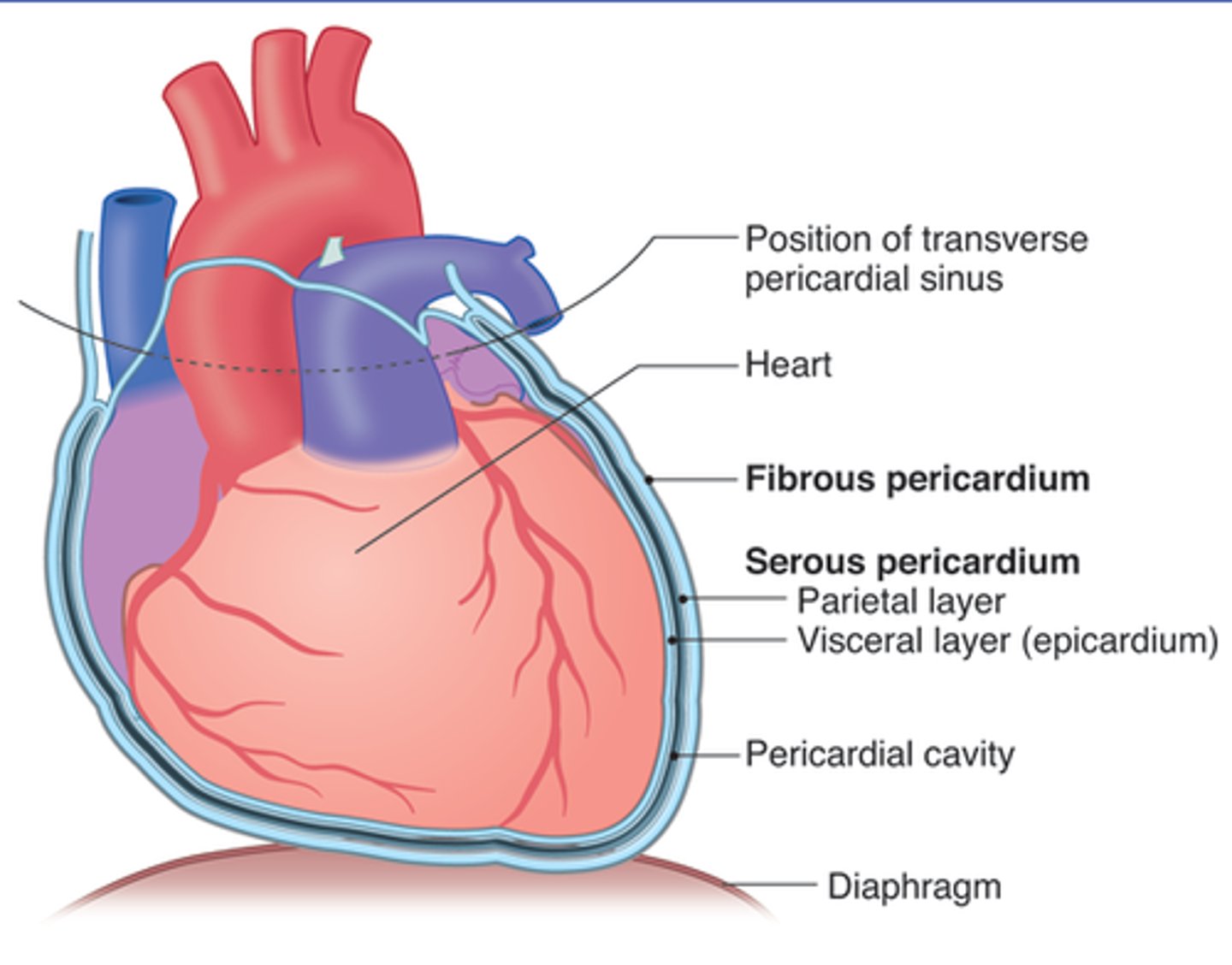
Serous Pericardium
Subdivided into a Parietal Layer which adheres to the outermost fibrous layer and a Visceral Layer which is also viewed as the outer surface of the heart wall.
A thin pericardial fluid lubricates the space between the visceral and parietal pericardium
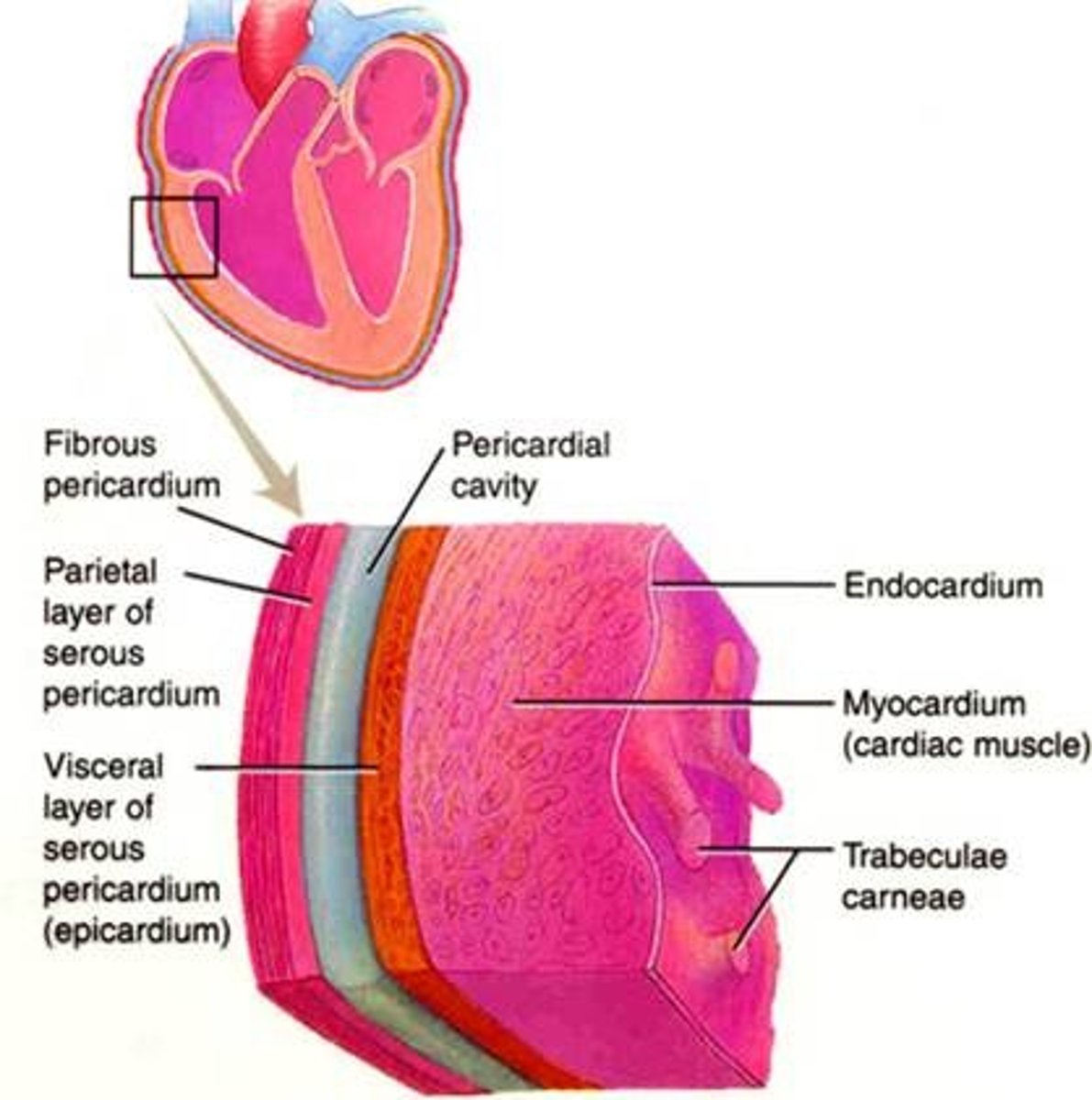
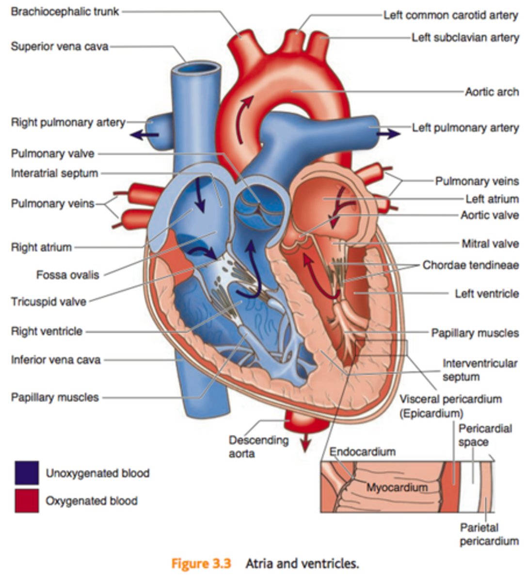
Layers of the heart wall
From superficial to deep they are:
Epicardium
Myocardium
Endocardium
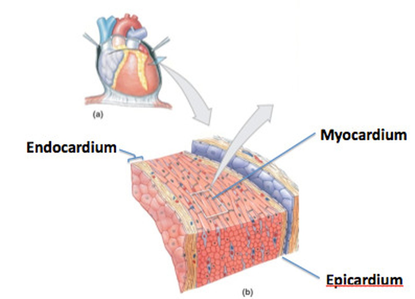
Epicardium
Thin, transparent outer layer of the heart wall, is also called the visceral layer of the serous pericardium.

Myocardium
thick middle layer, is composed of cardiac muscle
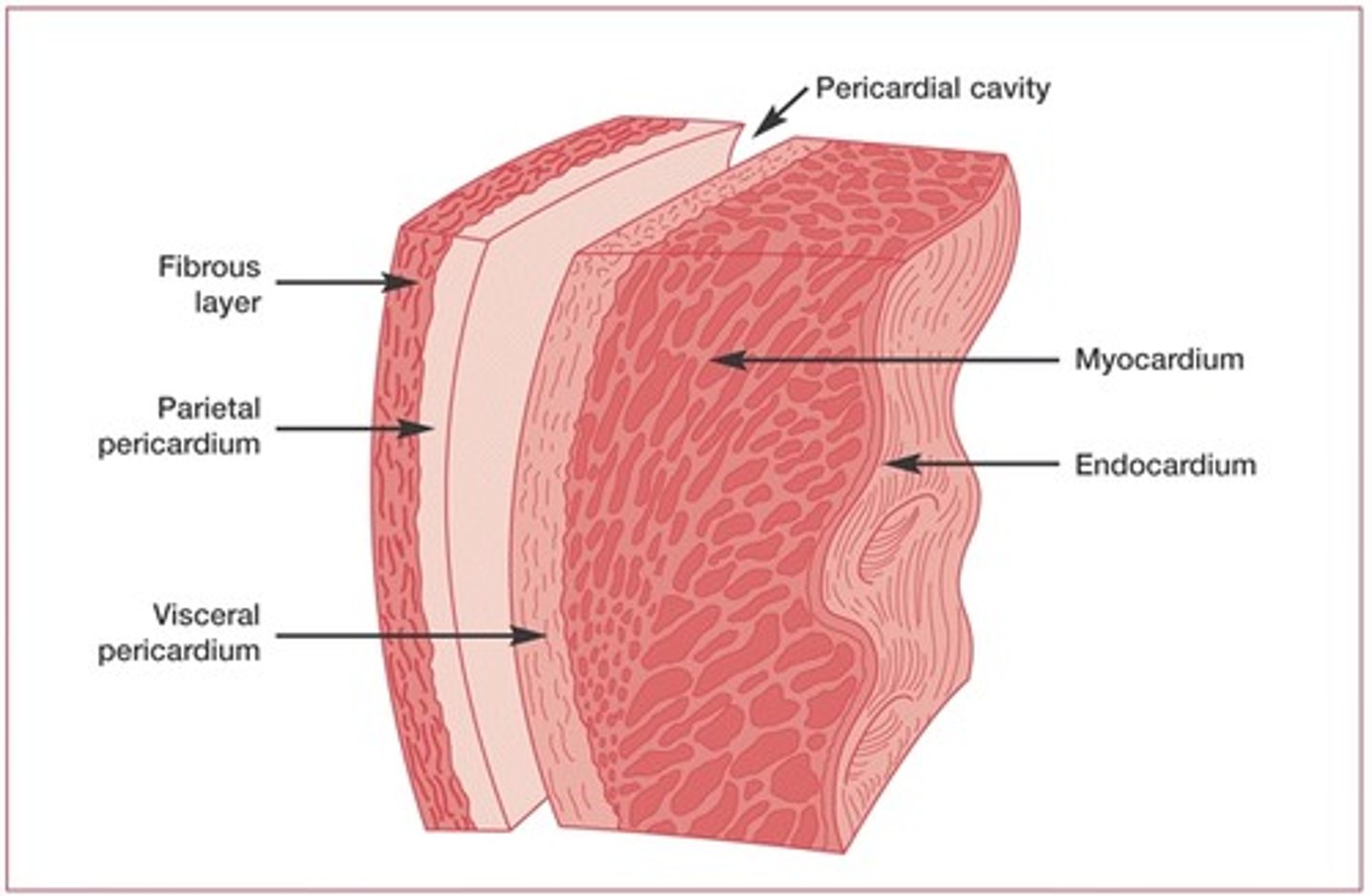
Endocardium
A simple squamous epithelium (known throughout the circulatory system as "endothelium")
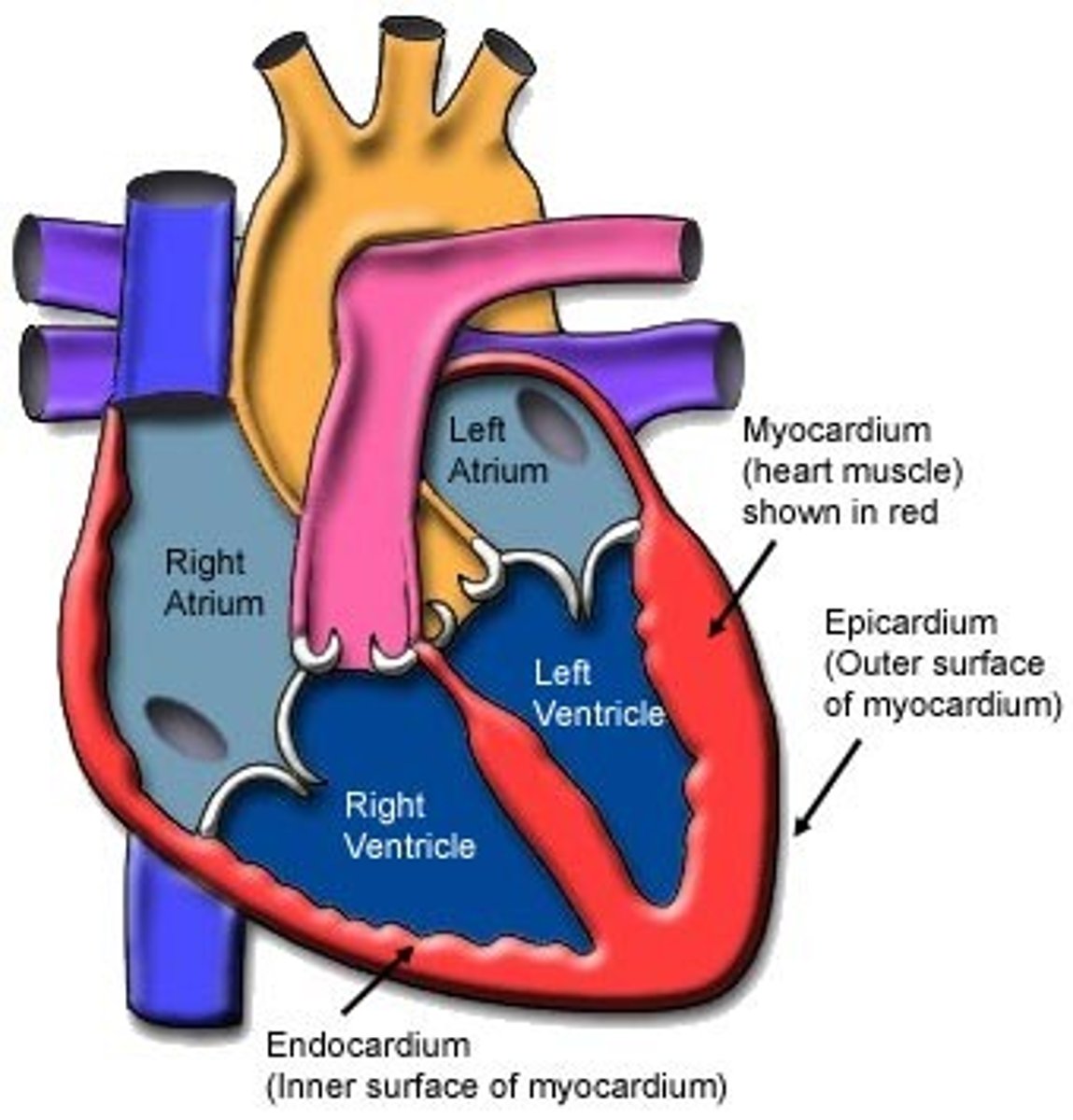
What are the 4 Chambers of the heart?
Right/Left Atria (upper 2 chambers)
Right/Left Ventricles (Lower 2 chambers)
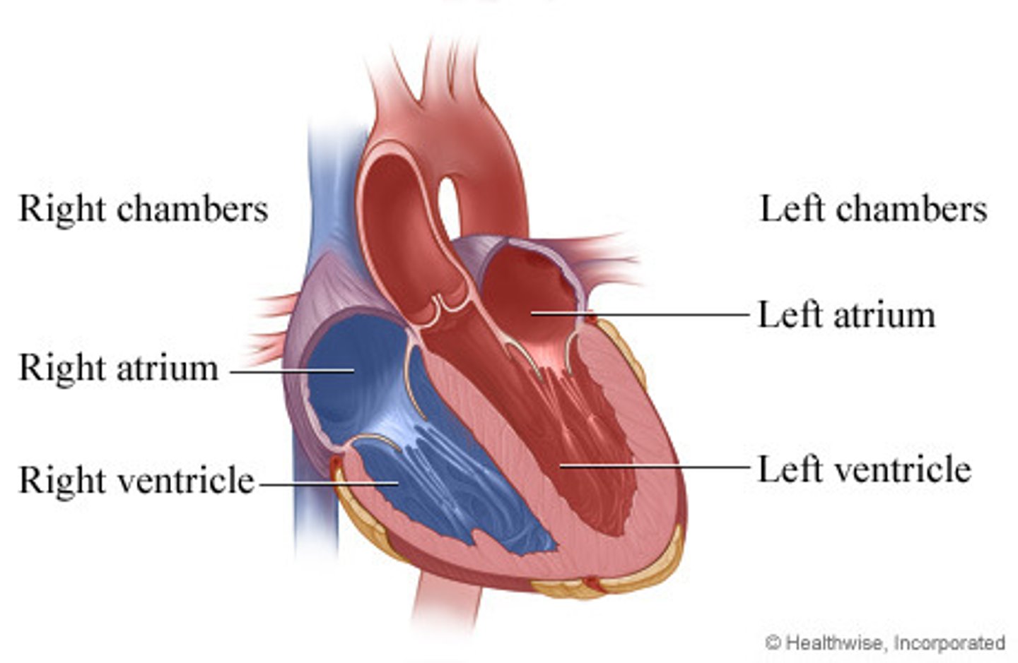
Right Heart
Consists of the right atrium and right ventricle, taking venous blood from the body and pumping it to the lungs for oxygenation
Left Heart
Consists of the left atrium and left ventricle, taking freshly oxygenated pulmonary blood and pumping it systemically (meaning to the body).
Top part of the heart
The "top part of the heart" is a weak pump consisting of the right and left atria. It loads the ventricles by giving an "atrial kick" before the ventricles contract.
Bottom Part of the Heart
The "bottom part of the heart" is a strong pump consisting of the right and left ventricles. It's the main pump for the pulmonary and systemic circuits
Atrial Kick
Responsible for only a 20% increase in the amount of blood ejected by the ventricles - important, but not essential.
"A-Fib" No atrial kick - blood still flows into the ventricles due to gravity.
Heart Valves
Blood always flows from an area of high pressure to an area of low pressure.
The flow of blood (dictated by differences in pressure, not muscles), operates the valves of the heart.
Atrioventricular valves: Open to allow blood to flow from the atria into the ventricles.
Outflow (semilunar): Open to allow blood to flow from the ventricles, into the outflow vessels.
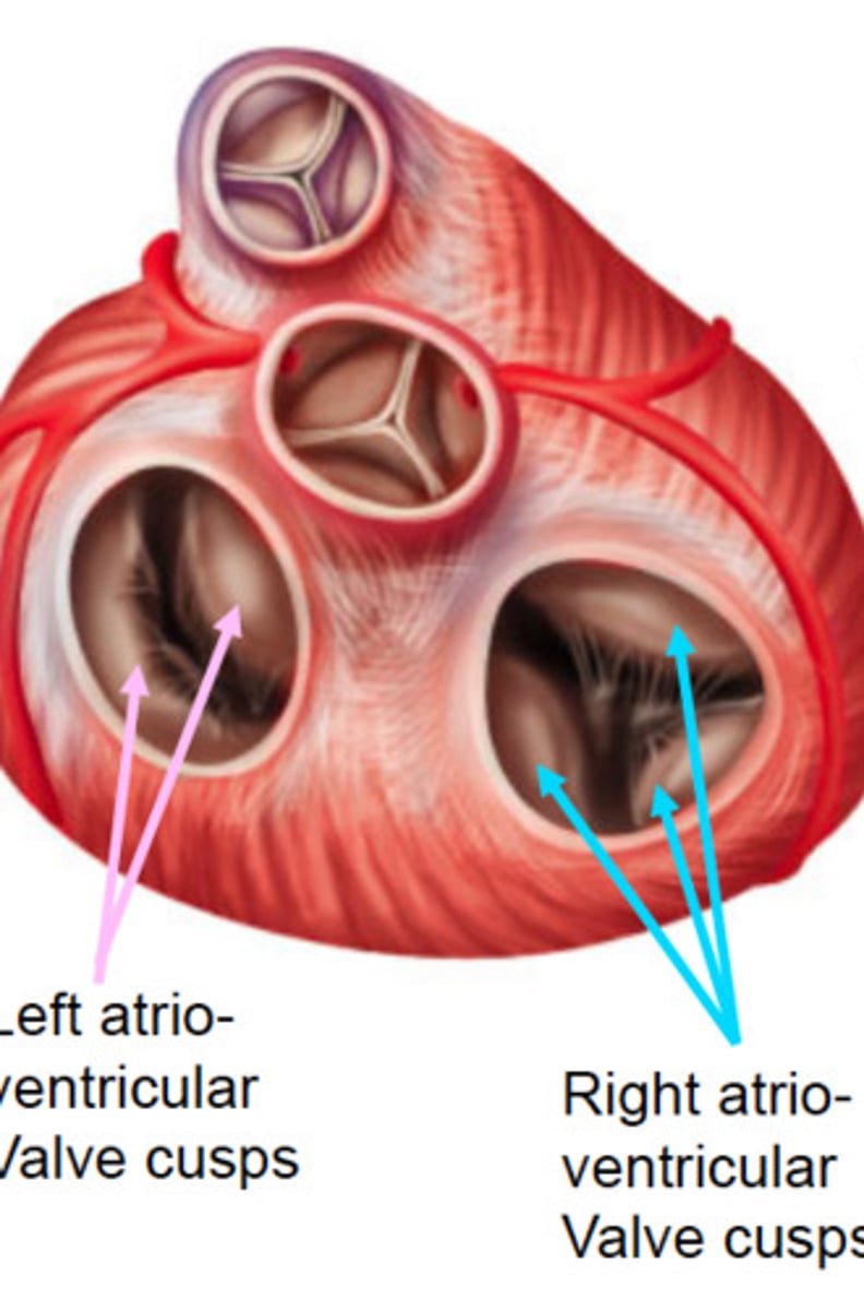
Atrioventrical valves (AV)
Positioned at the entrance to the ventricles:
The right AV valve (also called the tricuspid valve because of its three leaflets or cusps) opens into the right ventricle.
The left AV valve (also called the bicuspid or mitral valve) opens into the right ventricle.
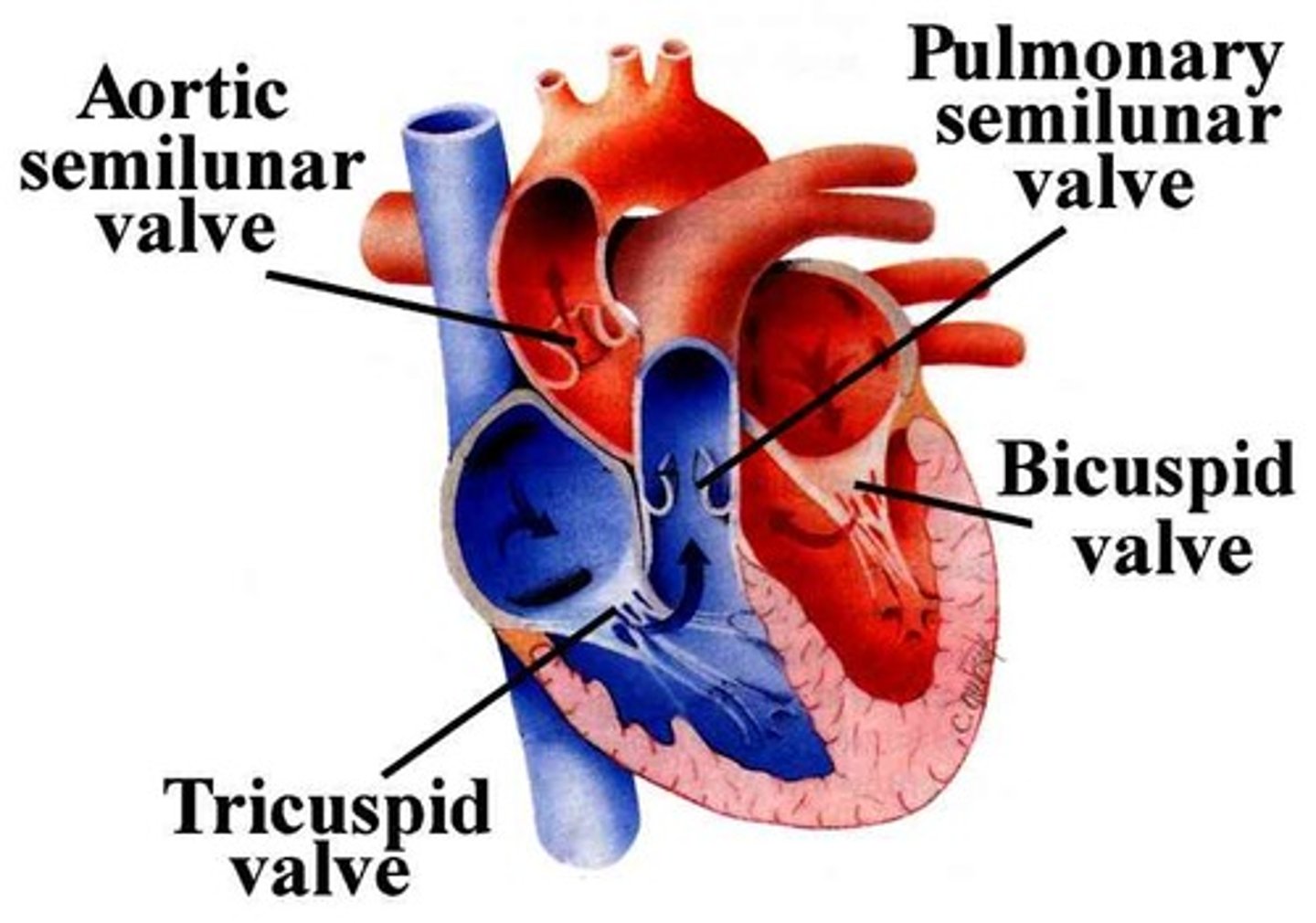
Outflow (Semilunar Valves)
The outflow valves are positioned at the entrance to the outflow vessels leading into the pulmonary and systemic circulation:
The right outflow valve (also called the pulmonary semilunar valve) opens into the pulmonary trunk.
The left outflow valve (also called the aortic semilunar valve) opens into the aortic arch.
