Knee Radiography: Projections, Anatomy, and Positioning Techniques
1/344
There's no tags or description
Looks like no tags are added yet.
Name | Mastery | Learn | Test | Matching | Spaced | Call with Kai |
|---|
No analytics yet
Send a link to your students to track their progress
345 Terms

Name this projection
AP knee

Where does the CR enter for this projection?
½" inferior to patellar apex

If the patient is sthenic (19-24 cm) how is the CR directed?
0 degrees (perpendicular to IR)

Label the image: A
femur

Label the image: B
patella
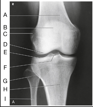
Label the image: C
lateral femoral epicondyle

Label the image: D
lateral femoral condyle

Label the image: E
Lateral tibial plateau

Label the image: F
intercondylar eminence

Label the image: G
head of fibula

Label the image: H
tibia

Label the image: I
fibula
What type of lateral projection is performed for the knee?
Mediolateral
How many degrees is the knee flexed for the lateral projection?
20-30 degrees
If there is trauma for a lateral knee, how should you modify the position?
Bend the knee no more than 10 degrees
What anatomy should be perpendicular to the IR in the lateral projection?
Femoral epicondyles and patella
Where does the CR enter for the lateral projection of the knee?
1" distal to the medial epicondyle
How is the tube angled for a lateral projection (degree and direction)?
5-7 degrees cephalic
What anatomical structure is in profile for the lateral projection of the knee?
Patella
How do you know if the knee has been under-rotated (lateral knee)
too much superimposition of the tibia and fibular head; the anterior surface of the medial condyle will appear farther from the patella
How do you know if the knee has been over-rotated (lateral knee)
tibiofibular joint open; the anterior surface of the medial condyle will appear closer to the patella
What anatomy should be evaluated for a true lateral position of the knee?
Femoral condyles superimposed posteriorly, open patellofemoral joint, and fibular head and tibia are slightly superimposed
How do you know if you used the proper angulation for the lateral projection of the knee?
The femoral condyles will be superimposed inferiorly
Why is the AP weight-bearing projection of the knee performed?
Useful for evaluating joint space narrowing and showing articular cartilage disease on the posterior surface of the femoral condyles
Where should the CR enter for the AP weight-bearing projection?
Perpendicular to MSP, entering ½" inferior to the patellar apex
Describe varus
occurs when the tibia turns inward, causing the knees to turn outward
Alternative names for varus
genu varum or "bowlegged"
Describe valgus
occurs when the tibia turns outward, causing the knees to turn inward
Alternative names for valgus
genu valgum or "knock-kneed"
Which oblique projection demonstrates the tibiofibular joint?
AP oblique medial rotation
Which oblique projection demonstrates the medial aspect of the knee joint?
AP oblique lateral rotation
Which oblique projection demonstrates the patella over the lateral femoral condyle?
AP oblique lateral rotation
Which oblique projection demonstrates the lateral aspect of the knee joint?
AP oblique medial rotation
Which oblique projection demonstrates the patella over the medial femoral condyle?
AP oblique medial rotation
How much should the leg be rotated for the AP oblique projection of the knee?
45 degrees

Name this projection.
AP oblique lateral rotation

What is demonstrated?
Medial aspect (distal femur, patella, medial tibial condyles, and fibula)

Label the image: A
femur

Label the image: B
patella

Label the image: C
medial femoral condyle

Label the image: D
lateral femoral condyle

Label the image: E
Lateral tibial plateau

Label the image: F
medial tibial plateau

Label the image: G
medial tibial condyle
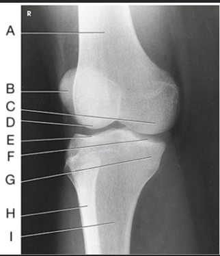
Label the image: H
fibula

Label the image: I
tibia

Name this projection
AP oblique medial rotation

What is demonstrated?
Lateral aspect (distal femur, patella, lateral tibial condyle, fibular head, and proximal tibiofibular joint)

What joint is demonstrated?
Proximal tibiofibular joint

Label the image: A
patella

Label the image: B
medial femoral condyle

Label the image: C
lateral femoral condyle

Label the image: D
medial tibial plateau

Label the image: E
Lateral tibial plateau

Label the image: F
medial tibial condyle

Label the image: G
lateral tibial condyle
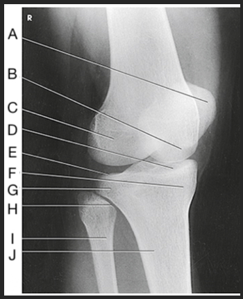
Label the image: H
tibiofibular articulation

Label the image: I
fibula

Label the image: J
tibia
If the patient measures 17 cm from ASIS to tabletop, how should the CR be directed for the AP oblique projection of the knee?
3-5 degrees caudad
If the patient measures 26 cm from ASIS to tabletop, how should the CR be directed for the AP oblique projection of the knee?
3-5 degrees cephalic
The Holmblad method is what type of projection?
PA axial IC fossa
The Holmblad method demonstrates which anatomy?
IC fossa and the posteroinferior surfaces of femoral and tibial condyles
How much is the knee flexed for the Holmblad method?
70 degrees (from anterior thigh to IR)
Where does the CR enter and exit for the Holmblad and Camp-Coventry methods?
Enters the popliteal fossa and exits the patellar apex
Which method places the tibia and fibula 40-50 degrees to the IR and has a CR that is perpendicular to the lower leg?
Camp-Coventry method
What is the method name for the AP axial projection of the IC fossa?
Béclére Method

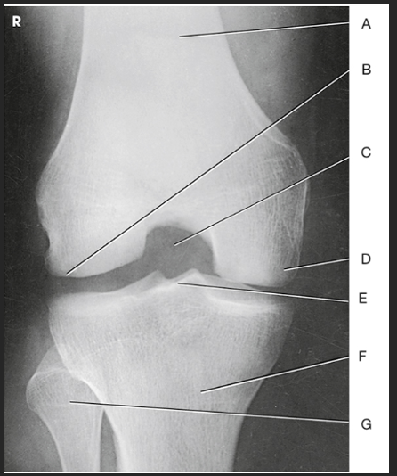
Name this projection.
AP axial IC Fossa

Name the method used.
Béclére Method
How do you know this image is a Beclere Method
Marker (line yourself up... only AP IC fossa) and is the smallest unilateral IC fossa (not elongated like Camp-Coventry and not as big as Holmblad)

Label the image: A
femur

Label the image: B
lateral condyle

Label the image: C
intercondylar fossa

Label the image: D
medial condyle

Label the image: E
intercondylar eminence

Label the image: F
tibia

Label the image: G
fibula
How does the IC fossa appear in the AP axial projection compared to the PA axial projection? (What sets the Beclere apart from the Holmblad/Camp-Coventry images?)
In Merrill's, the Beclere method makes the IC fossa look smaller. However, because we no longer have 10x12cassettes, we are forced clinically to have a larger OID in the AP axial projection, making the image look magnified compared to the PA axial projections
What are all three projections of the IC fossa looking for?
An open IC fossa (where the patellar apex is not superimposing the fossa)
Why is the PA projection of the patella preferred over the AP?
PA has the least amount of OID
How should the patella be positioned in relation to the IR for the PA patella projection?
Parallel
What type of fracture is best demonstrated in the PA projection of the patella?
Stellate
Where does the CR enter for the PA projection of the patella?
Perpendicular to the mid-popliteal area exiting the patella
Which projection of the patella is used to rule out a fracture before performing the tangential views?
Lateral patella
How many degrees is the knee flexed for the lateral projection of the patella?
5-10 degrees
Where does the CR enter for the lateral patellar projection?
Perpendicular to the mid-patellofemoral joint
What type of fracture is best demonstrated in the lateral projection of the patella?
Transverse fracture
Name the methods used for the tangential projection of the patella.
Settegast/sunrise and Merchant method
How is the patient positioned for the unilateral sunrise view?
Supine or prone with knee flexed until the patella is ⟂ to IR
How do you modify the CR if the patella is not perpendicular to the IR for Settegast Method?
Angle 15°-20° cephalic
The bilateral patellar projection uses what type of ancillary equipment?
An axial viewer device
What SID is used for the bilateral projection of the patellae?
72"
How many degrees are the knees flexed for the bilateral patellar projection?
40 degrees
The tangential projection of the patella places what anatomy in profile?
The patella

Label the image: A
popliteal surface

Label the image: B
adductor tubercle

Label the image: C
medial epicondyle

Label the image: D
medial condyle

Label the image: E
lateral condyle

Label the image: F
lateral epicondyle