CS Exam 2
 Call Kai
 Learn
Learn Practice Test
 Spaced Repetition
 Match
Match Flashcards
 Knowt Play
1/210

There's no tags or description
Looks like no tags are added yet.
Name | Mastery | Learn | Test | Matching | Spaced | Call with Kai |
|---|
No analytics yet
Send a link to your students to track their progress
211 Terms
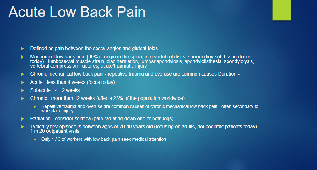
What is acute low back pain defined as?
Define:
Mechanical low back pain
Chronic Mechanical
Acute
Subacute
Chronic
Radiation consider “__?
Typical First Episode?
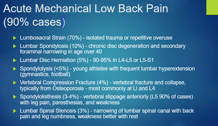
List out the causes of Acute Mechanical Low Back Pain
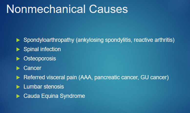
WHat are non mechanical causes?
What is the difference between Simple and Complicated MSK Low Back pain?
Simple = no red flags
Systemic disease and referred pain ruled out
Conservative therapy x 6 weeks (Home Exercise Program?)
List out the red flags for complicated Low BAck Pain
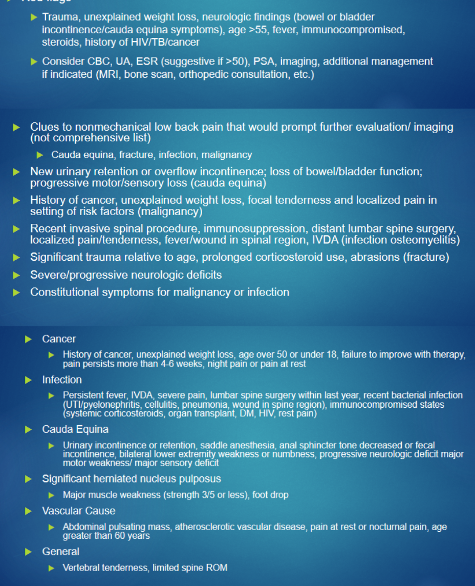
For Acute Low Back pain w/ Radicular Symptoms past knee (4%), Describe the difference criteria for conservative management vs urgent evalulation
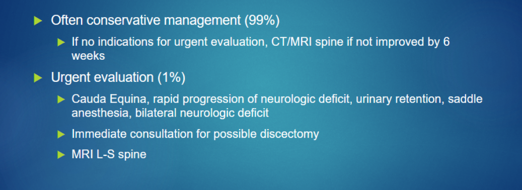
How would you know if the acute low back pain is due to lumbar stenosis?
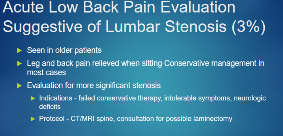
What are the six steps to a MSK exam for acute low back pain
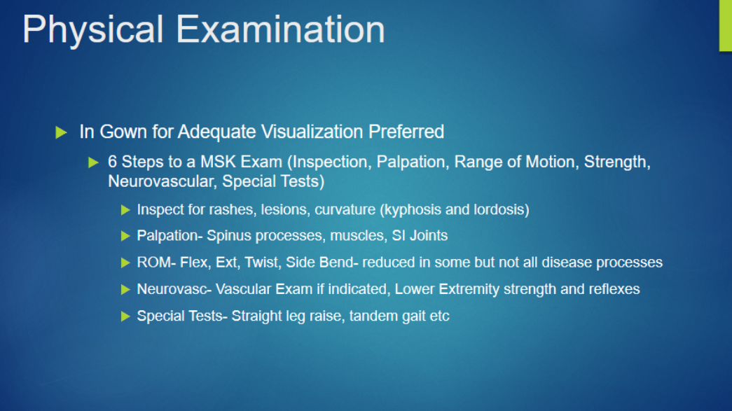
Describe the Imaging used for acute low back pain
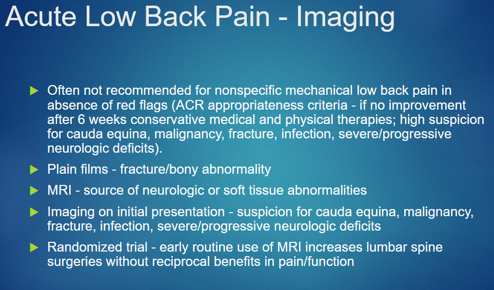
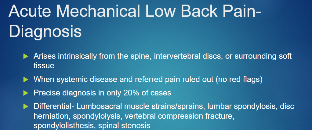
Describe how you could diagnose Acute Mechanical Low Back Pain;
Differentials?
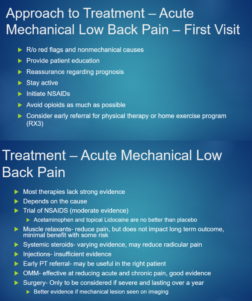
List out some treatments for acute mechanical low back pain;
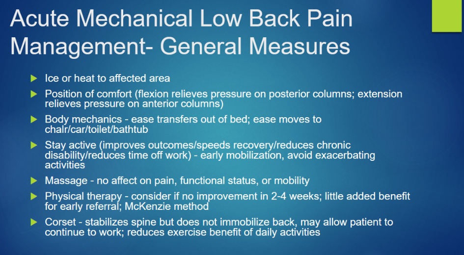
Describe Management of low back pain
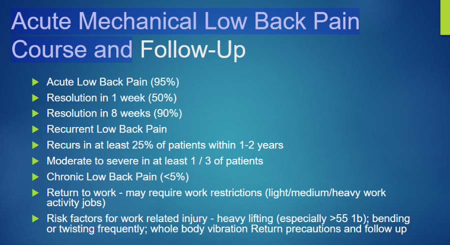
Describe the aftermath/resolution of Low back pain
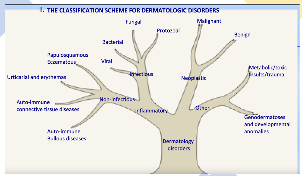
Describe the classification of Dermatological Disorders
What are the seven functions of the skin
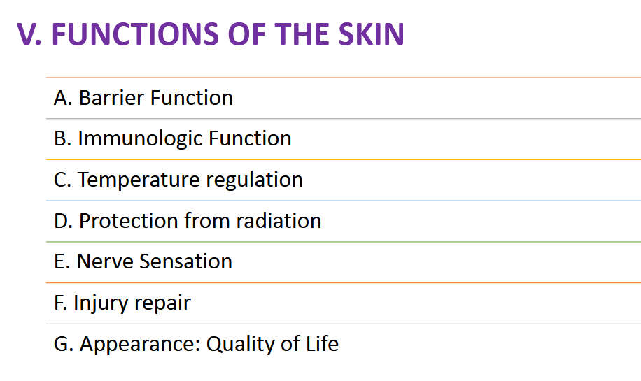
List the Diseases Associated w/ irregularity in each of the 7 functions of skin
Barrier function: ATOPIC DERMATITIS , (ECZEMA)
Immunologic function: MYCOSIS FUNGOIDES
a type of skin lymphoma. Dysfunction of cutaneous lymphocytes increases risk for bacterial and fungal skin colonization and subsequent infection.
Temperature regulation: ERYTHRODERMA (Exfoliative dermatitis)
>90% of the skin is erythematous (red) and/or inflamed. Extensive cutaneous vasodilation can cause hypothermia and high-output cardiac failure leading to organ failure.
Protection from radiation: ALBINISM: Higher risk of MELANOMA
Sensation: chronic stasis ulcer on the lower extremity of a patient with peripheral neuropathy related to diabetic neuropathy.
Injury repair: POST –RADIATION TREATMENT FOR SQUAMOUS CELL CARCINOMA
Appearance: Quality of life:
vitiligo
HIV-associated lipoatrophy, characterized by loss of fat throughout the face.
Describe the three layers of the skin; function/structure
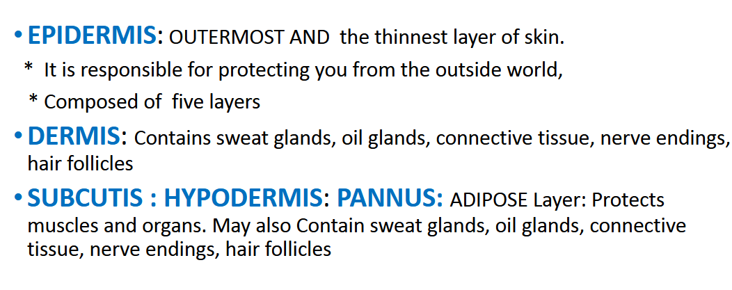
What is the function of the epidermis
Describe the five layers of the Epidermis; Structure/Function
Stratum corneum
Corneal layer Made of desquamating keratinocytes.
Thick outer layers of flattened keratinized non-nucleated cells provide a barrier against trauma and infection
Stratum lucidum
Lucid or Clear layer: Present only in the palms of the hands and soles of the feet.
The keratinocytes that compose the stratum lucidum are dead and flattened.
These cells are densely packed with eleiden, a clear protein rich in lipids, derived from keratohyalin, which gives these cells their transparent (i.e., lucid) appearance and provides a barrier to water.
Stratum granulosum
Granular layer: The keratinocytes produce lipids
(the mortar) in the granular cell layer and secretes it into the extracellular space between the keratinocyte forms a water barrier that Keeps water in the skin
Spinous layer
Center of epidermis.
Has a “spiny” appearance due to desmosomal junctions which hold the keratinocytes together.
Stratum basale
Basal layer
The basal layer is the source of epidermal stem cells.
Cell division occurs here.
Keratinocytes start in the basal layer and move upwards.
Melanocytes are located here
What are the four Diseases associated w/ the epidermis
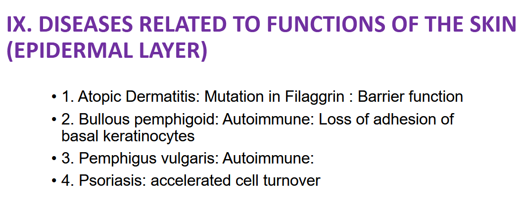
Describe Bullous pemphigoid:
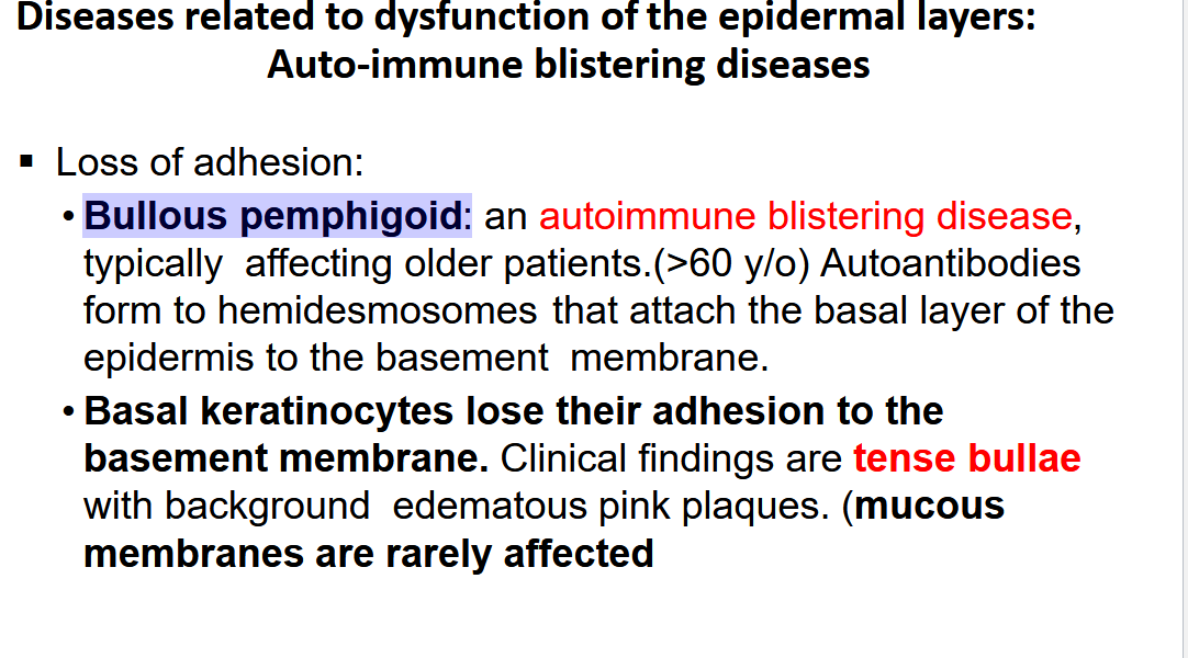
Describe Pemphigus Vulgaris
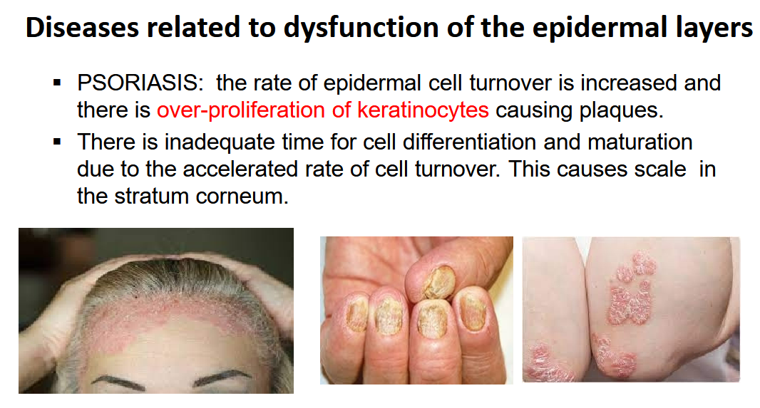
Describe Psoriasis
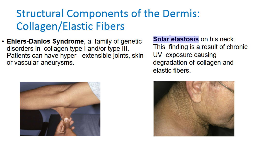
Describe Ehlers-Danlos Syndrome and Solar elastosis
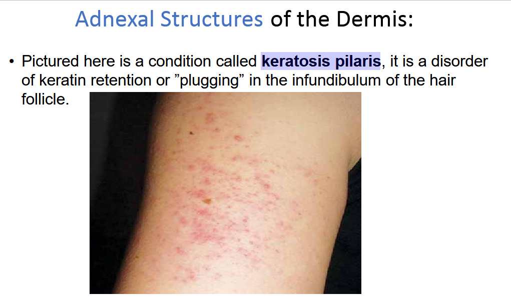
Describe keratosis pilaris
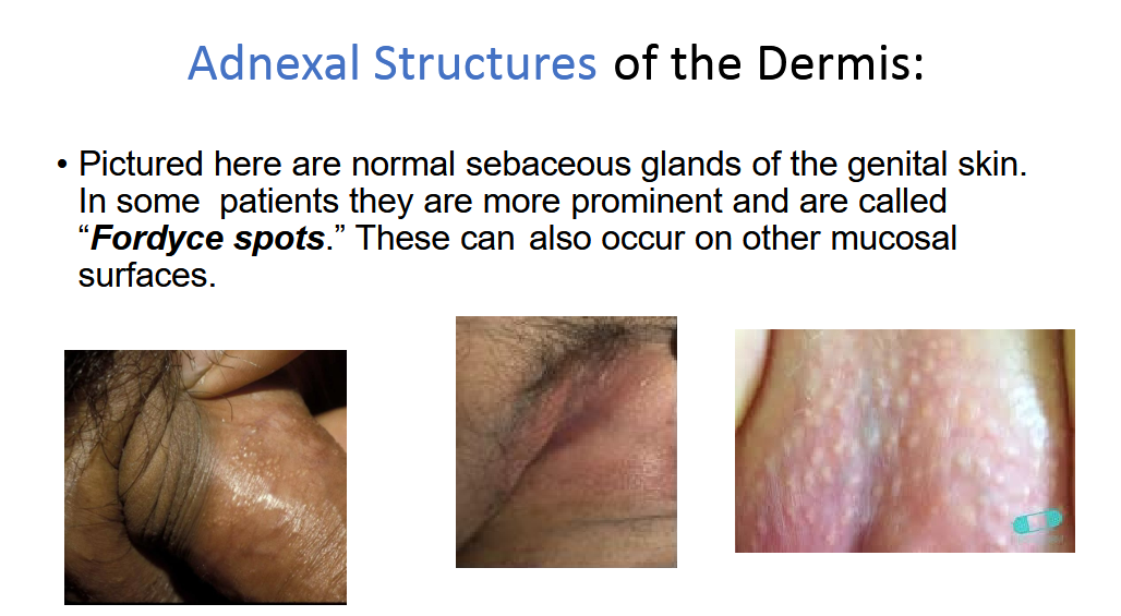
What are Fordyce spots?
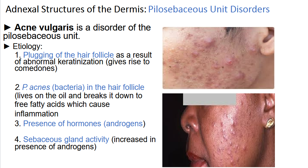
Describe Acne Vulgaris
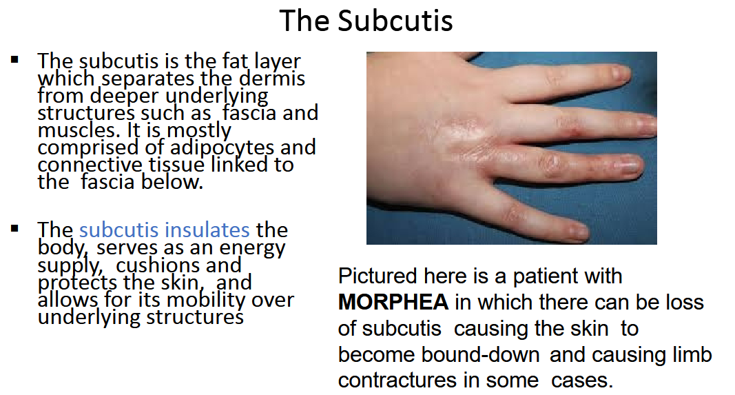
Describe Morphea
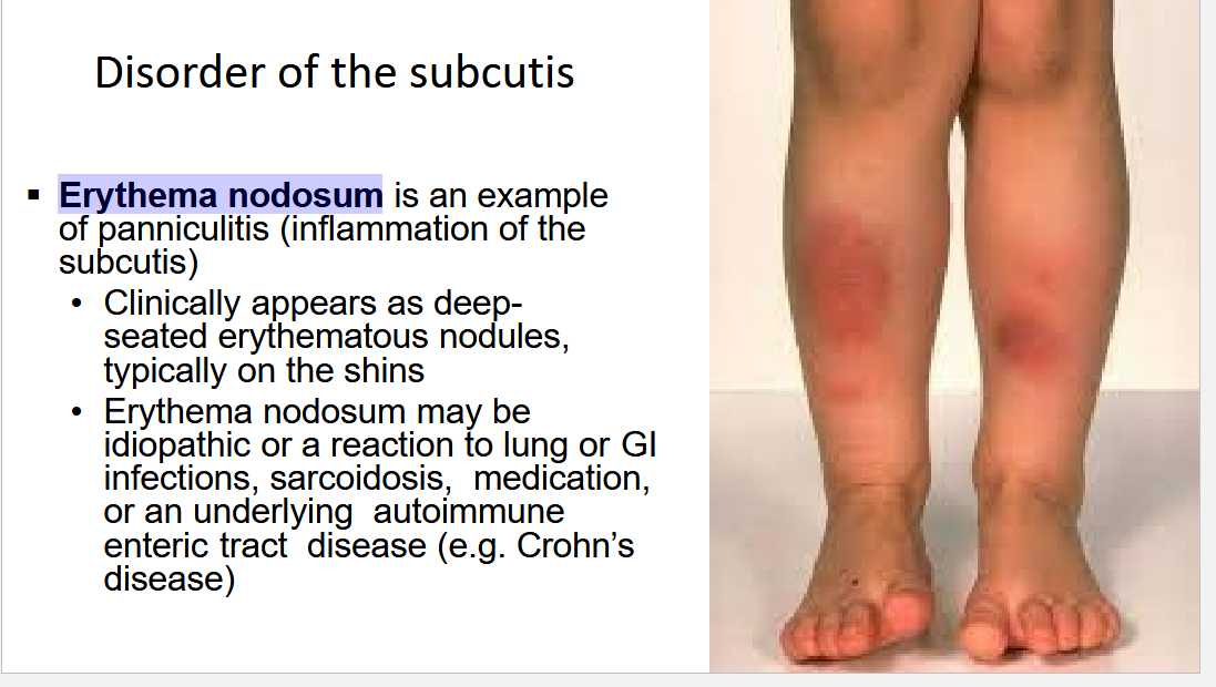
Describe Erythema nodosum
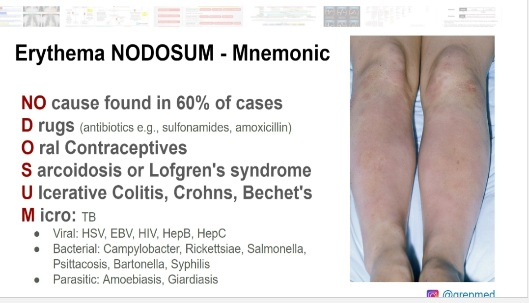
Erythema NODOSUM mneumonic
List out Lymph node terminology and description
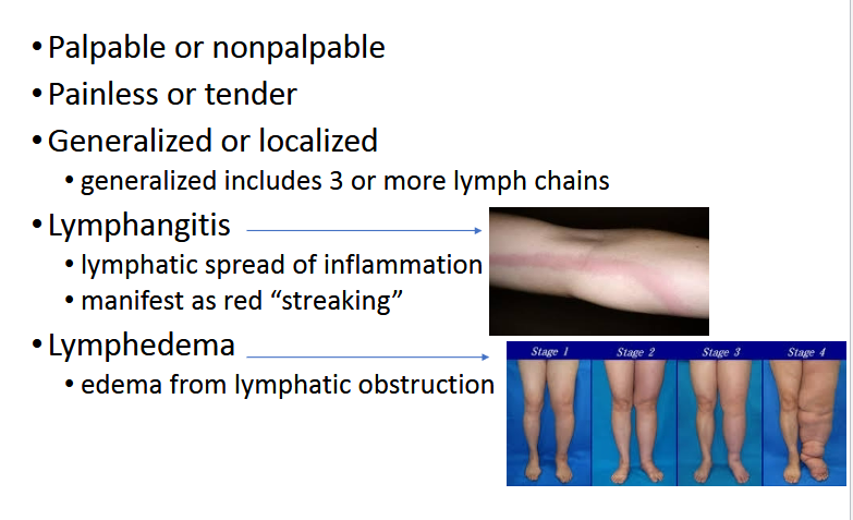
Describe the Upper Extremety Lymphatic System
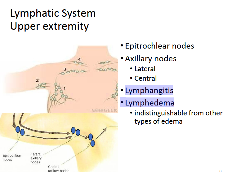
What is the difference between Lymphangitis and Lymphedema
Lymphangitis is an active infection; Lymphedema is a fluid retention problem, often a long-term consequence of other issues, including past infections or cancer treatment
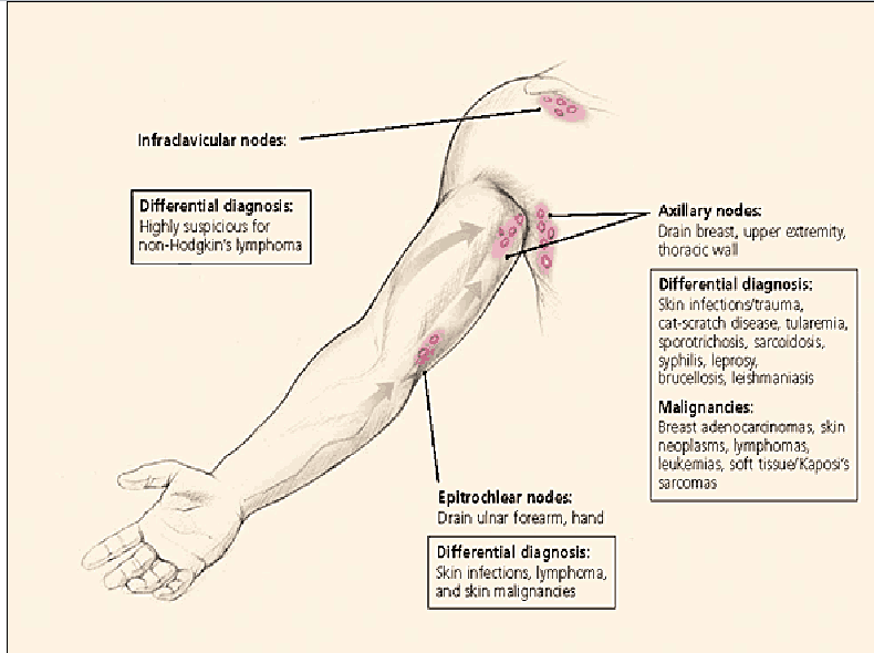
What are the Differential DIagnosis of:
Infraclavicular nodes
epitrochlear Nodes
Axillary nodes
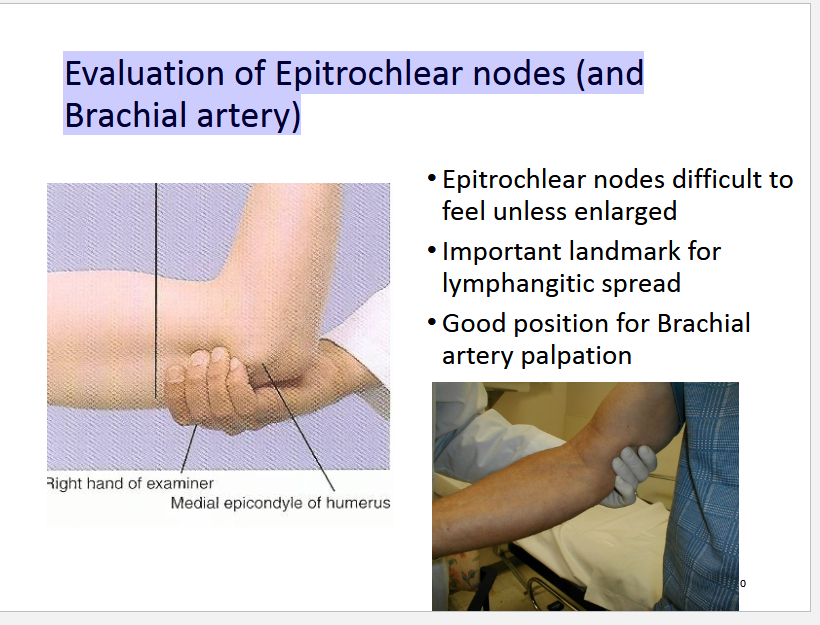
Describe Evaluation of Epitrochlear nodes (and
Brachial artery)
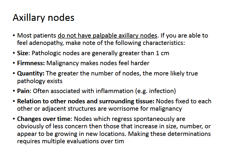
Describe Axillary Nodes:
Size
Firmness
Quantity
Pain
Relation
Changes over time
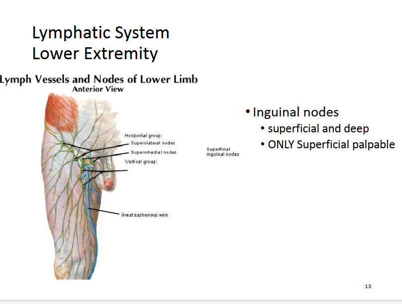
Describe the Lymphatics of the Lower Extremeties
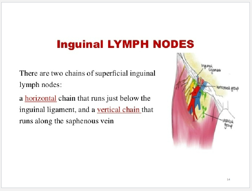
What are the two chains of superficial Inguinal Lymph Nodes?
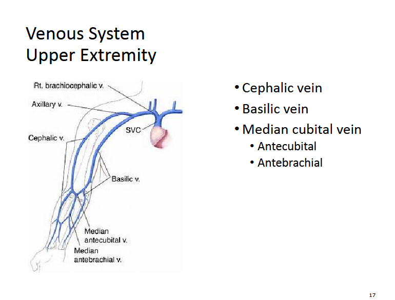
Describe the Venous System of the upper extremity
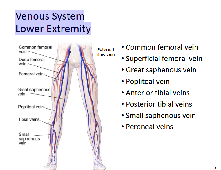
Describe the Venous System Lower Extremity
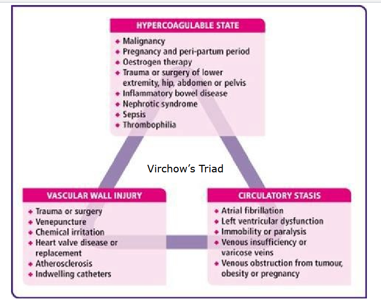
What is Virchow’s Triad
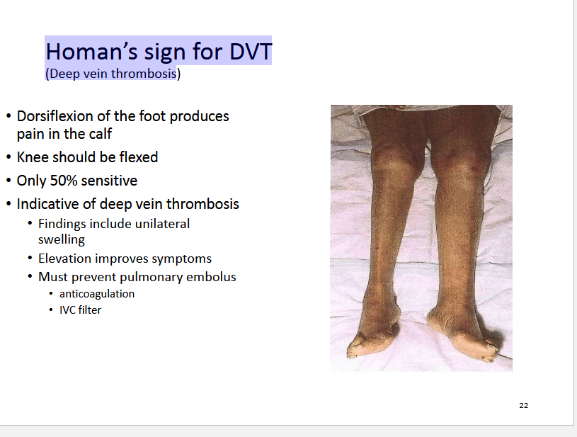
What is Homan’s sign for DVT
(Deep vein thrombosis
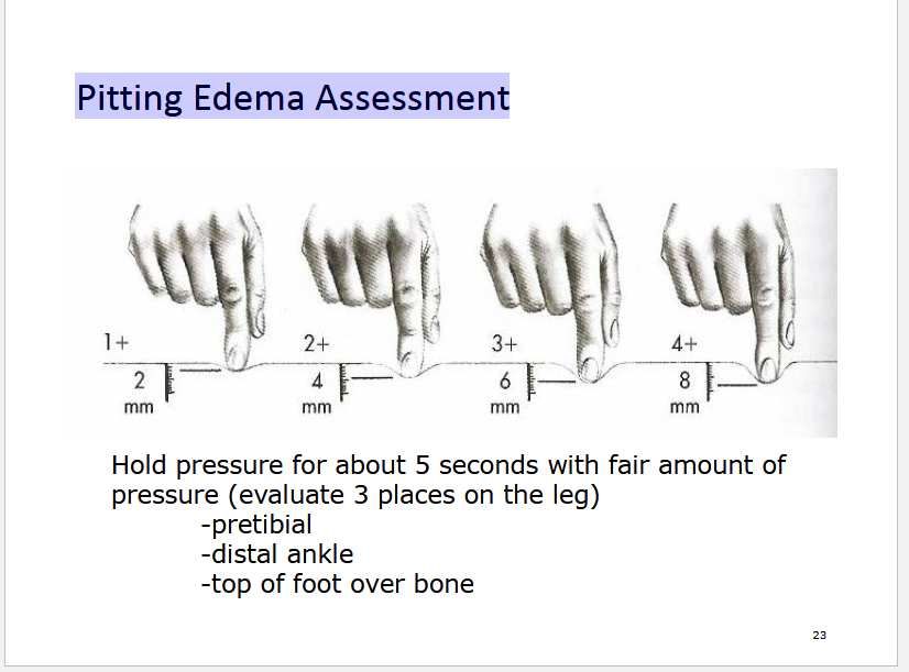
Describe Pitting Edema Assessment
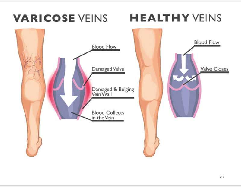
Describe difference between varicose and healthy veins
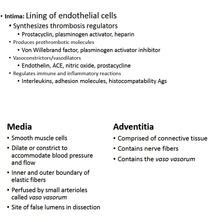
Describe the function of the layers of blood vessels
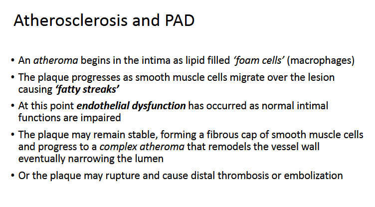
Describe the progression of Artheroslerosis and PAD
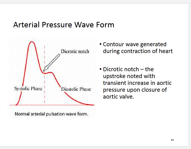
What is the Dicrotic Notch?
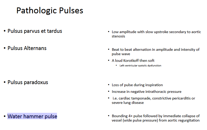
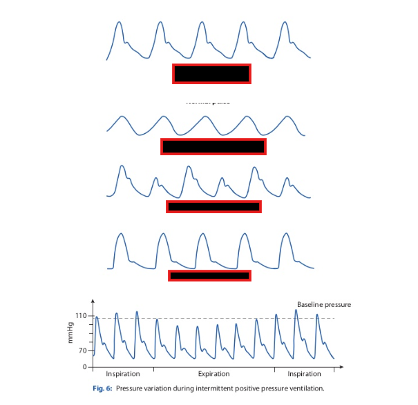
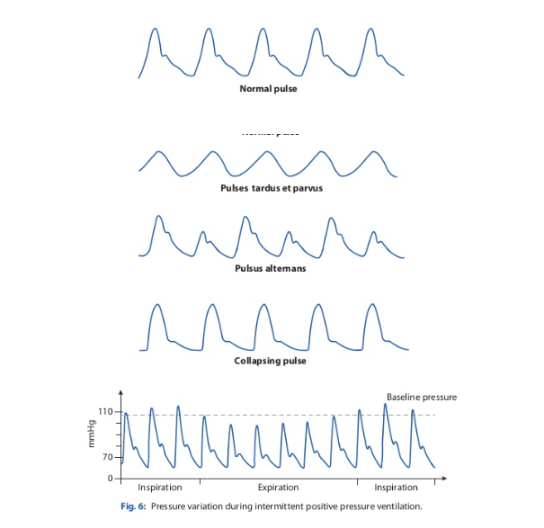
Describe these Pathologic Pulses:
Pulsus parvus et tardus
Pulsus Alternans
Pulsus paradoxus
Water hammer pulse
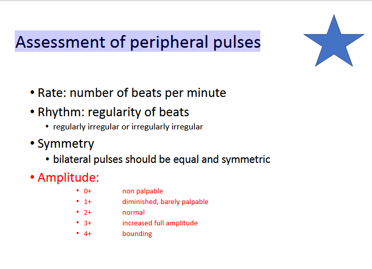
Describe how to do Assessment of peripheral pulses
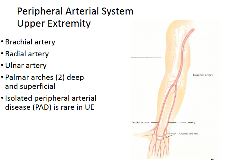
List the Peripheral Arterial System Upper Extremity
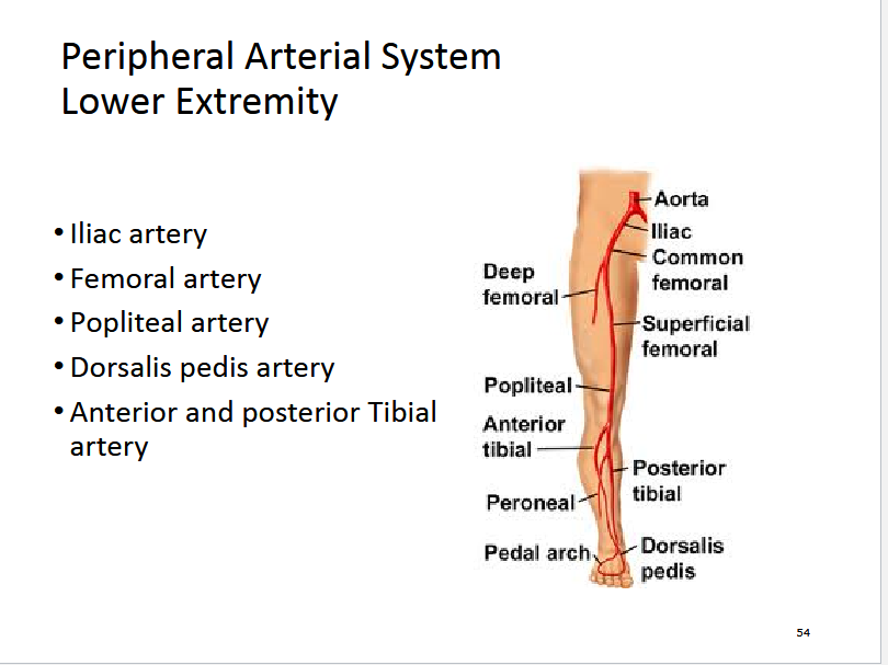
List the Peripheral Arterial System Lower Extremity
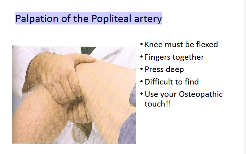
How would you Palpation of the Popliteal artery
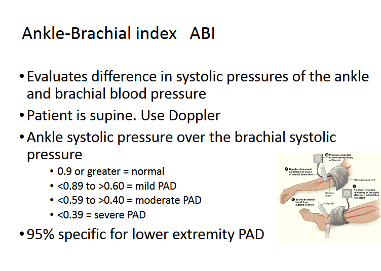
Describe how to determine Ankle-Brachial index
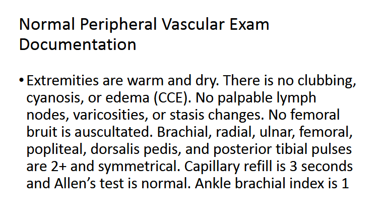
Describe Normal Peripheral Vascular Exam Documentation
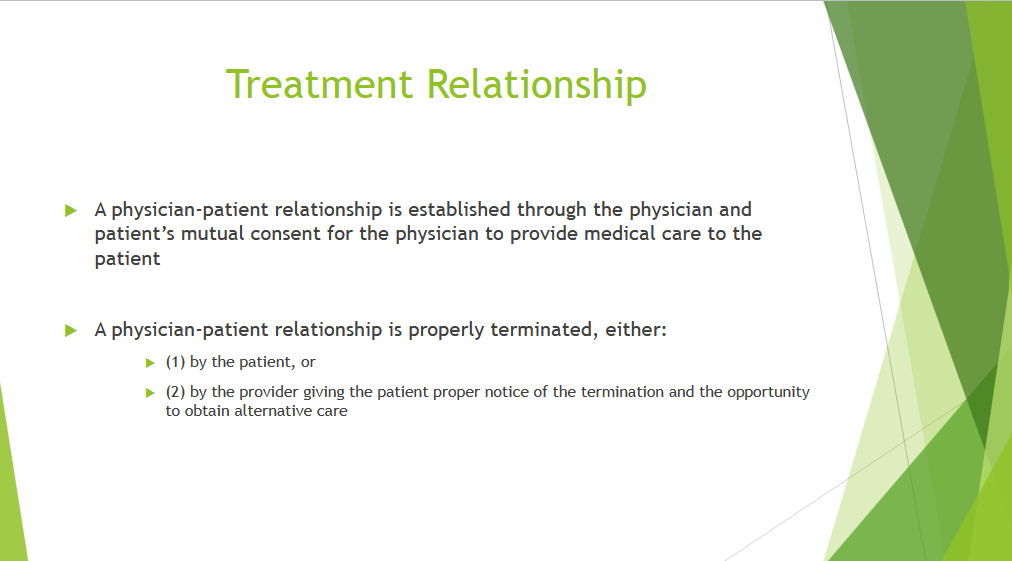
Describe how a patient-provider relationship is terminated?
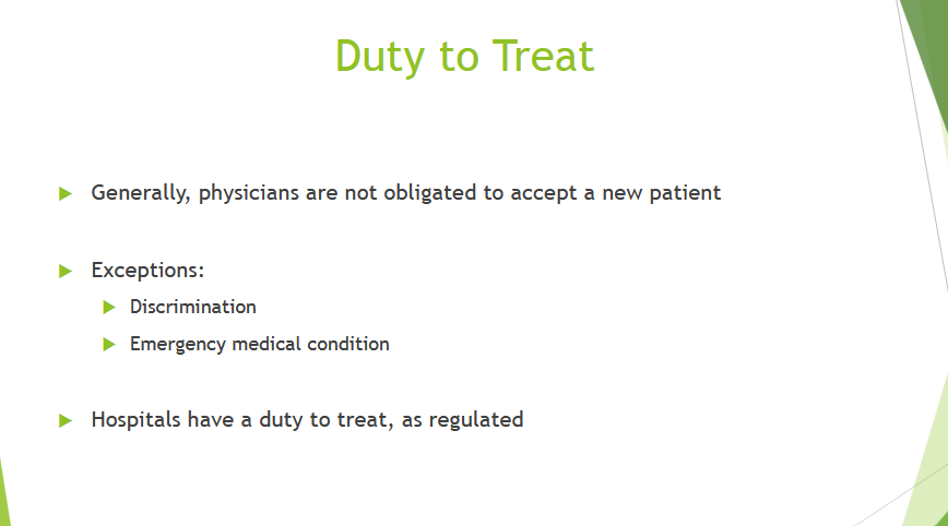
Describe the “duty to treat”
Describe Fiduciary Duty

Describe Confidentiality: Duty to Patient

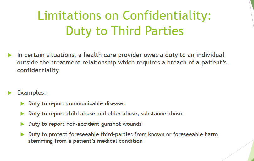
Describe Limitations on Confidentiality: Duty to Third Parties
Describe Autonomy

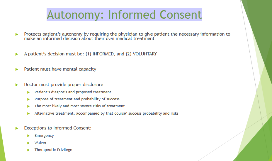
Describe Autonomy: Informed Consent
Describe Justice

Describe Beneficence and Non-Maleficence
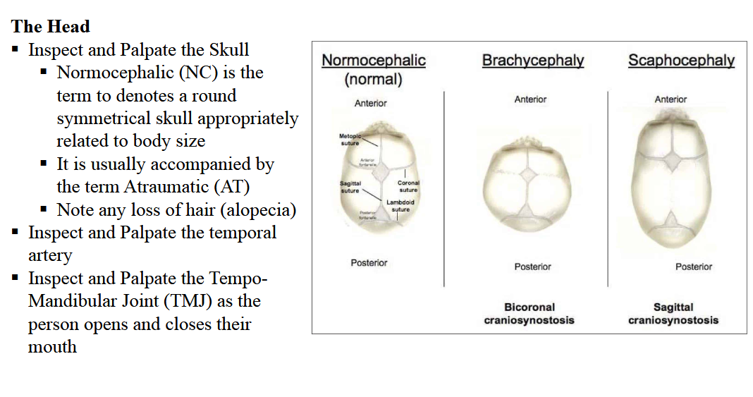
Describe how you would examine the Head
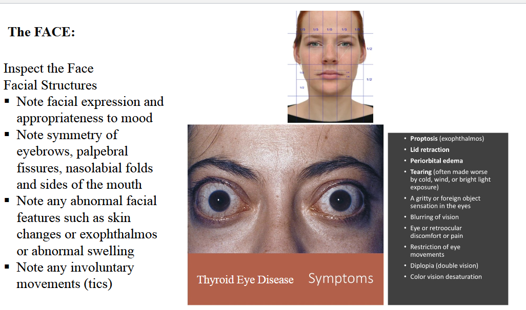
What sort of things are you looking for when inspecting the face
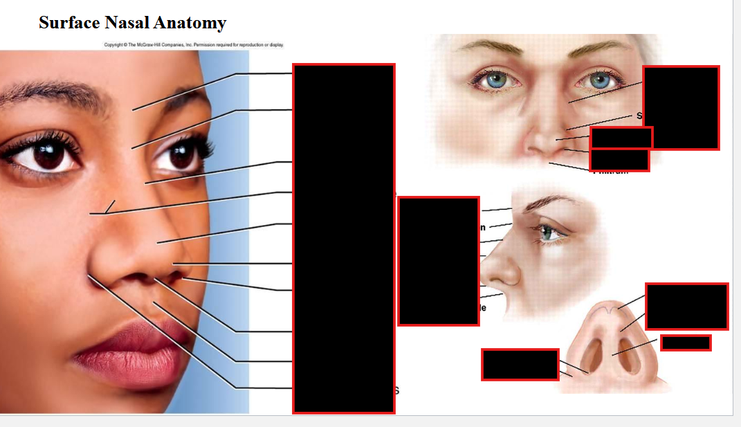
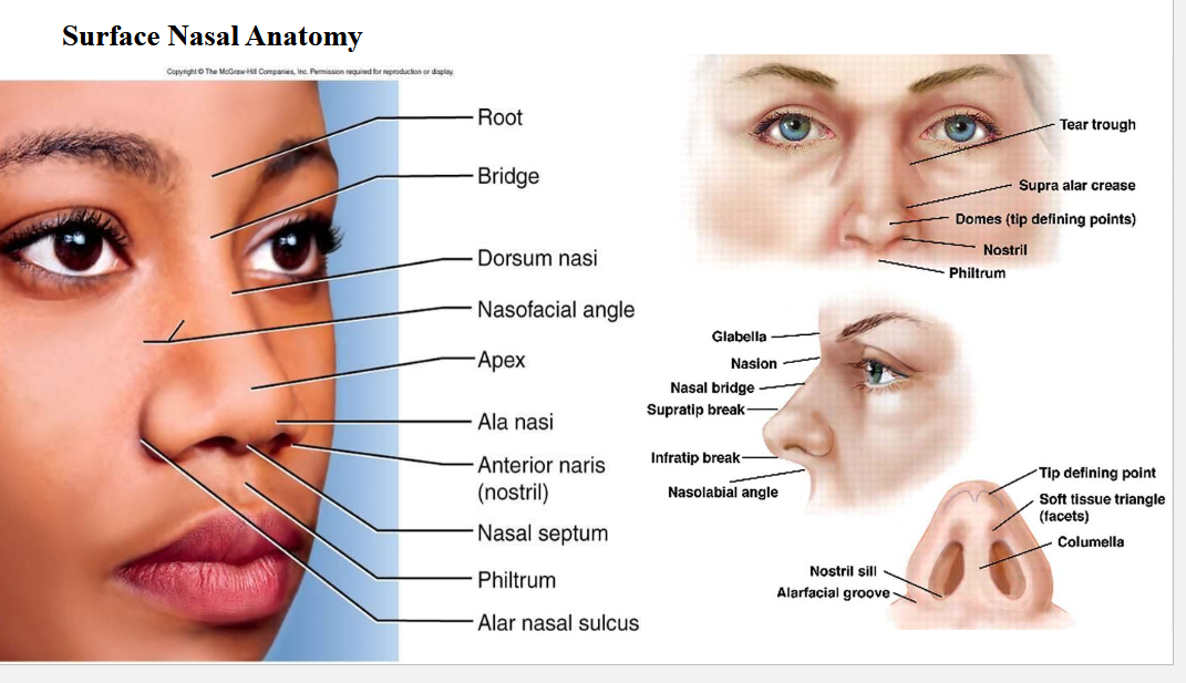
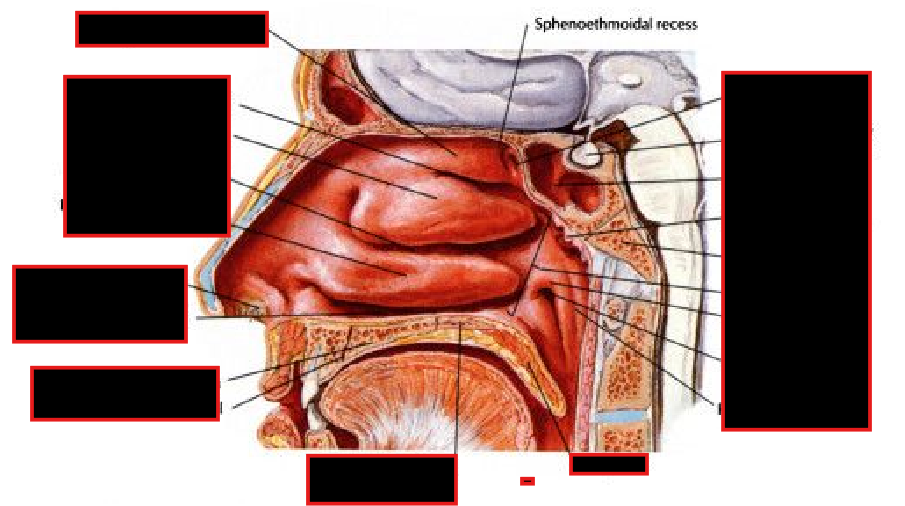
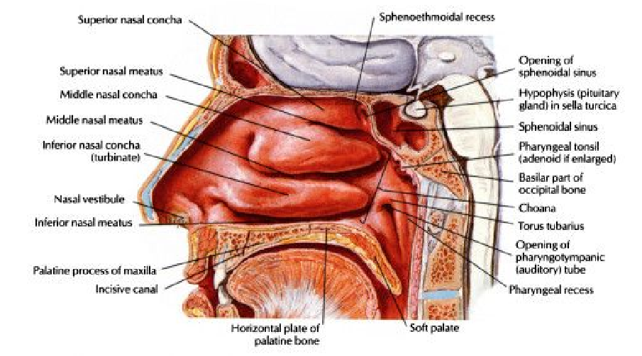
Describe the Anatomic Locations of the Sinuses
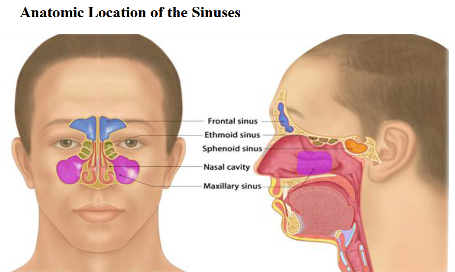
SINUS REVIEW
• Maxillary Sinus
• Ethmoid Sinus
• Frontal Sinus
• Sphenoid sinus
• Superior Turbinate
• Middle Turbinate
• Inferior Turbinate
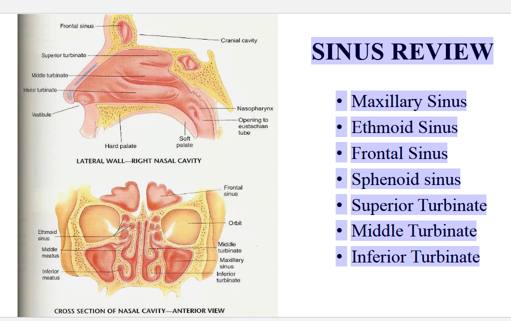
Describe how to do Sinus Palpation
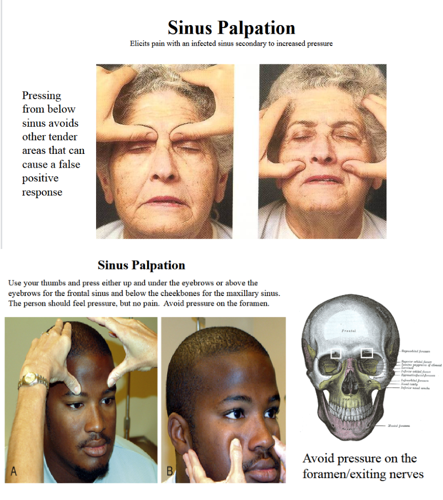
Describe What goes through the:
Infraobital foramen
Supraorbital foramen
Mental Foramen
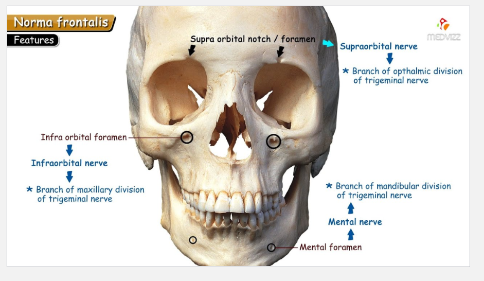
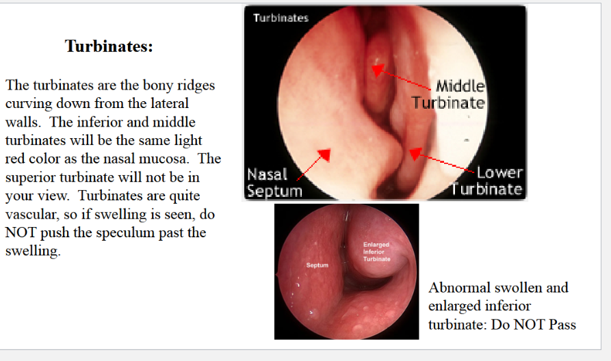
What are the Turbinates
Describe what you are looking for when inspecting/Palpating the Ear
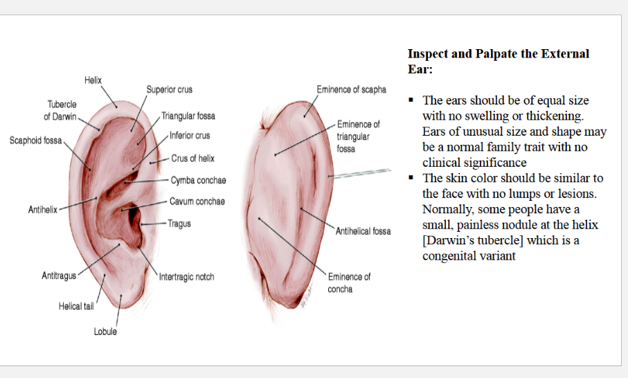
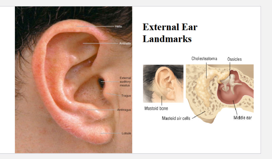
Describe the External Ear Landmarks
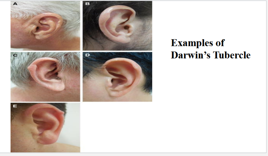
What is a Darwin’s Tubercle
Describe how you would examine the ear
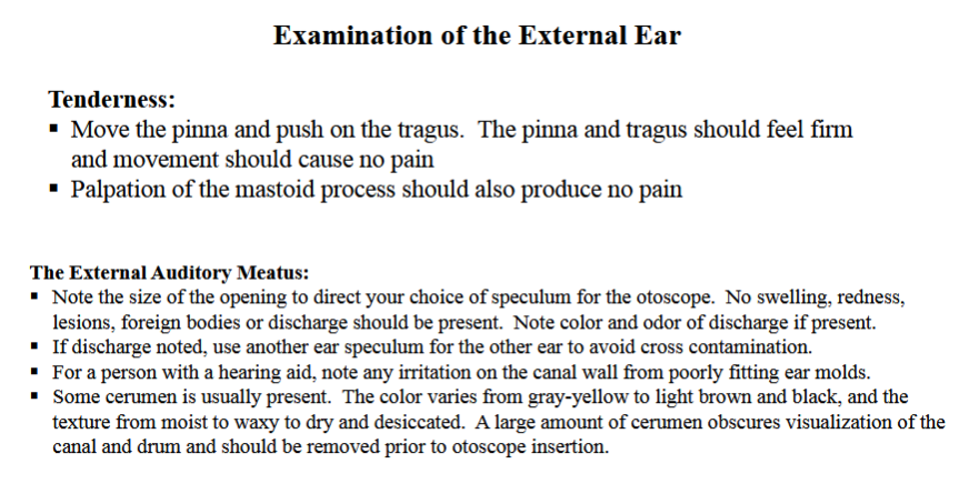
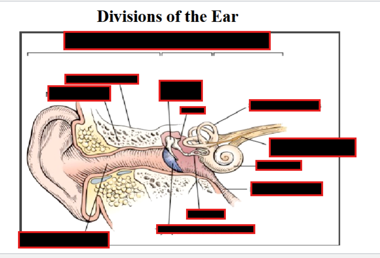
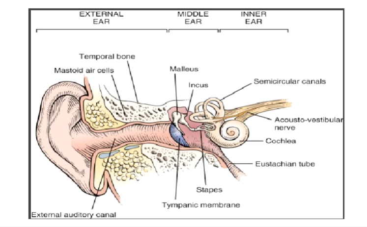
Explain how you would use the otoscope
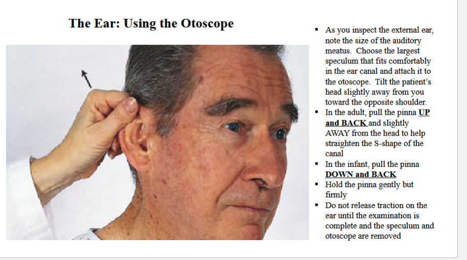
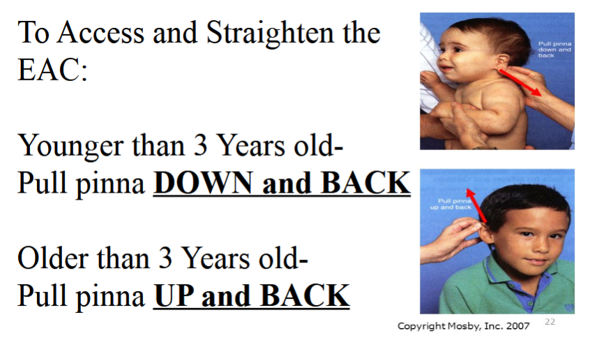
Describe the modification to using the otoscope for Kids (younger/older then 3)
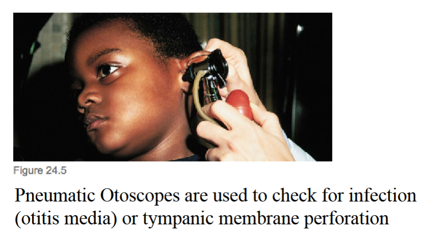
What are Pneumatic otoscopes used for?
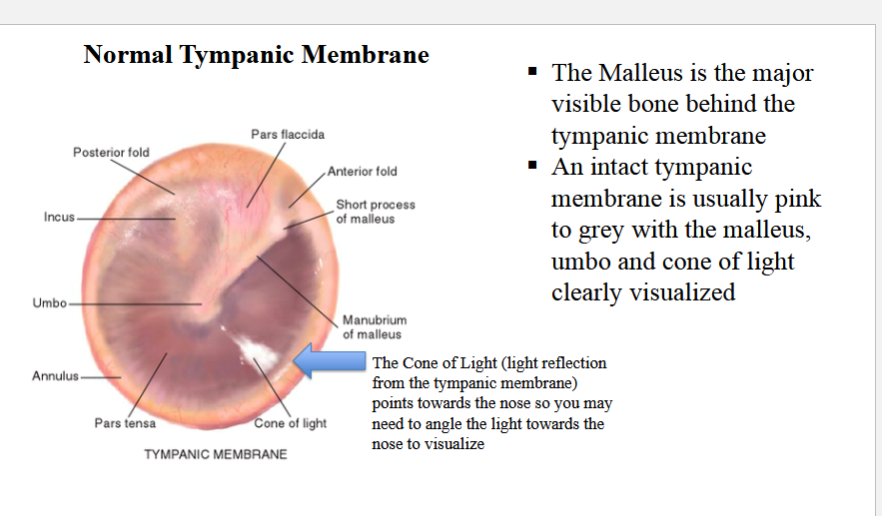
Describe what a Normal Tympanic Membrane Looks like?
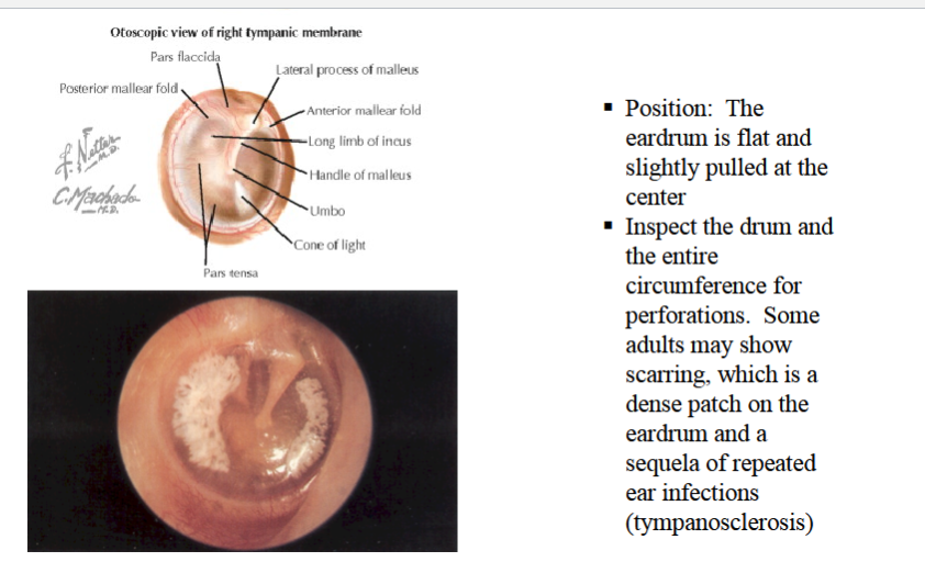
What does tympanosclerosis look like?
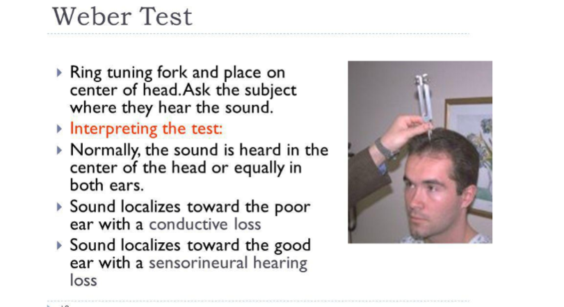
Describe the weber test
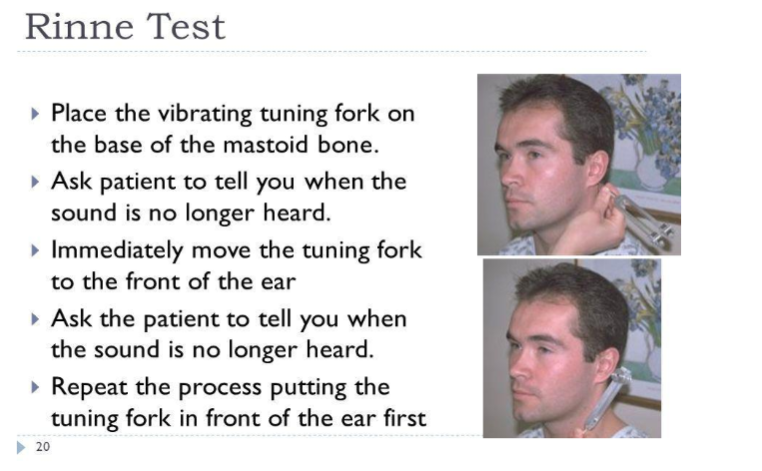
Describe the Rinne Test
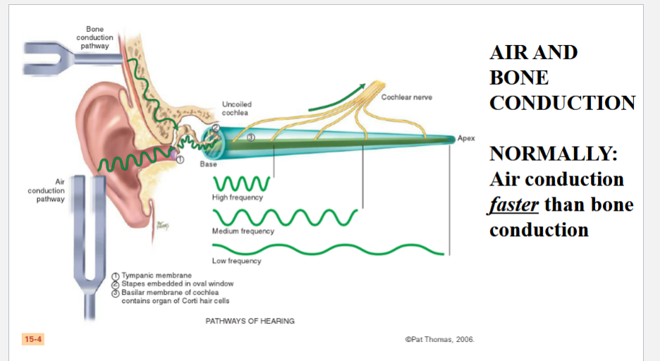
Describe the difference between Air/Bone conduction
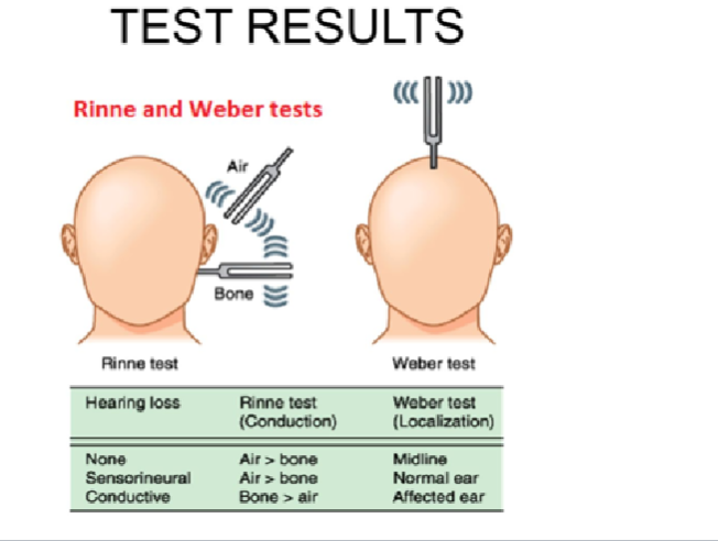
How do you interpret the results of the Rinne/Weber Test
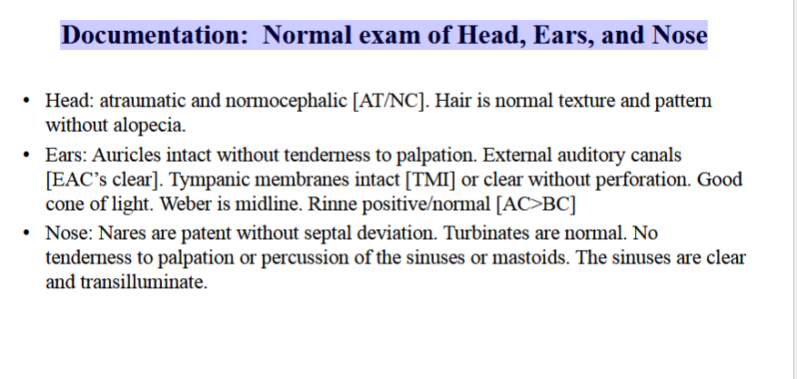
Describe the Documentation: Normal exam of Head, Ears, and Nose
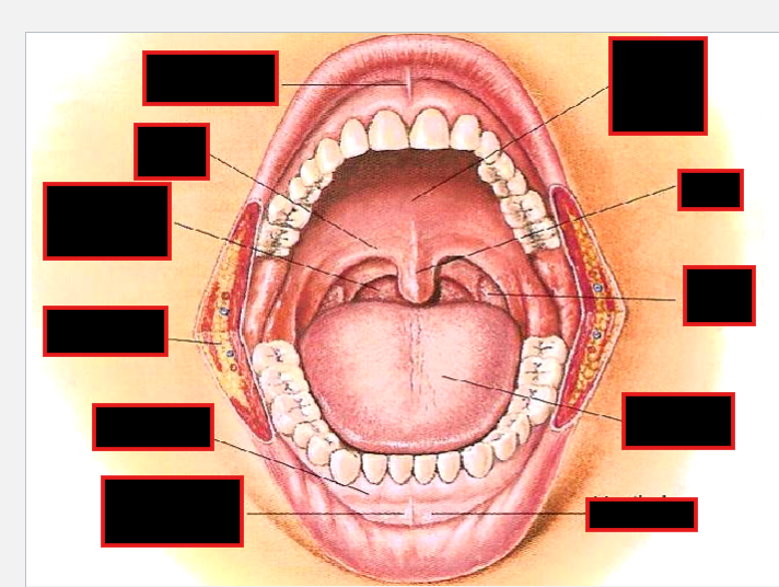
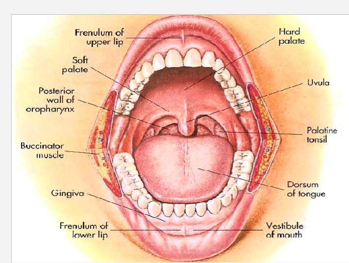
How would you inspect the throat?
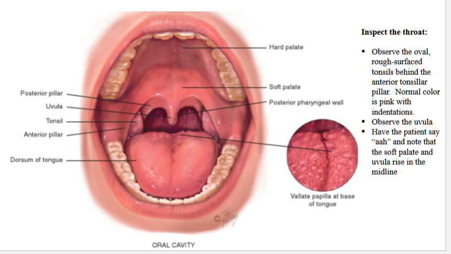
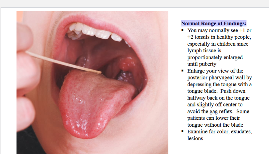
Describe the normal range of findings for throat exam
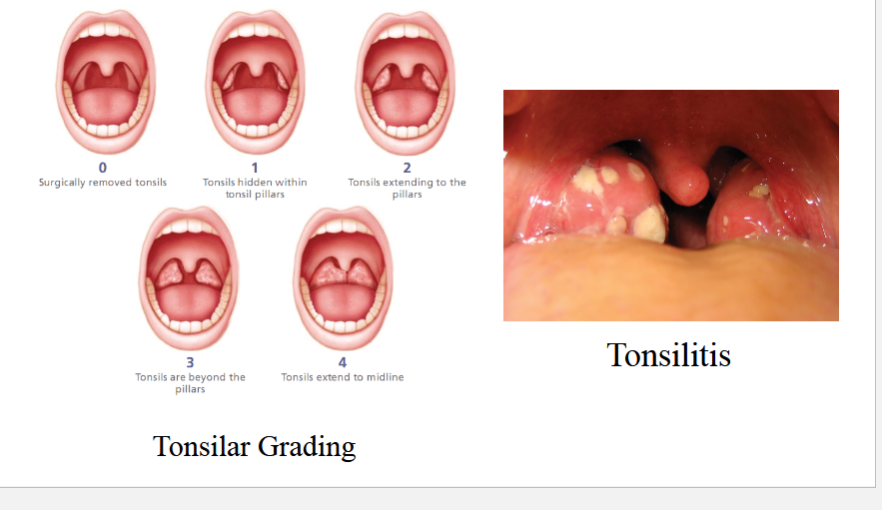
Describe Tonsilar Grading
Describe how you would inspect the mouth
Inspect the Mouth:
Begin with the anterior structures and move posteriorly
Use a tongue blade to retract structures and a bright light for optimal
visualization
Inspect the Lips:
Notice color, moisture, cracking or lesions
Retract the lips and note the inner surface as wel
Normally the gums look pink with a stippled surface. The gum margins should be tight and well defined.
With a gloved hand, hold the tongue with a cotton gauze pad for retraction and inspect both sides of the tongue. Look for
any white patches or lesions and if present, palpate checking for induration.
Inspect carefully the entire U-shaped area under the
tongue behind the teeth. Oral malignancies are likely to develop here. Note any white patches, nodules or ulcerations. If lesions are present or for any person older than 50 or with a positive history of smoking or alcohol use, use your gloved hand to palpate the area. Place your other hand under the jaw to stabilize the tissue and the ‘capture’ and abnormalities.
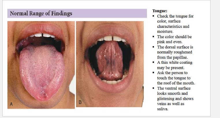
How would you examine the Tonque
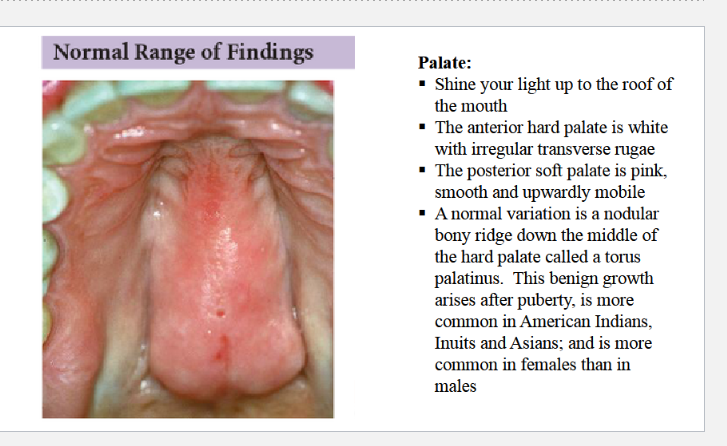
How would you examine the Palate
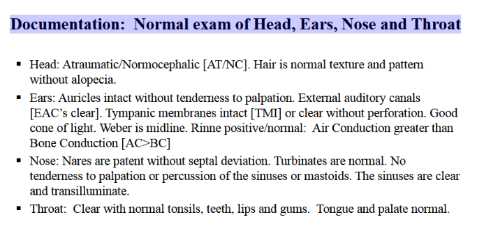
Describe Documentation: Normal exam of Head, Ears, Nose and Throat
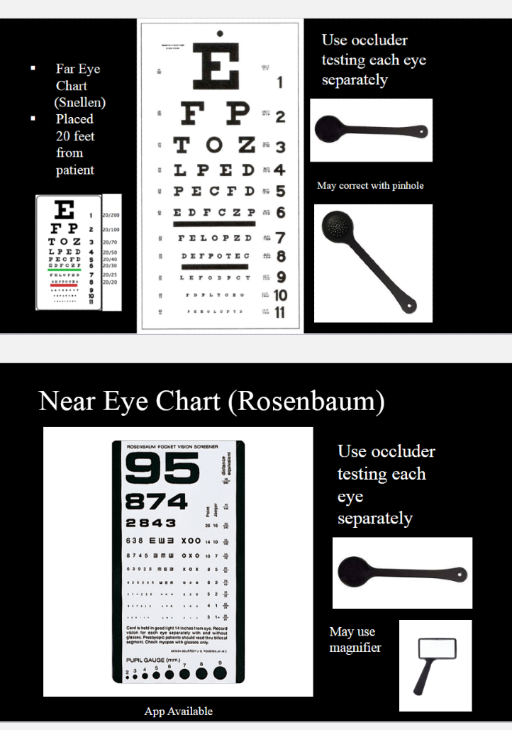
Describe how you would test for Visual Acuity (VA)
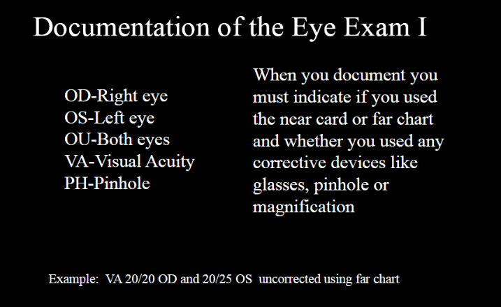
How would you document Visual Acuity (eye exam)
What happens if the patient is unable to read ANY of the chart (illiterate)
CF: Count Fingers @ ?? feet
HW: Hand Wave @ ?? feet
LP: Light Perception
NLP: No Light Perception
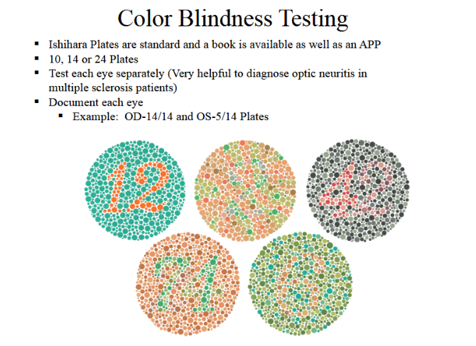
How would you test for color blindness