emt wk 3
 Call Kai
 Learn
Learn Practice Test
 Spaced Repetition
 Match
Match Flashcards
 Knowt Play
1/176

There's no tags or description
Looks like no tags are added yet.
Name | Mastery | Learn | Test | Matching | Spaced | Call with Kai |
|---|
No analytics yet
Send a link to your students to track their progress
177 Terms
outer layer of bone
cortical/compact bone
core of long flat bones and found under the cortical bone at the end of long bones (and purpose)
spongy bone - impact absorption & houses red bone marrow (where red blood cells are made)
center of long bone shaft + its purpose
medullary cavity = holds/stores bone yellow marrow
near end of bone and purpose
epiphyseal line = location of growth plates (children)
different types of bones (human body)
long bones (long shaft and rounded heads) —> ex) femur, humerus, etc
short bones (cube-ish shaped with thin layer of compact bone surrounding spongy interior) —> ex) wrist/ankles
flat bones (thin and generally curved) —> ex) sternum and skull
sesamoid (bones embedded into tendons) —> ex) patella
irregular (irregular shape/complicated) —> ex) spine/pelvis
what are joints and what are the types?
joints: where bones come together
fibrous (held together w dense and connective tissue) - no mvmt
cartilaginous (connected by cartilage w some movement)
synovial (capsule of synovial fluid and allowing for movement — have articular cartilage @ end of bone making up the joint) = most common joint type (knees, elbows, shoulders, etc)
types of synovial joints
types based on movement they allow:
gliding joints
hinge joints
pivot joints
saddle joints
ball and socket joints
cartilage
firm, flexible connective tissue that lines the end of bones + provides shape/support in other areas of body
ligament (can it regenerate?)
fibrous connective tissue connecting bone to bone & provide support for joints
(do not regenerate if damaged)
tendons (can it regenerate?)
fibrous connective tissue connecting muscle to bone
(can heal slowly from damage)
fascia
bands/sheets of connective tissues surrounding muscles & other internal organs — compartments are sections in body containing muscles and nerves which r surrounded by fascia
what is a fracture?
broken bone
what is disclocation?
joint disruption so bone ends no longer are in contact
subluxation
incomplete disruption of a joint
sprain
injury (stretching/tearing) to ligaments, articular capsule, synovial membrane, or tendons (only those crossing joints)
strain
stretching/tearing of muscle (and tendons) causing pain and swelling
sprain vs strain
sprain = stretching/tearing ligaments (bone to bone)
strain = stretching/tearing muscle or tendons (muscle to bone)
amputation
severing extremity from body
open vs closed fractures
open fractures = overlying skin is broken (bleeding, route for infection)
closed = overlying skin is together
displaced vs non-displaced fractures
displaced = moved from normal position and produce deformity by shortening, rotation, or angulation
non-displaced = no deformity
types of fractures (can be displaced or not)
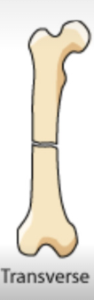
transverse
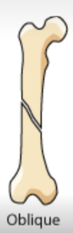
oblique
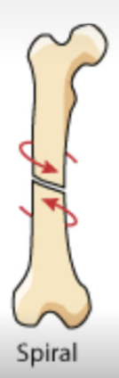
spiral
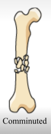
comminuted
avulsion
impacted
fissure
greenstick
transverse fracture
straight across bone
oblique fracture
angular
spiral fracture
broken from twisting
comminuted fracture
break into several pieces
avulsion fractures
fragment of bone is separated from main mass
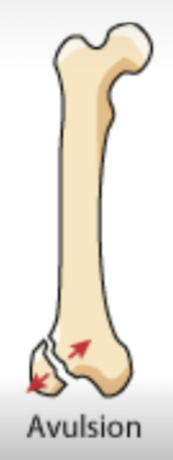
impacted fractures
bone fragments are driven into each other
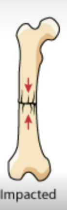
incomplete fractures
bone is still partially joined
pathological fractures
caused by weakness/diseased bone (from underlying condition)
dislocation
may or may not pop back in (depending on patient’s movement) & may have deformity
findings:
swelling
tenderness when palpated
locked joint
potential impaired circulation or numbness
can also damage supporting tissues and joint capsules too
sprains symptoms
(similar to non-displaced fractures)
point tenderness over injured ligaments
swelling and ecchymosis (discoloration from bruising)
pain preventing normal joint movement
joint instability (too much motion)
strain
“pulled muscle”
violent muscle contraction or excessive stretching
no deformity, minor swelling
possible increased pain w passive movement (when third party moves body part)
Compartment Syndrome
SYMPTOMS: pain out of proportion to findings during physical exam, muscle weakness, “pins and needles”, pallor, numbness, & pain is aggravated by passive stretch of muscles in compartment
develop within 3-48 hrs
from increased pressure in compartment containing muscles/nerves —> fractures, crush injuries, burns can cause
fascia does NOT stretch well —> minor bleeding can still raise pressure in compartments and begin process —> restricted blood flow to tissues —> permanent damage
what to do if patient has orthopedic emergency
splint patient
take to trauma center for surgical intervention
assess CSM on extremities (before AND after apply splint)
apply cold packs to reduce swelling
what to do when about to splint
expose entire area for assessment
splint in position of deformity IF…
distal pulses are present and there are no life threats
what to do if lose distal pulse when splint
apply some manual traction to realign (put injury in correct anatomical position) —> splint!
what/where to immobilize when fracture on shaft of long bone
immobilize JOINTS above and below bone
what/where to immobilize when injury is on/around joint
immobilize BONES above and below joint
hazards of improper splinting
nerve, tissue, blood vessel compression
reduced distal circulation
injury aggravation
nerve, tissue, blood vessel, muscle injury bc of excessive mvmt of bone/joint
delayed transport of patient w life threatening injuries
why do you usually not drive code 3 for musculoskeletal injuries
dangerous + aggravate pain from injury
what is one of only times u drive code 3 w musculoskeletal injuries?
pulseless limb (even after manual traction)
treatment for clavicle/scapula injuries
splint w sling and swathe (make sure hand is NOT dangling and it is in position of function (curved))
acromioclavicular (AC) joint injury
AC joint connects outer end of clavicle and acromion process (of scapula) —> ligament tear
AC separation (“AC separation”) = common in football/hockey when fall/land on point of shoulder
—> can lead to end of clavicle sticking out + tenderness over joint
clavicle/shoulder injury —> sling and swathe!
shoulder dislocations
(glenohumeral joint = where head of humerus meets glenoid fossa of scapula)
when head of humerus is no longer in correct anatomical position in regards to the glenoid fossa at the glenohumoral joint
symptoms:
significant pain and guarding
humeral head can protrude anteriorly (usually shoulder dislocations are anterior)
potential numbness on outer part of shoulder and maybe hand (from axillary nerve compression)
treat w SLING AND SWATHE
what to do if arm cannot be brought to chest when trying to splint them (shoulder dislocation)?
pad space between arm and chest w pillow or roll of blankets
humerus fracture
can happen anywhere along humerus
elders = increased likelihood of proximal fractures from a fall
youth = increased likelihood of midshaft fracture from violent injury
treat w SLING AND SWATHE
elbow injuries
distal fractures (more common in children) —> can cause deformity and severe swelling
locked joint w pain (esp if try moving)
—> immobilize above and below injury + apply sling and swathe + pad for comfort
forearm fracture
common in very young and old — esp if fall onto outstretched arms
—> splint (immobilize wrist) & sling (patient comfort)
wrist/hand injuries
splint w forearm and hand immobilized in a sling for comfort
pelvic fractures
very painful + life-threatening, can lead to severe blood loss, maybe deformity
—> use pelvic binder (or sturdy pants)
hip dislocation
hip is usually very stable, so takes a lot to dislocate (MVC, fall into a standing position)
hip dislocation is usually posterior —> can compress sciatic nerve —> impaired leg/foot sensation
signs:
severe hip pain
strong resistance to joint mvmt
tenderness in lateral/posterior aspect (when palpate)
femoral head can be felt/palpated in muscles of buttock
(unlike hip fracture) dislocation could lead to internal rotation
what to do when hip dislocation
—> do NOT try to reduce dislocated hip
splint dislocation
place patient supine on backboard
support affected limb w pillows
secure limb to backboard w long straps
more common “hip” fracture
proximal femur fracture (more common in elderly)
shortened/externally rotated lower limb
—> assess pelvis to rule out pelvic injuries
femoral shaft fracture
can be anywhere on shaft —> pain, deformity, angulation, muscle spasms and external rotation @ fracture site
up to 1 L of blood loss —> can present w shock symptoms (just from upper leg alone)
risk of vascular/nerve damage if bone fragments injure vessels/nerves
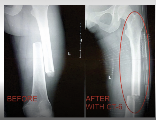
what to do when mid shaft femur fractures
can use traction splint (traction down femur by constantly pulling on ankle and pushing against pelvis) to reduce pain and decrease bleeding
when are you unable to use traction splints
if have or suspect knee, hip, pelvis, lower leg, foot, ankle injuries
—> also (depending on area), contraindication when have open mid shaft fracture bc concern for infection
injuries of knee ligaments
—> stable femur and tibia w splint & use padding (comfort) during transport
—> (if LOCKED knee) use blankets/pillows under knee when in stretcher
knee dislocation
check CSM bc increased risk of damaging popliteal artery by compression or laceration —> apply traction
dislocated patella
usually lateral displacement (bulge) & lack of patella in correct position
—> splint injury (under a lot of tension so might relocate during splinting)
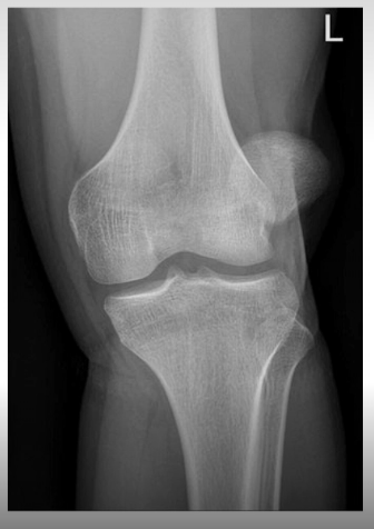
tibia/fibula fractures
tibia has higher likehood of creating open fracture (breaking through skin) bc so close to surface
can have knee, mid shaft, or ankle pain
what to do differently when splint ankle/foot injury
leave toes exposed to assess for circulation (CSM)
what is orbit and what is its purpose?
round bony structure/socket that eye lies in —> help protect eye from injury
6 bones form major structure of face
nasal bone
2 maxillae (upper jawbones)
2 zygomas (cheek bones)
mandible (jawbone)
the exposed part of the ear is comprised of __ covered by skin
cartilage
what is ur pinna?
external, visible part of the ear
what r ur earlobes?
fleshy portion under ur ears
what is ur tragus?
small, rounded, fleshy bulge (cartilage flap) immediately anterior to ear canal (think of what u push when trying to not hear smth)
what is ur mastoid process?
prominent bony mass ~1cm behind external ear opening
motion of the mandible occurs @ _____
temporomandibular joint (hinge joint)
angle of the mandible
below ear and anterior to mastoid process
major blood vessels in the neck
carotid arteries + jugular veins
globe of eye keeps its shape bc of ___
the pressure of fluid within its two chambers:
vitreous humor = clear, jellylike fluid near BACK of eye
aqueous humor = clear, watery fluid in front of eye lens
penetrating trauma to eye can cause __ humor to leak out
aqueous —> but can make more w time
NOTE: aqueous humor is fluid in the anterior chamber (in front of lens) vs vitreous humor is fluid in the posterior chamber (behind the lens)
retina is connected to the ___
optic nerve
conjunctiva
delicate membrane that covers surface of eye
sclera
white part of eye (tough, fibrous tissue extending over globe to keep eye structure and protect layers/stuctures underneath
lacrimal glands
(tear glands) —> keep conjunctiva and inner surface of eyelids MOIST
how does blinking work?
sweeps fluid from lacrimal glands to clean eye surface
tears drain onto inner side of eye through 2 lacrimal ducts into nasal cavity
cornea
clear transparent membrane in front of eye —> allows light to enter
pupil
eye opening which allows light into back of the eye
iris
circular muscle and surrounding tissues behind cornea (adjusts size of pupil to control how much light gets in)
iris is pigmented (diff eye colors!)
lens
behind iris and focuses image onto retina
the brain interprets ___ as vision
optic nerve impulses
possible injuries to face and neck
primarily around airway obstruction:
bleeding in mouth —> can clot
aspirating vomit = obstruction
dislodged teeth or dentures
swelling from soft tissue
maybe also brain/spine injuries
soft tissue injuries (face/neck)
face/neck = very vascular, so…
more swelling than in other parts of body
penetrating trauma —> severe bleeding
blunt injuries —> vessel wall break —> hematoma (bruising)
general head/neck injuries management
priority: maintain open airway
recovery position to empty airway
→ if on backboard, can tilt to one side to allow blood/vomit to drain out of airway
control external bleeding w direct pressure (BUT not too much pressure if possible skull fracture)
CALL ALS
what to do w avulsed tissue
(avulsed = detached tissue)
if still attached to body —> put avulsed tissue in “normal” position if possible and cover with DRY, STERILE DRESSING
if separated from body —> wrap avulsed tissue in sterile dressing, put in plastic bag, keep it COOL (do NOT put directly on ice) —> (can maybe still graft tissue onto body later)
facial fractures (and when to suspect)
suspect if sustained direct blow to nose or mouth
signs/symptoms:
bleeding in mouth
cannot swallow or talk
loose/absent teeth
loose/movable bone fragments
patient says closing jaw “just doesn’t feel right” — irregular bite
—> take spinal precautions (assess for c-spine / brain injuries) bc need a lot of force to fracture these bones
—> TAKE OFF JEWELRY BC POSSIBLE SWELLING (esp in first few hours-day)
mandible, maxillary and dental injuries
(more common - usually from MVCs, assaults)
if suspected mandible fracture (since need a lot of force to fracture mandible) —> also check if other facial trauma and/or c-spine injuries
signs/symptoms:
facial swelling
instability when feel facial bones
misaligned/fractured/avulsed teeth
dental injuries
prevent tooth from becoming airway obstruction —> remove loose dentures/dental bridges
maintain direct pressure to stop bleeding (tooth being violently displaced out of socket)
handle teeth by the CROWN, not the ROOT
put avulsed teeth into sterile saline (bring when transport)
notify facility abt avulsed tooth bc re-implantation is necessary within narrow time window (20 min -1 hr after trauma)
cheek injuries (think impaled object + bleeding treatment)
if impaled object in cheek causes airway compromise —> RARE instance where can remove object
if bleeding cheek —> provide direct pressure from inside AND outside (make sure not too much gauze so no airway obstruction)
what to do for eye injury
do a proper eye exam:
check if entire circle of iris is visible
check for PERRL (pupils are equal, round, react equally when exposed to light)
check if both eyes move together (when follow moving finger)
check if eye discoloration (eye, iris bleeding, redness)
check if any obvious foreign matter
asking/assessing patient abt clarity of vision or sensitivity to light
check if any previous eye surgeries/injuries
foreign objects in eye
(even a grain of sand) can cause redness and inflammation of the conjunctiva —> conjunctivitis
—> patient might feel like object is still there after remove bc scratched eye (possibility)
patient difficulty keeping eyes open bc bright light irritates
if SMALL object —> use SALINE to flush it out (start from inner corner and flush outward - to prevent get in other eye)
if LARGE object —> can NOT take out so stabilize (bandage around BOTH EYES —> to prevent other eye from moving - eyes move together)
chemical eye burns
chemical —> flush eyes out with water or sterile saline (inside to outside)
—> send saline through nasal cannula (lie over nose bridge) bc can drip into both eyes and flush out
—> force patient’s eyelids open
if alkaline solution or strong acid, irrigate eyes >=20 minutes bc can penetrate eyes deeply
thermal eye burns
can burn eyelid off (bc ppl close eyes from reflex)
—> cover BOTH eyes with moist, sterile dressing
—> eye shield (prevent as much light getting into eyes as possible)
infrared rays, sunlight CAN damage retina (permanent damage)
intense lights CAN cause superficial burns (like sunburn but to eye)
stop burn
cover eyes
prevent light from entering
get to higher level of care
eye laceration
if laceration on eyelid —> direct pressure (but not too hard)
if laceration on globe —> do NOT put direct pressure on eye
can hinder blood supply to back of eye —> permanently damage retina
can squeeze out more fluid —> make worse
—> put them SUPINE (minimize fluid loss) + moist, sterile dressing (cover BOTH) + protective shield
blunt trauma to eye
can cause “black eye” or severely damaged globe (range)
orbit fracture (ex - blowout fracture = lose tension around eye from broken bone and no longer support eye)
fractured bones can trap eyes so no move right, double vision, vision issues
retinal detachment (cloudy vision, flashing lights) —> cover both eyes, send to hospital
hyphema - bleeding into anterior chamber of eye —> can impair vision = LIFE-THREATENING
iif eye is displaced out of socket —» do NOT move back into place (LEAVE it there, cover BOTH eyes w moist/sterile dressing, transport as fast as possible, keep SUPINE)
eye injuries AFTER head injury (aka how to tell if head injury by looking at eyes (??))
findings that suggest possibility of head injury:
anisocoria (unequal pupil sizes)
eyes pointing in different directions
eyes cannot follow moving finger
bleeding under conjunctiva (obscure sclera)
protrusion/bulging of eye
if unconscious patients —> keep eyes closed so no dry up
blast injury
foreign objects —> stabilize, cover BOTH eyes, transport
do NOT force eyelids open if swelling/hematoma bc can increase pressure
give CLEAR, VERBAL directions