APEX: ANS Anatomy and Physiology
1/60
There's no tags or description
Looks like no tags are added yet.
Name | Mastery | Learn | Test | Matching | Spaced | Call with Kai |
|---|
No analytics yet
Send a link to your students to track their progress
61 Terms
The entire CNS is made up of what things?
Spinal cord
Medulla
Pons
Midbrain
Thalamus
Cerebral Hemispheres
What are the 2 divisions of the autonomic nervous system?
SNS
—fight or flight
PNS
—rest + digest
In both the SNS and PNS:
—preganglionic neurons are what type of fibers?
—postganglionic neurons are what type of fibers?
Preganglionic—Type B,lightly myelinated
Postganglionic — Type C Unmyelinated
Where does the SNS preganglionic neurons originate?
Where do the cell bodies lie?
"Thoracolumbar"
From T1-L2
Cell bodies are located within the LATERAL horn of the spinal cord grey matter from the above spinal regions
—Intermediolateral nucleus (Lamina of Rexed 7)
Where does the PNS preganglionic neurons originate?
"Craniosacral" Division
Cranial nerves 3,7,9,.+10 and S2-S4
Cell bodies in the lateral horn of spinal cord grey matter (LR7)
In the autonomic nervous system: (answer short / long)
—The SNS preganglionic are __________ and post ganglionic are_____________
—The PNS preganglionic are ____________and post ganglionic are _____________
SNS:
—pre—short
—post —long
PNS:
—Pre—long
—post—short
Where does the somatic nervous system preganglionic neurons originate?
What is the difference between the autonomic and somatic neurons when it comes to number of neurons.
From the Ventral horn of the spinal cord (grey matter) from each spinal section.
—Lamina of rexed 8 or 9
With the somatic (motor) nervous system, there is only ONE neuron from the spinal cord to its target effector— the NMJ (lower motor neuron)
In the autonomic nervous system, there is a preganglionic and post ganglionic neuron.
In the SNS, the axons of nerves exit the ___(ventral/dorsal)__ root and seperate shortly after to form the __________________.
Why is it called this?
Where are these present?
Ventral
White Rami.
—white rami are myelinated
Only present from T1-L2 because all preganglionic SNS fibers originate here.
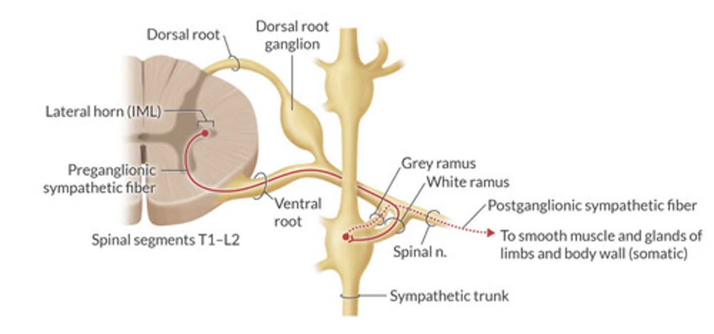
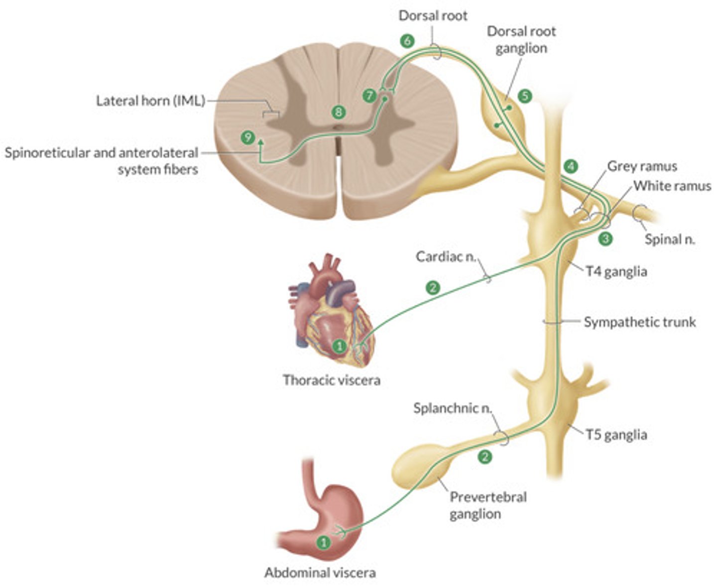
Describe the SNS route (route 1)from spinal cord to effector on same spinal level
Exit spinal cord
—> Enter into white rami communicans
—>Synapse in sympathetic ganglionic trunk
—>post ganglionic neuron (c fiber) exits via grey rami
—>exits spinal nerve to synapse with effector on same spinal nerve level.
How many pairs of sympathetic ganglia are there?
3 Cervical
—superior, middle, and inferior
11-12 Thoracic
3-5 Lumbar
3-5 Sacral
1 Coccygeal
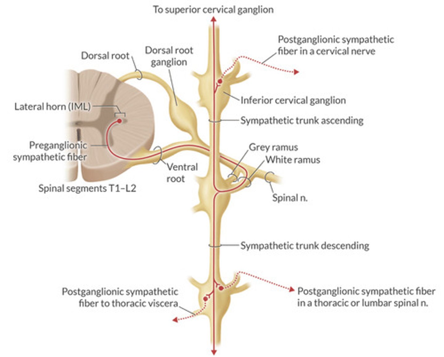
Describe the SNS route (route 2)from spinal cord to ascend/descend to target effectors
Exit spinal cord
—>preganglionic neuron enters into white rami
—>ascends or descends in sympathetic trunks
—>synpases on spinal level of where it will exit
—>post ganglionic neuron exit through grey rami
—>synapse on effector.
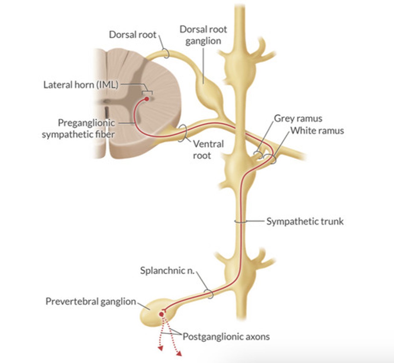
Describe the SNS route (route 3) from spinal cord to pass through splanchnic nerves and synapse on abdominal/pelvic visceral effectors:
Exit spinal cord
—>enter into white rami
—>ascend or descend and exit via splanchnic nerve
—>enter into prevertebral ganglion to synapse with post ganglionic neuron
—>exit into plexuses that surround aorta and arterial network.
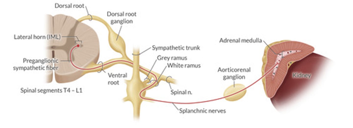
Describe the SNS route (route 4)that exits the spinal cord and synapses with the adrenal medulla
Exit spinal cord
—>enter into white rami
—>exit via splanchnic nerves
—>enter into aorticorenal ganglion
—>exit to synpase in adrenal medulla with chromaffin cells
THERE IS NO POST GANGLIONIC NEURON
What is the stellate ganglion?
Blockade of this provides what symptoms?
Fusion of the Inferior cervical ganglion and the T1 ganglion
V-PAM
—vasodilation of upper extremities
—Ptosis
—Anhidrosis
—Miosis
—enopthalmus
—nasal congestion
This occurs on the IPSILATERAL side.
Which cranial nerves have a parasympathetic component?
3, 7, 9, 10
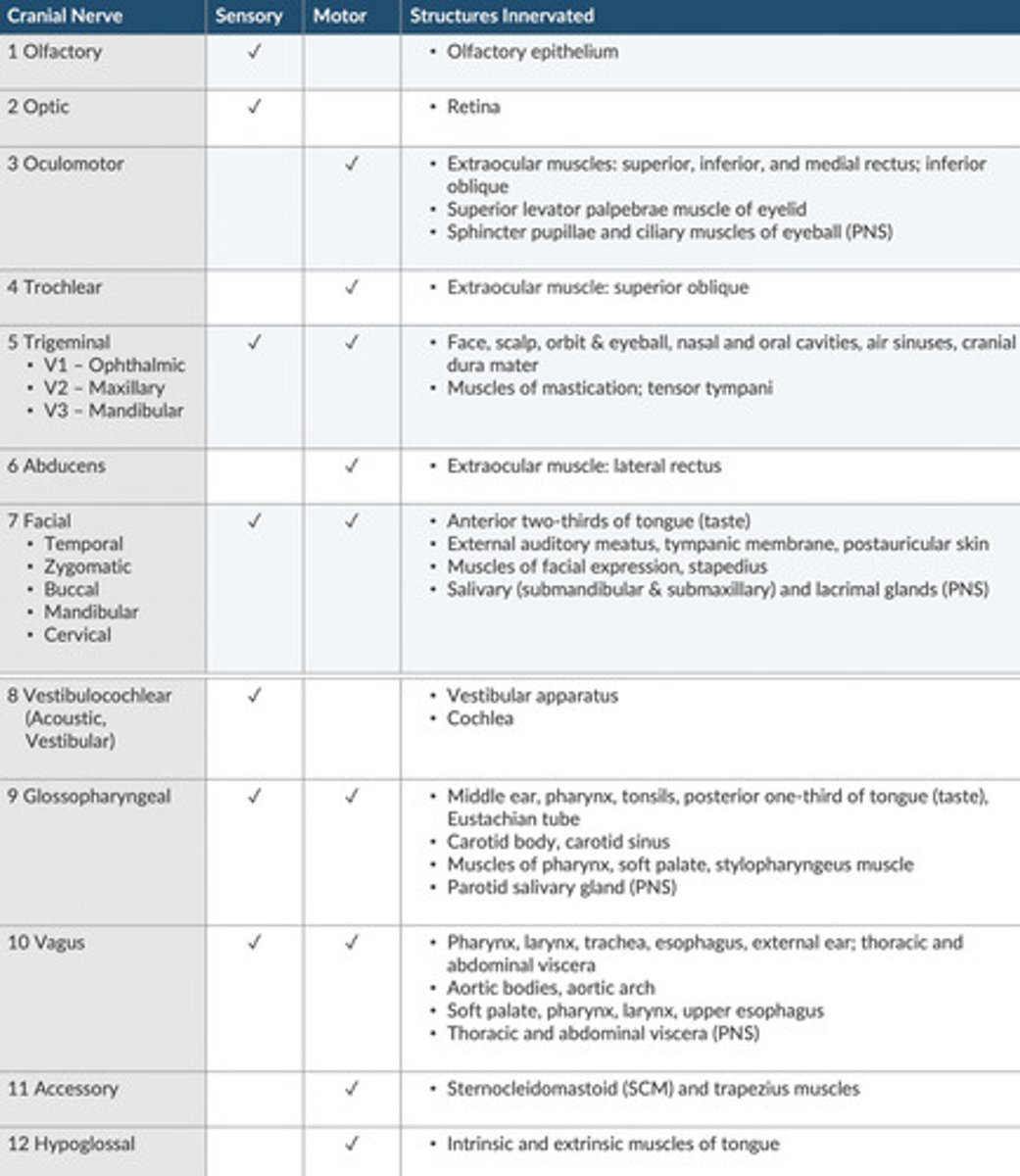
Describe the cranial nerve innervations andwhich ones are sensory/motor: (chart)
Approximately ____% of PNS outflow is through the _________ nerve.
75%
Vagus
The vagal PNS preganglionic efferent fibers originate in which 2 brainstem nuclei?
Dorsal Motor nucleus of vagus
—heart, bronchi, GI tract.
Nucleus Ambiguus
—pharynx, larynx, palate
Where is the origin of the only visceral PNS innervation that does not come from CNX?
Do these travel through white rami?
S2-S4 Lateral horn
(LR7)
NO—white rami only exists T1-L2
Visceral afferent fibers are involved in certain reflexes (circulation, respiration, digestion...etc) and transmit pain.
What type of fibers are these?
T or F:
The afferent pathways from any structure are typically along the same route as the efferent fibers that supply that structure.
Usually Type C fibers (unmyelinated) or A-Delta (light myelination)
TRUE
Which cranial nerves handle sensory afferents from baroreceptors and chemoreceptors from the aortic arch and carotids?
CN 9-from carotids via Herings nerve
CN10-from aortic arch
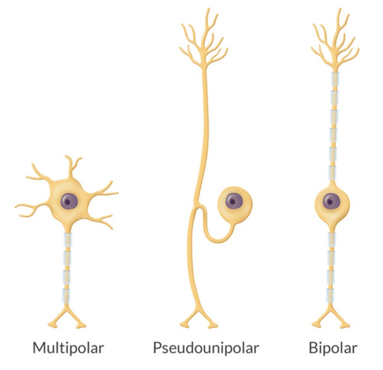
Most sensory afferent fibers are ____(bi/multi/psuedo)____ polar neurons.
What do the other types of neurons do?
PSUEDOPOLAR neurons
—Mostly Axon—from the periphery to the CNS connected by a cell body.
(Bipolar neurons are sensory of the eye and ear. Most other neurons are multipolar)
Explain the pathway of sensory afferent neurons from the viscera to the CNS:
Sensory Receptors in viscera are stimulated and travel toward spinal nerves in splanchnic nerves.
—they do not synapse in ganglia (sympathetic chain or Paravertebral)
Pass through white rami to join spinal nerve —>DRG
Enters spinal cord through dorsal root and synapses with secondary neuron in dorsal grey matter.
Second order neuron crosses to other side and projects to thalamus (3rd order)
T or F:
Most visceral pain information is conducted in sympathetic nerves
What is the exception and clinical relevance of this?
TRUE
2 Exceptions:
—receptors distal to midway of sigmoid colon and inferior to pelvic pain line travel via PNS pelvic nerves.
Relevance:
Caudal epidural block will anesthetize cervix and vagina but will have little effect from uterine body(superior to pelvic pain line)
—injection into the lumbar epidural space is the most common approach and will block both of these.
Describe visceral pain:
Felt in the region of affected origin
Vague, Deep, and often accompanied by sweating or nausea.
Ex: inflammation/ulcer in GI tract, obstruction in intestine, stretch of solid organ(liver, kidney, pancreas)
What is referred pain?
How is it transmitted and why does it happen?
Noxious stimuli is perceived as pain arising from somatic portion of body wall (skin, bones, muscles)
Transmitted via SNS sensory fibers and is typically referred to same segmental levels of spinal cord as original structure.
—brain falsely interprets this because it shares the same pathway in common with original structure.
What is Parietal pain?
(Viscerosomatic pain)
Somatic Pain arising directly over the affected organ
—Ex: pain on palpation in RLQ with appendicitis
Noxious stimuli from parietal membranes travel along somatic pain pathways.
Describe the PNS transmission of an AP:
—what is released from the preganglionic neuron and on what receptor?
—what is released from the postganglionic neuron and on what receptor?
Preganglionic
—releases ACh to bind with Nicotinic (N) receptors on post ganglionic neuron in the autonomic ganglia.
Postganglionic:
—releases Ach to bing with Muscarinic receptors on target effectors.
What causes the release of Ach?
How is it eliminated?
Ca+ dependent exocytosis.
(Mg is an antagonist of Ca+
—this explains why Mg can cause weakness.)
Elimination:
—Acetylcholinesterase in the synaptic cleft is hydrolyzed to Acetate and Choline
—acetate is sent to liver to be excreted and Choline renters the nerve terminal and used again for Ach synthesis.
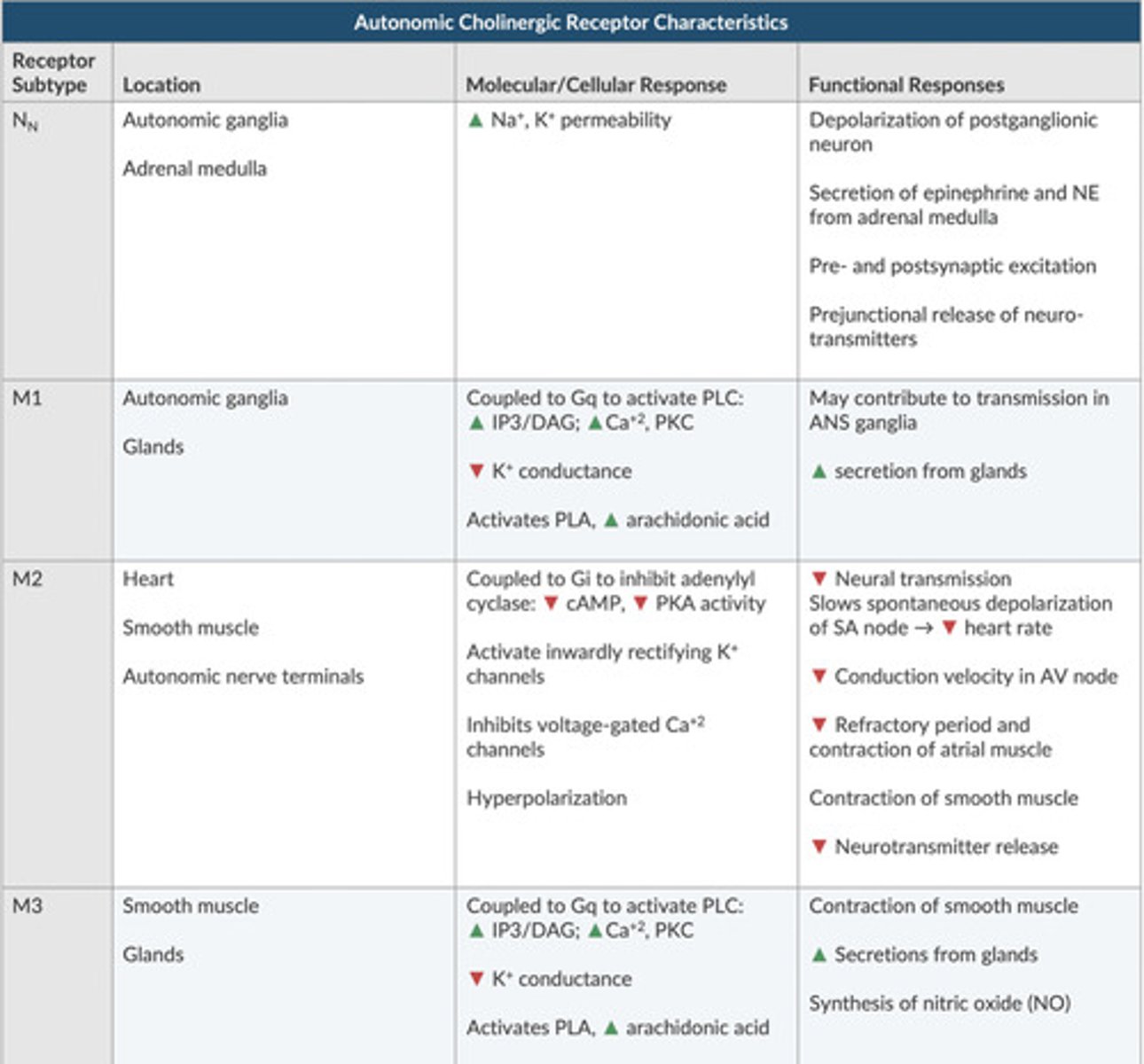
What are the different types of Muscarinic receptors and where do they work?
M1-M5: (M1-M3 important)
—M1-found in autonomic ganglia and some glands
—M2-Found in heart (cardiac receptors) and presynapically as negative feedback loop to limit Ach release
—M3-(glandular and smooth muscle) prominent in exocrine glands and smooth muscle. These mediate bronchoconstriction
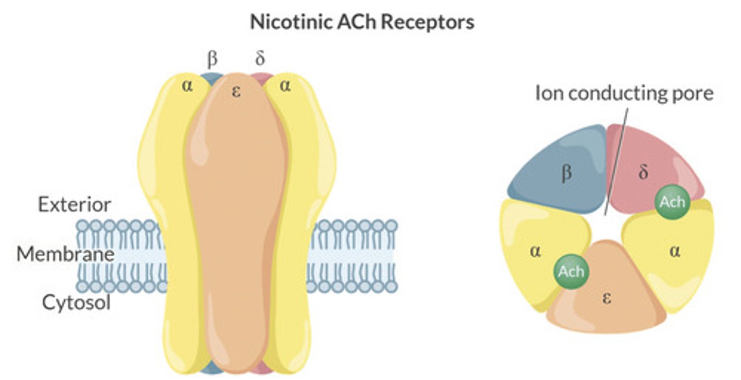
What causes the stimulation of nicotinic receptors at post ganglionic neurons?
2 Ach molecules must bind with the Alpha subunit on the N receptors to initiative confirmational change.
Nicotinic receptors are pentameric complexes with 2 binding sites: see picture.
What are the g-protein types assosciated with each Muscarinic receptor:
What is the result of activation?
M1, M3, M5: coupled with Gq
—activates phopholipase C
—>Increased IP3 and DAG, Increased Ca+
—>Protein Kinase C Activation
M2, M4: coupled with Gi
—inhibits adenylyl cyclase
—>decreased cAMP
—>decreased Protein Kinase A
Know the autonomic Recpetors and their responses to stimulation (chart)
All preganglionic neurons in the SNS use __________ as a neurotransmitter.
What is the receptor on the post ganglionic neuron?
Ach.
Nicotinic (N)
—2 Ach molecules must bind to the 2 alpha subunits on the N receptor to cause conformational change.
Describe the synthesis of NE and other catecholamines
Phenylalanine —> Tyrosine
—by Phenylalanine Hydroxylase
Tyrosine —> L-DOPA
—by Tyrosine Hydroxylase
L-DOPA—>Dopamine
—by Aromatic L-amino acid decarboxylase
Dopamine ->NE
—by dopamine decarboxylase
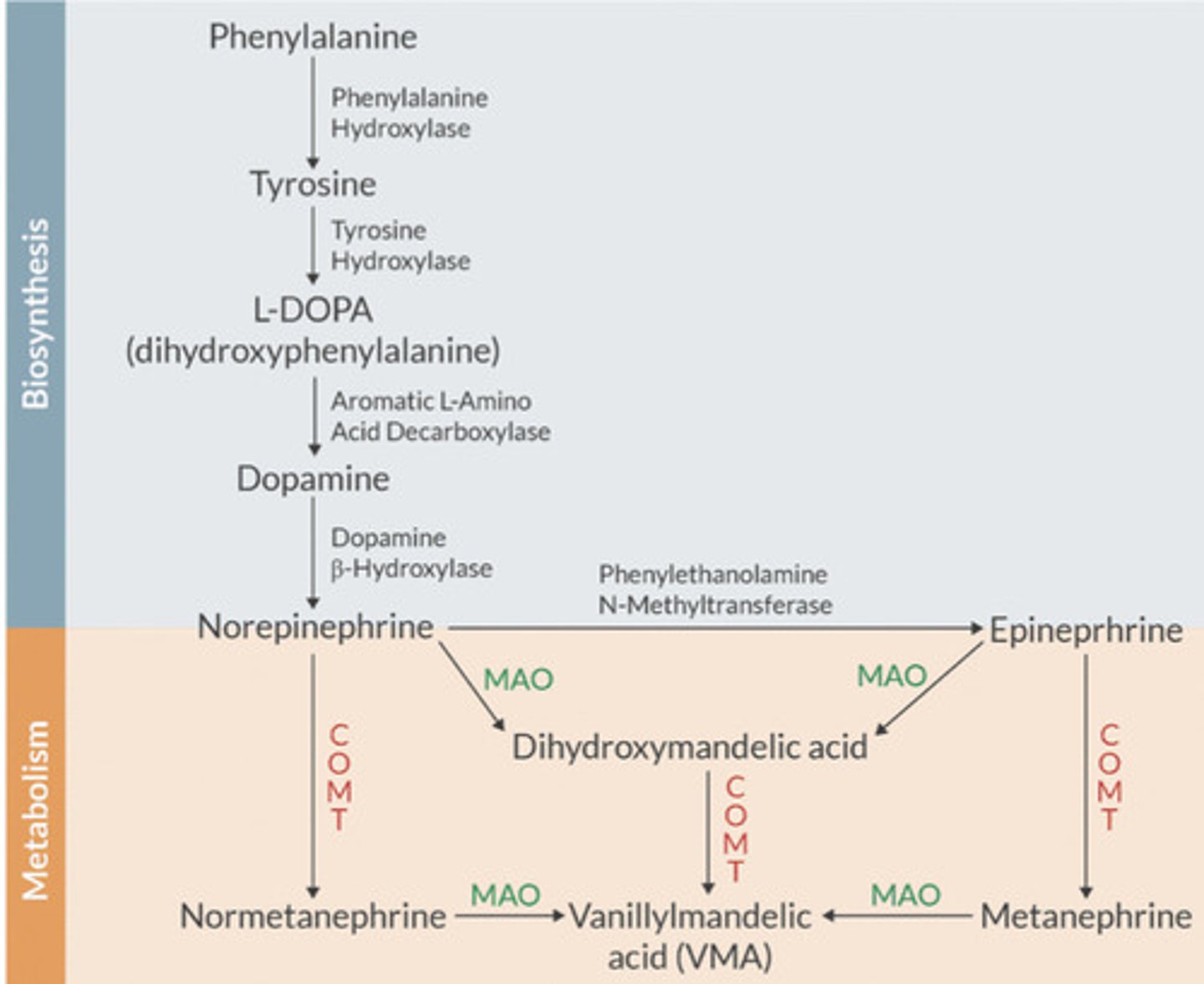
Most postganglionic SNS neurons release ________ as a neurotransmitter.
Norepi
What are the 2 intrinsic muscles of the eye and what do they cause? ?Are they innervated by the PNS or SNS? What receptor?
Radial Muscle: A1 receptor
—SNS releases NE on A1 receptor
—This causes contraction of the radial muscle which causes mydriasis (pupil dilation)
—Horners syndrome paralyzes this muscle soothe pupil constricts in response (Part of PAM)
Circular Muscle: M3 receptor
—PNS releases Ach on Mreceptor
—This causes contraction of the circular muscle which causes miosis (pupil constriction)
The upper eyelid is raised by the _________ muscle.
What muscle maintains the eyeball in a forward position in orbit?
Levatator Palpebrae superior muscle.
—innervated by CN3
Orbital is muscle
—innervated by superior cervical ganglion.
—enopthalamus—recession of eyeball in orbit—caused by blockade of this (Seen in Horners)
Describe the autonomic control of the heart:
—what is it innervated by PNS and SNS
—what are the receptors
—what do they cause?
SNS innervation by Cardiac accelerator Fibers (T1-T4)
—Ne released and binds to B1 receptors
—causes increased chrono/dromo/ino/lusitropy
PNE innervation by vagus nerve (CNX)
—Ach released on Muscarinic receptors
—causes decreased chrono/dromotropy (NOT inotropy)
—Ach has little to no influence on ventricular contractility (inotropy)
The autonomic nervous system is activated mainly by centers located in the _____________, ____________, and ______________.
What is the most important integrator of autonomic and endocrine function?
What is the major lower brainstem command center for visceral control?
Spinal Cord, Brainstem, and Hypothalamus
Hypothalamus.
Nucleus Tractus Solitarius
What is the reticular formation?
a set of interconnected nuclei that are located throughout the brain stem
Important in CV, respiratory, and central pattern generators.
The _______________ contains second-order neurons that receive all input from peripheral chemoreceptors, baroreceptors, and non-nociceptive input from every organ in the thorax and abdomen.
Which cranial nerves are involved in these visceral afferents?
Nucleus Tractus Solitarius
CN 7, 9, +10
What are examples of cortical control of autonomic output?
Fear/Panic—initiates fight or flight
Emotional Distress/Pain—vasodilation, hypotension (vasovagal)
Seizure—can induce cardiac arres/death from massive sympathetic output.
Sleep Deprivation—can lead to loss of thermoregulatory and CV control
Cognitive—sexual arousal
Nervous—diarrhea
What is the Baroreceptor reflex?
Where are the afferents?
Where do the efferent go?
Negative feedback loop that controls BP around a set point.
—Short Term (resets/adapts in 1-3 days)
Afferents:
—aortic arch (CNX) and carotid bifurcation (CNIX + Herings nerve)
—travels to NTS for interpretation and response (release of glutamate).
—can either increase SNS output or decrease SNS output
Efferent signals sent to heart and blood vessels
—increase/decrease in HR and vasodilation/constriction
How do you know if the baroreceptor response is preserved under anesthesia?
What anesthetic agents impair or preserve the baroreceptor reflex?
If preserved—HR will increase/decrease in setting of BP changes.
If not—Hr will not change
Impair:
—Volatile anesthetics
—Propofol
—beta blockers/CCB, ACE-I, PDE inhibitors
Preserve:
—Thiopental
—NTG/nitroprusside/hydralazine
—phenylephrine
What is the Bainbridge reflex?
Increase in HR caused by an INCREASE in venous return.
—stretch receptor activation from increased volume.
—increase in HR is greatest at low baseline rates.
Prevents damming of up of blood flow in veins, atria, and pulmonary circulation.
What is the Bezold Jarish Reflex?
What are the stimulus, afferent pathway, control center, efferent pathway, and response?
Bradycardia, Hypotension, and Coronary Artery Dilation in response to noxious ventricular stimulus.
—Stimulus: MI, low venous return, thrombolytics
—Can be seen with spinal/epidural or regional in sitting position.
Sensor: chemo/mecanoreceptors in the LV wall
Afferent: nonmyelinated C fibers in vagus
Control Center: NTS and medullary CV nuclei.
Efferent: Vagus
Response: decreased HR (SA node) and Decreased Dromotropy (AV node)
What is the strongest drive at the peripheral chemoreceptors?
Where are they located and describe the efferent response:
Hypoxia
Carotid (CNIX+Herings) + Aortic Arch (CNX)
—afferent travels to the NTS
—Respiratory Response to hypoxia—increase in RR and TV
—CV response—activation of PNS (decreased HR and contractility)—persistent hypoxia leaves to increased SNS = increased CO.
SUBanesthetic concentrations (<0.1MAC) of volatiles blunt this reflex.
What are the 9 types of adrenergic receptors?
What G-protein types are they coupled with and what is the response?
Alpha 1: A1a, A1b, A1d
—Gq/11
—Activates Phospholipase C
Alpha 2: A2a, A2b, A2c
—Gi
—Inhibits adenylyly Cyclase
Beta: B1, B2, B3
—Gs
—Stimuates adenylyl cyclase
What occurs with a vasovagal response?
Physiologic stress, or peritoneal stretching/distension
Trigger causes vagal afferents to CNS and hypothalamus which causes massive PNS outflow and abolition of SNS outflow.
—massive vasodilation and hypotension (failure of baroreceptor response)
—bradycardia and decrease SV—>profound decrease in MAP
—global cerebral ischemia—if lasts 10sec will lose consciousness.
Can precipitate CV collapse and cardiac arrest.
What is the oculocardiac reflex?
What is the afferent and efferent limbs?
Five and Dime Reflex (Afferent CN V....Efferent CNX)
Stimuli: traction of extraocular muscles (medial rectus), strabismus surgery, or ocular pressure.trauma.
—retrobulbar block can CAUSE or PREVENT the OCR.
Response: decreased SA and AV node activity.
—bradycardia hypotension, Junctional rhythm, AV block, asystole. W
What factors worsen the Oculocardiac reflex?
Treatment?
Hypoxemia, Hypercarbia, Light anesthesia.
Treatment:
—release traction
—100% Fio2, ensure ventilation and deepen anesthetic.
—Admin anticholinergic (atropine or glycopyrrolate)
What is the CNS ischemic reflex?
When is it activated and at its most intense?
Blood flow to medullary vasomotor centers decreased to cause ischemia
—causes a massive activation of SNS.
Intense vasoconstriction and increase in BP
—so intense some peripheral vessels may become totally occluded
—Renal blood flow may be compromised.
One of the most powerful activators of all SNS systems.
—response is not significant until MAP<50mmHg
—greatest degre when MAP 15-20mmHg.
Adrenal medulla:
—preganglionic SNS neuron synapses directly with Nicotinic (N) receptors on the adrenal medulla (chromaffin cells) to stimulate Epi (80%) and NE (20%)release
—Adrenal medulla acts as a post ganglionic receptor.
Sweat glands:
—postganglionic SNS neuron releases Ach that binds with Muscarinic (M3) receptors at the sweat glands.
Vessels in Skeletal Muscle:
—post ganglionic SNS release Ach and bind on M3 Muscarinic receptors in blood vessels to cause dilation.
What is Cushings Reflex?
Increased ICP causes physiologic response:
—increased BP (SBP>DBP..wide pulse pressure)
—baroreceptor reflex from HTN causes bradycardia
—Irregular Respirations.
Where does the body send afferent signals about temperature?
Skin, core organs, and CNS detect temp and send afferents to pre optic area in hypothalamus
Efferent impulses to effectors:
—blood vessels—constriction/dilation
—brown fat and skeletal muscle for shivering/thermogenesis
—sweat glands to provide evaporative heat loss
—piloerection (effective in animals but not humans)
What is sweating controlled by and what can they be blocked by?
What can shivering be blocked by?
Cholinergic fibers (SNS nerve—>releases Ach—>Muscarinic receptors)
Can be blocked by muscarinic antagonists
—Glyco...atropine.
Shivering blocked by NMBs.
T or F
All General anesthetics impair thermogenesis reflexes.
TRUE
What is the valsalva reflex?
Forced expiration against closed glottis
—increased Intrathoracic pressure—>decreased venous return + CO/BP
Baroreceptor reflex increases HR
Upon glottic opening, increase in venous return—>increased CO/BP
—this causes reflex decrease in HR to baseline.
What are the 2 versions of the Mass reflex?
Fear, exercise or other stress
—causes massive coordinated output to all end organs
—cease of PNS outflow
—diffuse physiognomy response instead of discrete effects.
Autonomic Dysreflexia / Hyperreflexia
—reflexes cause reflexes of many/or all spinal portions.
—This causes strong flexor spasm, evacuation of colon/bladder, HTN, and profuse sweating
Which receptors mediate dilation of coronary arteries and skeletal muscle vessels?
Beta 2