Kin 411: Cerebellum (All 17, 18, 19)
1/125
There's no tags or description
Looks like no tags are added yet.
Name | Mastery | Learn | Test | Matching | Spaced | Call with Kai |
|---|
No analytics yet
Send a link to your students to track their progress
126 Terms
Which side of the hemisphere is non dominant? Main fx?
Usually the right hemisphere is non dominant
Main fx:
Visual spatial analysis
Spatial attention
What is the role of the non dominant hemisphere.
What area of the brain exactly plays that role? What inputs does it use to carry out that role?
The parietal area of non dominant (right) hemisphere plays a role as a control center to attend to critical information in the env and filter out non-relevant info in the entire spatial field.
It uses: visual, sensory and motor inputs
What is neglect?
Damage to right parietal association cortex or frontal cortex causes inability to attend to/focus on/identify sensory information coming from the contralateral side.
Neglect often happens on one side of the body.
What is hemi-spatial neglect? What causes it?
Ignoring or failing to attend to activity on one side of the spatial field (visual, sensory, motor)
It is caused by a stroke in the non-dominant (right) parietal cortex
Damage to right (non-dom) hemisphere parietal association cortex, extending to frontal cortex, causes what and where
Causes hemi-spatial neglect (inability to attend to important sensory info or movements) on the contralateral side
What is common about strokes that cause hemineglect?
Strokes can be of different sizes but they’re all located at the parietal/frontal association cortex leading to hemineglect
How is the attention field divided in a normal brain
Right parietal cortex/hemi:
Attends to entire space but more attention on contralateral (left) space
Left parietal cortex/hemi:
Limited attention
All attention focus on right spatial field
Together they cover the entire attention field on a normal brain
Non dom ____ cortex damage, also called ___ hemi lesion, causes severe ___ neglect
parietal; right hemi lesion; left neglect
Describe what happens during a right hemisphere lesion
Complete neglect of the left spatial field
i.e. Lose ability to attend to info (visual, sensory, motor field)
Right spatial field still present, albeit less so, as left hemisphere can still somewhat attend to it
Does left hemisphere damage show neglect on any side? Why or why not? Is there a difference between the strength of attention for either field?
No, left hemisphere damage does not show any neglect on right spatial field, or any field for that matter.
This is because right hemisphere has a specialization for attention and can attend to the whole field
Attention is stronger for the left spatial view but still able to attend to the right field
Hemi-neglect always appears in which visual space? Which visual side of the body?
Hemi-neglect appears in the left visual space, on the left side of the body
Damage to both hemisphere leads to ?
Lead to neglect of the entire visual field altogether
Damage to left (dominant) hemisphere leads to?
No neglect, as right hemisphere is intact and it has specialization to attend to the whole field.
What are the types of neglects?
Sensory neglect: Visual, tactile, or auditory neglect
Motor-intentional neglect
Combination of sensory and motor neglect
Conceptual Neglect
What’s key to remember about sensory neglect?
It is NOT an inability to receive raw sensory input. PCML, somatosensory cortex is still intact. They’re able to focus on a specific area individually when tested
It is the inability to attend to sensory input when they need to “span their attention” ie when testing both sides together
What is Motor-intentional neglect - what can pt do, what can’t they do, and what causes it?
Pt able to do unilateral movements when focusing individually
Inability to do motor movements bilaterally
i.e when asked to move both sides, will neglect movement on left side, only do movement on the right side
due to damage on right (non dom) hemisphere leading to left hemineglect
free
free
Conceptual Neglect
Damage to right (non dom) parietal cortex leading to:
Representational neglect
Anosognosia
Hemiasomatognosia
Representational neglect: is a form of? caused by? leads to?
Form of conceptual neglect due to damage to right parietal cortex
Leads to: attending to right side conceptual memories only e.g. remembering the right side buildings on the map
Anosognosia
No awareness of hemi-neglect or -plegia
Neglect or lack of awareness of the actual neglect itself OR that they have a particular injury
e.g. stroke causes hemi-neglect to the left side of the body but pt not aware that they have Sx/deficits on the left side of the body
Hemiasomatognosia
Disowning limbs: disowning left side limbs (extreme conceptual neglect)
Dressing Apraxia
Apraxia is usually due to loss of skilled movement due to damage to the left hemisphere
BUT, dressing apraxia is due to RIGHT parietal lesion
Pt forgets to attend to the left side of their body while dressing
This is a symptom of neglect, NOT a symptom of loss of skilled movement
Alien Hand
Damage to Supplementary Motor Area or corpus callosum on the right (non dominant) hemisphere
Left hand is “out of control” i.e. acts autonomously, mischievously
Theory behind Alien Hand
Two consciousness on each side of the brain:
Dominant consciousness keeps non-dominant consciousness in check
When we hurt the corpus callosum, the right hemisphere can do whatever it wants since it’s not being checked by left (dom) hemisphere
Cerebellum location with respect to skull
Sits nestled in the posterior fossa in the base of skull
Covered by the tentorium cerebelli, which separates it from the inferior occipital lobe
General cerebellum location
Posterior to the brainstem
It makes up the dorsal wall of the CSF filled 4th ventricle
It is nestled in the posterior fossa where it is separated from the occipital and temporal lobe by the tentorium cerebelli
Cerebellum acts like a mini brain. ____ matter on the ___ surface making the folds. The ___ folds are called _____.
_____ matter projects ____ as tracts
Grey matter on the outer surface
The outward folds are called folia
White matter project inwards as tracts
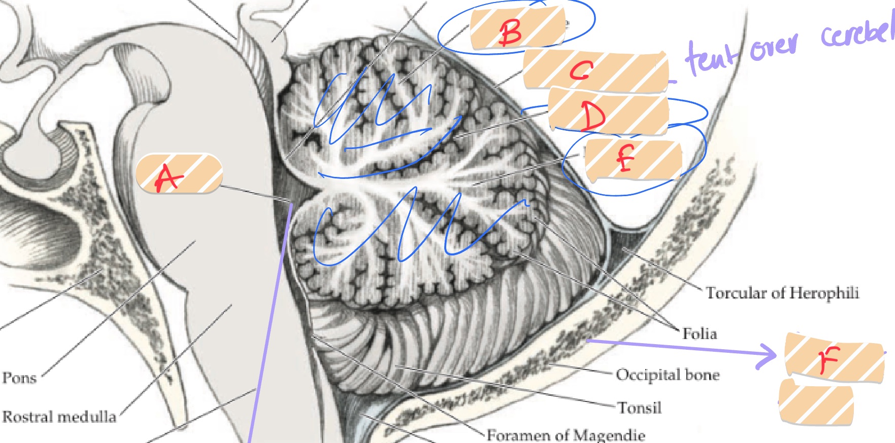
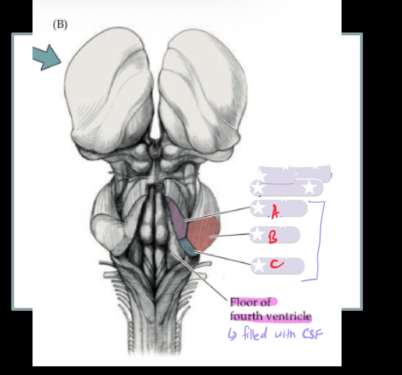
Label A-F
A- Fourth Ventricle
B- Anterior Lobe
C- Tentorium cerebelli
D- Primary fissure
E- Posterior Lobe
F- Posterior fossa
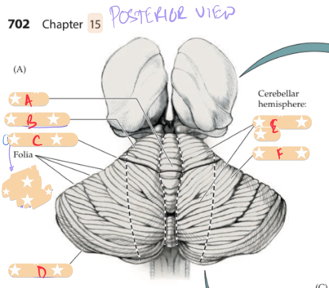
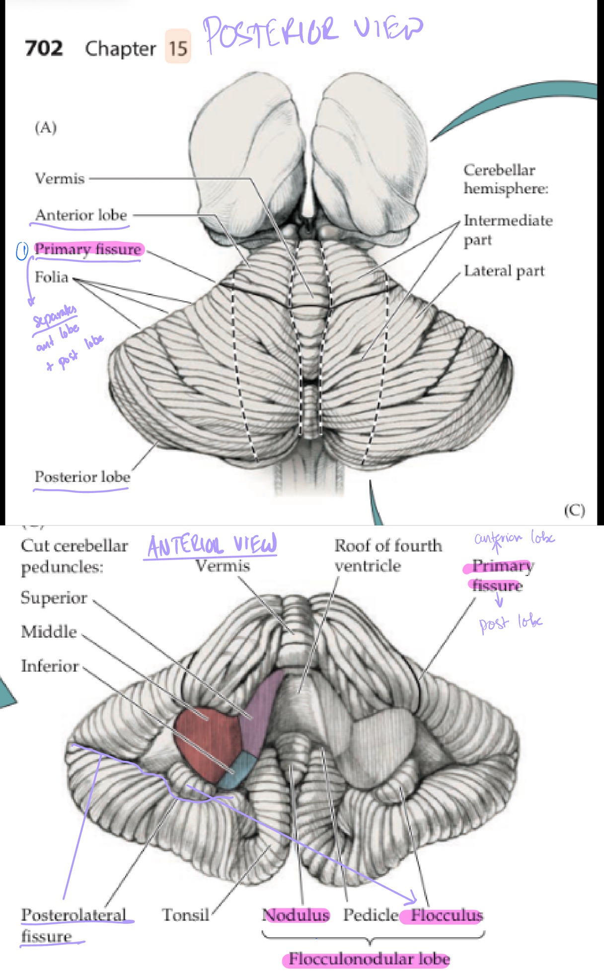
Label A-F
A- Vermis
B- Anterior lobe
C- Primary fissure
D- Posterior lobe
E- Intermediate zone
F- Lateral zone
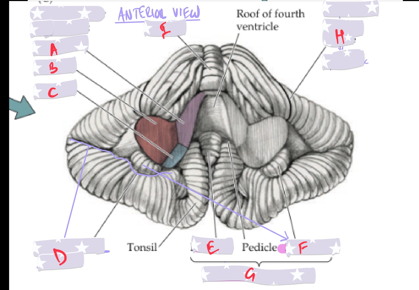
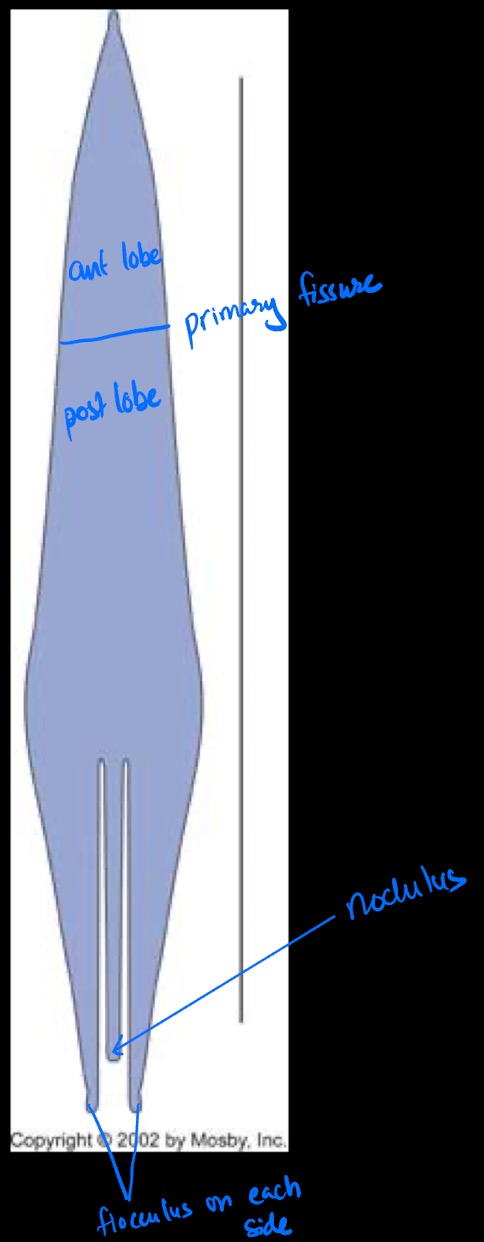
Label A-H
A- Superior cereb ped
B- Middle cereb ped
C- Inferior cereb ped
D- Postero-lateral fissure
E- Nodulus
F- Flocculus (floculli on both sides)
G- Flocculonodular lobe
H- Primary fissure
Cerebellum anatomy
Cerebellum has 3 major lobes separated by 2 key landmarks (fissures):
Anterior lobe
Primary fissure separates anterior and posterior lobe
Posterior lobe
Postero-lateral fissure separates inferior posterior lobe from the flocculo-nodular lobe
Flocculo-nodular lobe
Primary fissure
Separates anterior and posterior lobe
Postero-lateral fissure fx. Which view is it visible from?
Only visible from anterior view
Separates inferior posterior lobe from the flocculo-nodular lobe
What is flocculo-nodular lobe made up of?
Nodulus in the center and 2 flocculi (bulges) on either side
How do you make a cerebellum?
Stretch it to look like an octupus
Anterior lobe top 1/3
Divided by primary fissure
Middle 1/3 is posterior lobe
Bottom tentacles has the nodulus in the middle with 2 flocculi on each side
What are folia?
They are outward folds of grey matter on the cerebellum.
They fold to connect to the white matter tracts that go inward
What are the 4 functional region of the cerebellum? Explain where they are located
Think of the posterior view of cerebellum like a moth
Vermis: central region (body of the moth)
Intermediate zone: middle 1/3 region
Lateral zone: lateral 2/3 region
Flocculo-nodular zone:
made up of flocculo-nodular zone
only seen from anterior view
Where are the 4 functional region of the cerebellum projecting to
To the deep cerebellar nuclei
How are the functional regions of the cerebellum associated with its nuclei?
Functional grey matter region of the cerebellum are aligned with their corresponding deep nuclei
Name the deep nuclei of the cerebellum and their associated fx region
Dentate nuclei = lateral zone
Interposed nuclei = Intermediate zone (memory trick: Both are I)
Fastigial Nucleus = vermis + flocculo-nodular zone
Dentate nuclei
Aligns with the lateral zone
Interposed nuclei
Made up of 2 smaller nuclei:
Embolus nucleus
Globus nucleus
Aligns intermediate zone
Fastigial nucleus
Along the midline
Aligns with the vermis and flocculonodular zone
How to remember Order of Nuclei in the cerebellum
From most lateral → most medial
Dont = Dentate
Eat = Emobolus
Greasy = Globus
Foods = Fastigial
*Emobolus + Globus = interposed nuclei
Where do these deep nuclei in cerebellum receive information from?
Deep nuclei in cerebellum receive info from the axons of the grey matter
Purkinje cells in the:
Lateral zone → dentate nuclei
Intermediate zone → interposed nuclei
Vermis + flocculonodular zone → fastigial nucleus
All information are carried to and from cerebellum via _____
Cerebellar peduncles
What are the cerebellar peduncles? Names? Fx? What level of the brain are they?
They are projections of white matter in and out of the cerebellum
Superior, middle, inferior peduncles
They carry info to and from cerebellum
They come out at the level of the pons giving it its bulge shape
What are the 3 cerebellar peduncles
Superior cerebellar peduncle
Middle cerebellar peduncle
Inferior cerebellar peduncle
Label A-C
A- Superior cereb peduncle
B- Middle cerebellar peduncle
C- Inferior cerebellar peduncle
Cerebellar peduncles are not to be confused with ?
Cerebral peduncles which are tracts of white matter running down the anterior surface of the midbrain
What are Purkinje cells
Neurons specific to the cerebellum, found in the exterior surface of the cerebellum and they project into the deeper nuclei
Fx of superior cerebellar peduncles
Superior cerebellar peduncles carry information from cerebellum and project OUT into the nervous system
Fx of middle and inferior cerebellar peduncles
Middle and inferior cerebellar peds is to carry inputs INTO the cerebellum
Describe path of info into and out of cerebellum
Info comes into cerebellum via inferior and middle cerebellar peduncles
Gets process in the cerebellum by grey matter and purkinje cells
Axons from purkinje cells project into the deep nuclei according how they are aligned (dont eat greasy food)
Deep nuclei does further processing of the info
Info is projected back out into the nervous system by the superior cerebellar peduncle
What are the 3 main input pathway to the cerebellum? What do they do?
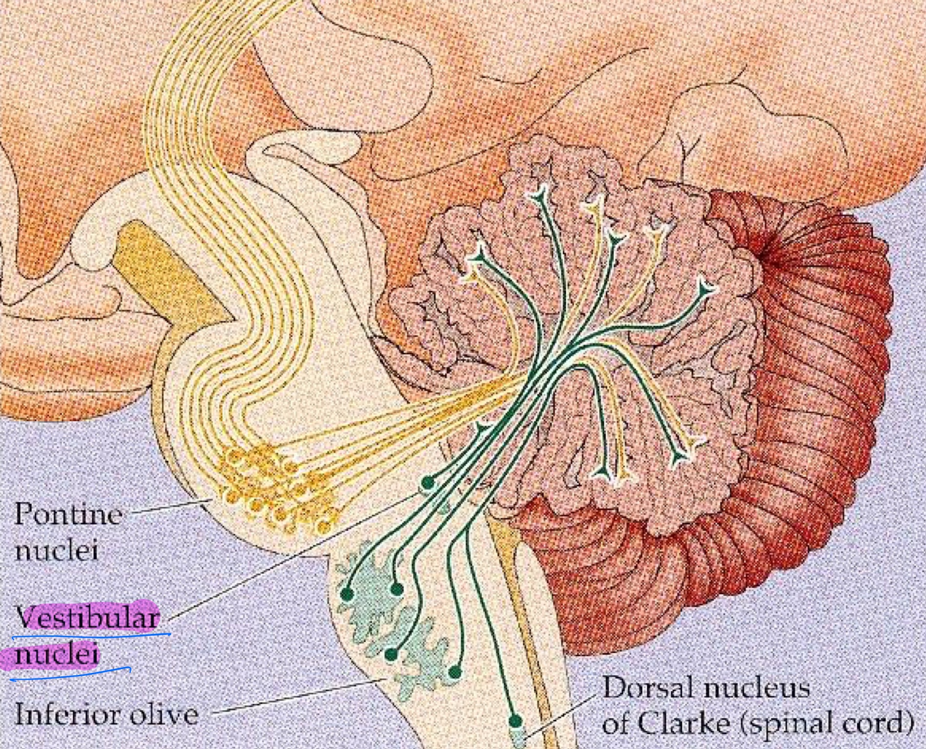
Pontocerebellar fibers
Spinocerebellar pathways
Dorsal spinocerebellar tract
Cuneocerebellar tract
Vestibular inputs
Inputs from these 3 control ongoing movement + coordinate movement
Input pathway: Coming from → Cells Projecting to Cerebellum → Entering thru which Cerebellar peduncle
Pontocerebellar fibers
Pontocerebellar fiber: Motor info coming from cortex → pontine nuclei → middle cereb peds
Input pathway: Coming from → Cells Projecting to Cerebellum → Entering thru which Cerebellar peduncle
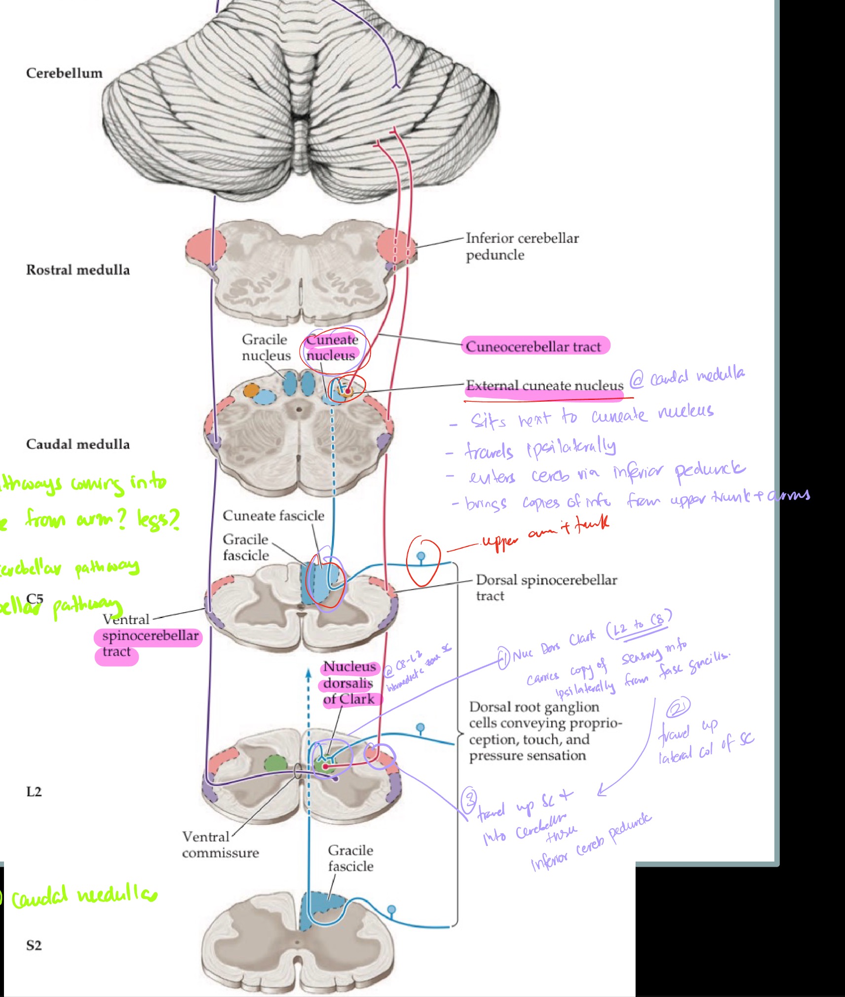
Dorsal spinocerebellar tract
Spinocerebellar Pathways: Proprioceptor legs → Nucleus Dorsalis of Clark (C8 - L2) [ipsi] → Enter Inferior cerebellar penduncle (ipsi)
Input pathway: Coming from → Cells Projecting to Cerebellum → Entering thru which Cerebellar peduncle
Cuneocerebellar Tract
Cuneocerebellar Tract: Proprioceptor/vibration/LT from arms → External cuneate nucleus (caudal medulla) [ipsi} → Inferior cerebellar penduncle (ipsi)
Input pathway: Coming from → Cells Projecting to Cerebellum → Entering thru which Cerebellar peduncle
Vestibular inputs
Vestibular inputs: Vestibular system → vestibular ganglia + vestibular nuclei (ipsi) → juxtarestiform body (subsection of inferior peduncle) ipsi
Where do inputs into cerebellum come via
Middle and inferior cerebellar peduncles
Inputs to the cerebellum: motor
One input coming into cerebellum is ongoing motor information from the motor cortex
Copy of motor info is sent from cortex to cerebellum via pontocerebellar pathway
Cortical pontine fibers (axons from the motor cortex) descend ipsilaterally synapsing onto pontine nuclei
Ponto-cerebellar fibers from pontine nuclei cross the midline @ pons
Enter cerebellum via middle cerebellar peduncle
How does signal of a motor plan to move the right hand travel from cortex to cerebellum
Signal to move right hand originates at the right cortex
Copy of that input from right motor cortex travel ipsilaterally to the right pontine nuclei
Axons from pontine nuclei decussate @ pons
Enter left cerebellum thru the middle cerebellar peduncle
Inputs to the cerebellum: Sensory
Afferent copy (external feedback) from periphery is carried into cerebellum, through the inferior cerebellar peduncle, via two Spinocerebellar Pathways:
Cuneo-cerebellar tract
Dorsal spinal-cerebellar tract
Both tracts resemble the PCML pathway
Cuneo-cerebellar tract
Sensory info coming from upper arm and trunk enter lateral part of the posterior column @ fasiculus cuneatus
Travels up to the caudal medulla and synapse onto Nucleus Cuneatus
Collaterals from Nucleus Cuneatus synapse onto External Cuneate Nucleus @ caudal medulla
Travel up ipsilaterally to enter the cerebellum via the Inferior Cerebellar Peduncle
Dorsal Spinal-cerebellar tract
Sensory info coming from lower leg enter the medial part of posterior column @ fasiculus gracilis
Collaterals from Fasiculus Gracilis synapse onto the Nucleus Dorsalis of Clark @ intermediate zone of grey matter of the SC, at the same level
NDC collaterals are found at every level from C8 → L2/L3
Axons from NDC travel to the lateral column of the SC, at the same level
Start traveling ipsilaterally up the Spinocerebellar tract
Enter the cerebellum via the inferior cerebellar peduncle
How is sensory info from lower leg sent to cerebellum?
Sensory info from leg/foot travel via the Spinocerebellar pathway:
Inputs from periphery, such as foot, travels to contralateral somatosensory cortex
A Copy of that goes to the ipsilateral cerebellum
Fasiculus gracilis, which receives the input from legs at the SC, synapses onto the Nucleus Dorsalis of Clark in the intermediate zone of the SC at the same level
NDC send axons to lateral column of SC at the same level
Start traveling ipsilaterally up the SC via the lateral column on the Dorsal Spinocerebellar tract
Enter cerebellum via inferior cerebellar peduncle
How is sensory info from arms/trunk sent to cerebellum?
Sensory info from arms/trunk travel via the Cuneocerebellar tract:
Inputs from periphery, such as arms, travels to contralateral somatosensory cortex
Copy of that goes to the ipsilateral cerebellum
Fasiculus Cuneatus, which receives the input from arms at the SC, travels up to caudal medulla and synapse onto Nucleus Cuneatus
Collaterals from Nucleus Cuneatus synapse onto the External Cuneate Nucleus @ caudal medulla
Axons then travel up ipsilaterally via the Cuneocerebellar Tract
Enter cerebellum via inferior cerebellar peduncle

Nucleus Dorsalis of Clark
Elongated nucleus found in the intermediate zone of grey matter on the SC
Travels from C8 to L2/L3
Collaterals from Fasiculus gracilis synapse on it
Carries copies of sensory info from legs + lower trunk
External Cuneate Nucleus
Sits @ caudal medulla, next to Nucleus Cuneatus
Collaterals from Nucleus Cuneatus synapse on it
Carries copies of sensory info from upper trunk + arms
free
free
Inputs to Cerebellum: Vestibular
Mention peduncle it enters thru*
2 connections carrying vestibular input into the cerebellum:
Primary vestibular sensory neurons (hair cells in otoliths)
Vestibular nuclei (Second order sensory neurons)
Both vestibular PSN and 2SN have DIRECT projections into the cerebellum via juxtaresitform body (a subsection of inferior cerebellar peduncle)
What is the cerebellum on one side receiving in total?
Copy of motor info from contralateral motor cortex via pontocerebellar fibers
Copy of sensory info from ipsilateral side via Spinocerebellar (legs) and Cuneocerebellar (arms) tract
^ Receives ongoing sensory info & compares it to the planned motor function from the cortex
Receive information from the vestibular system via primary neurons (hair cells) and second order neurons (vestibular nuclei)
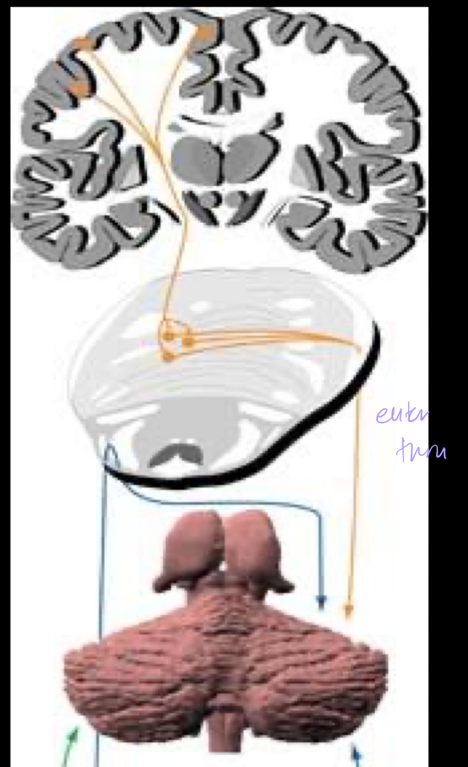
Outputs from the cerebellum travel via ? Where are outputs sent?
Information from the cerebellum leaves via the Superior Cerebellar Peduncle
Most output will be sent to the cortex via the thalamus
What is the golden rule of cerebellar outputs?
Cerebellar outputs always control the ipsilateral side of the body
It does this either via:
double crossing → ipsilateral control
no crossing → ipsilateral control
Main cerebellar output pathways
Region of cerebellum → Deep nuclei axons project out via → Cerebellar peduncle → Main Output
Lateral, intermediate, vermis, inferior vermis and flocculonodular lobe
Lateral region → Dentate → Superior peduncle → Contralateral Thalamus
Intermediate region → Interposed → Superior peduncle → Contralateral Thalamus
Vermis → Fastigial N → Uncinate fasciculus (superior peduncle) + Juxtarestiform body (inferior peduncle) → Vestibular N + Reticular formation
Inferior vermis + flocculonodular lobe → Verstibular nuclei + fastigial nuclei → Juxtarestiform body (inferior peduncle) → MLF (eye movement)
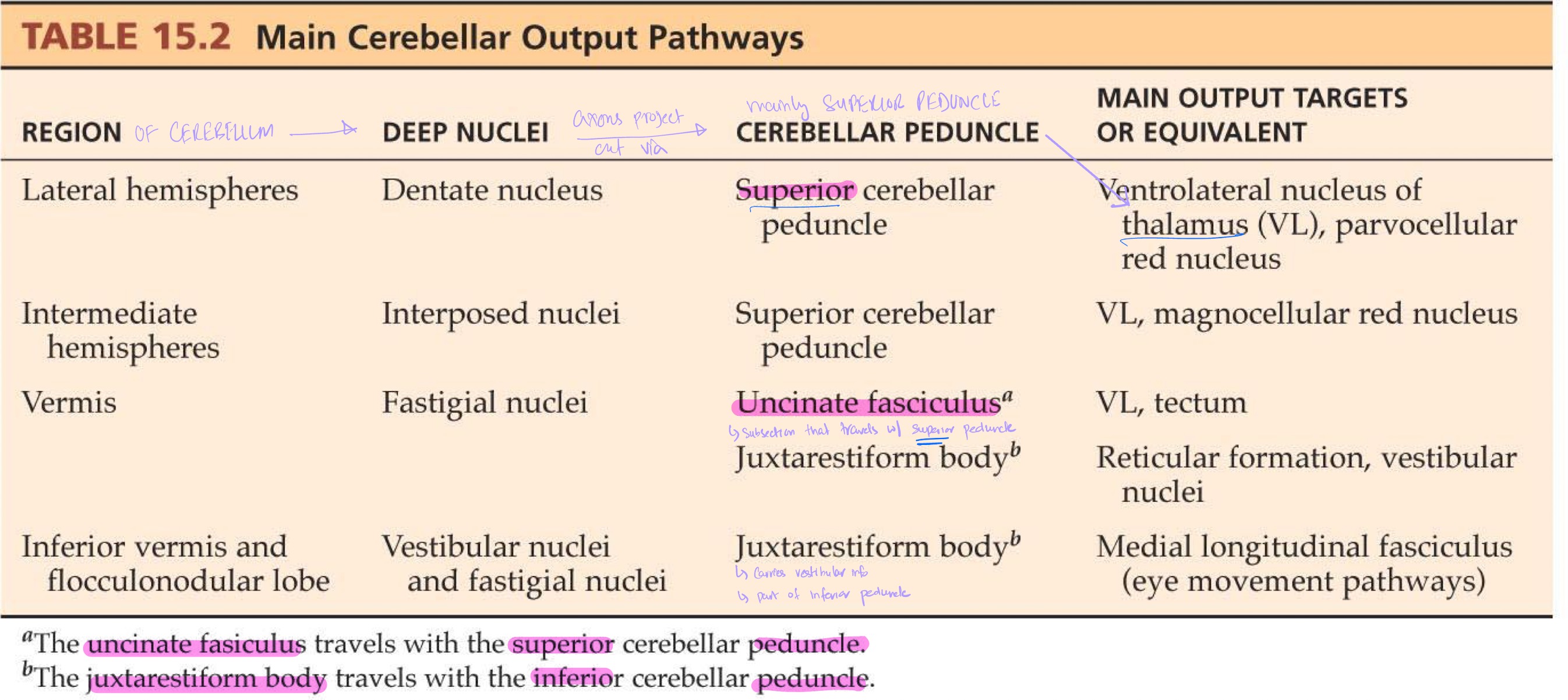
__ fasciculus travels with the _ cerebellar peduncle
Uncinate fasiculus → superior peduncle
__ body travels with the _ cerebellar peduncle
Juxtarestiform body → inferior peduncle
What is the lateral cerebellum responsible for?
Lateral region involves motor planning for the LCST ipsilateral to the cerebellum
Describe the output path from lateral cerebellum
Lateral cerebellum is responsible for motor planning:
Cerebellum receives sensory information from the body ipsilaterally
Lateral GM regions of cerebellum take this information and projects into the dentate nucleus for processing
Dentate fibers travel out of cerebellum via Superior Peduncle
Dentate fibers cross the midline @ midbrain at red nucleus
Travel to the contralateral thalamus
Cell bodies from contralateral thalamus project to motor cortex (PreMC, PMC, SMA)
Motor cortex sends out motor commands via LCST which then decussate and reach the limbs ipsilateral to the cerebellum that sent the info

Cerebellum plans movements for parts of the body ___ to itself. It does so by communicating with the ___ motor cortex
ipsilateral to itself; contralateral motor cortex
What is the intermediate region of the cerebellum responsible for
Intermediate region/Interposed nuclei:
Control distal limbs ipsilateral to cerebellum
via contralateral motor cortex
Influence arms ipsilateral to cerebellum
via contralateral red nucleus → Rubrospinal tract (RST)
Output from intermediate zone for distal limb control
Cerebellum receives sensory information from the body ipsilaterally
Intermediate zone of cerebellum projects into the interposed nuclei
Interposed N project out via Superior Peduncle
Cross the midline @ midbrain
Reach the contralateral thalamus
Cell bodies from thalamus project to motor cortex (PreMC, SMA, PMC), contralateral to the cerebellum
Control distal limbs ipsilaterally
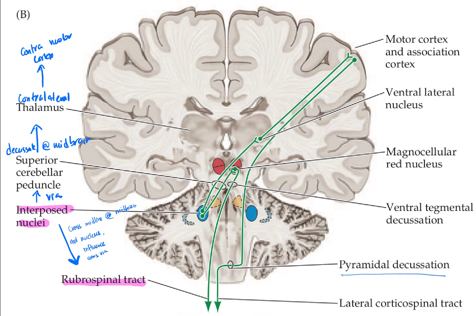
Output from intermediate zone for arm control
Cerebellum receives sensory information from the body ipsilaterally
Intermediate zone of cerebellum projects into the interposed nuclei
Interposed N project out via Superior Peduncle
Cross the midline @midbrain and reach the contralateral red nucleus
Cell bodies from red nucleus travel down Rubrospinal Tract
Control arms ipsilaterally

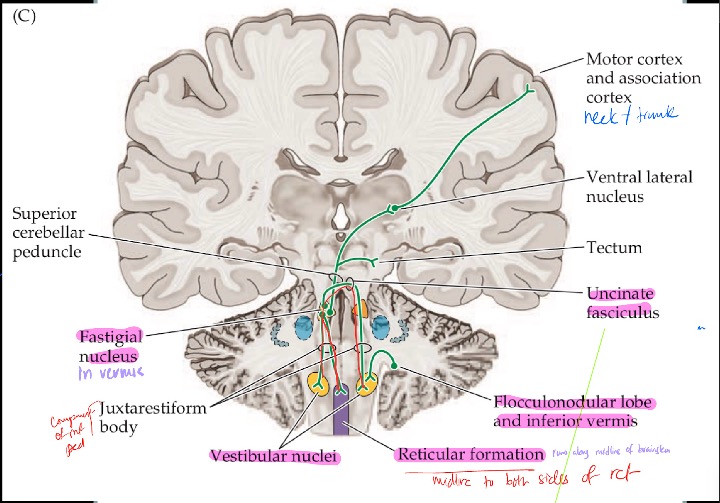
Describe the outputs via the vermis region of the cerebellum
Fastigial Nucleus to Contralateral thalamus to:
Influences Axial muscles (trunk/neck motor areas) bilaterally via ACST
Fastigial Nucleus to Vestibular nuclei + Reticular formation bilaterally
Influence balance reflex and general tone
Inferior vermis + flocculonodular lobe DIRECTLY to Vestibular nuclei ipsilaterally (no deep nuclei)
Influence Balance + eye movement (VOR)
How do Inf vermis + flocculonodule lobe project out of the cerebellum? What does this allow?
There is a bidirectional connection between vestibular system and cerebellum
This is via Inf vermis + flocculonodular lobe connecting with vestibular system on the same side
This allows for cerebellar control over all vestibular reflexes (VOR, VCR)
What is the uncinante fasciculus?
Fibers that are from the fastigial nuclei on one side that cross the midline, before they go out via juxtarestiform body, to the other vestibular nucleus
Where are the 2 body representations in cerebellum?
Anterior lobe
Posterior lobe
What is the somatotopic organization of the cerebellum?
Two body representations —- one on anterior and posterior lobe each
Heads of the two bodies are pointed to one another with anterior lobe having an upside man
Somatotopic organization of the anterior lobe
Trunk and neck region of muscles are along the vermis
Distal limbs (feet + arms) are along intermediate zone
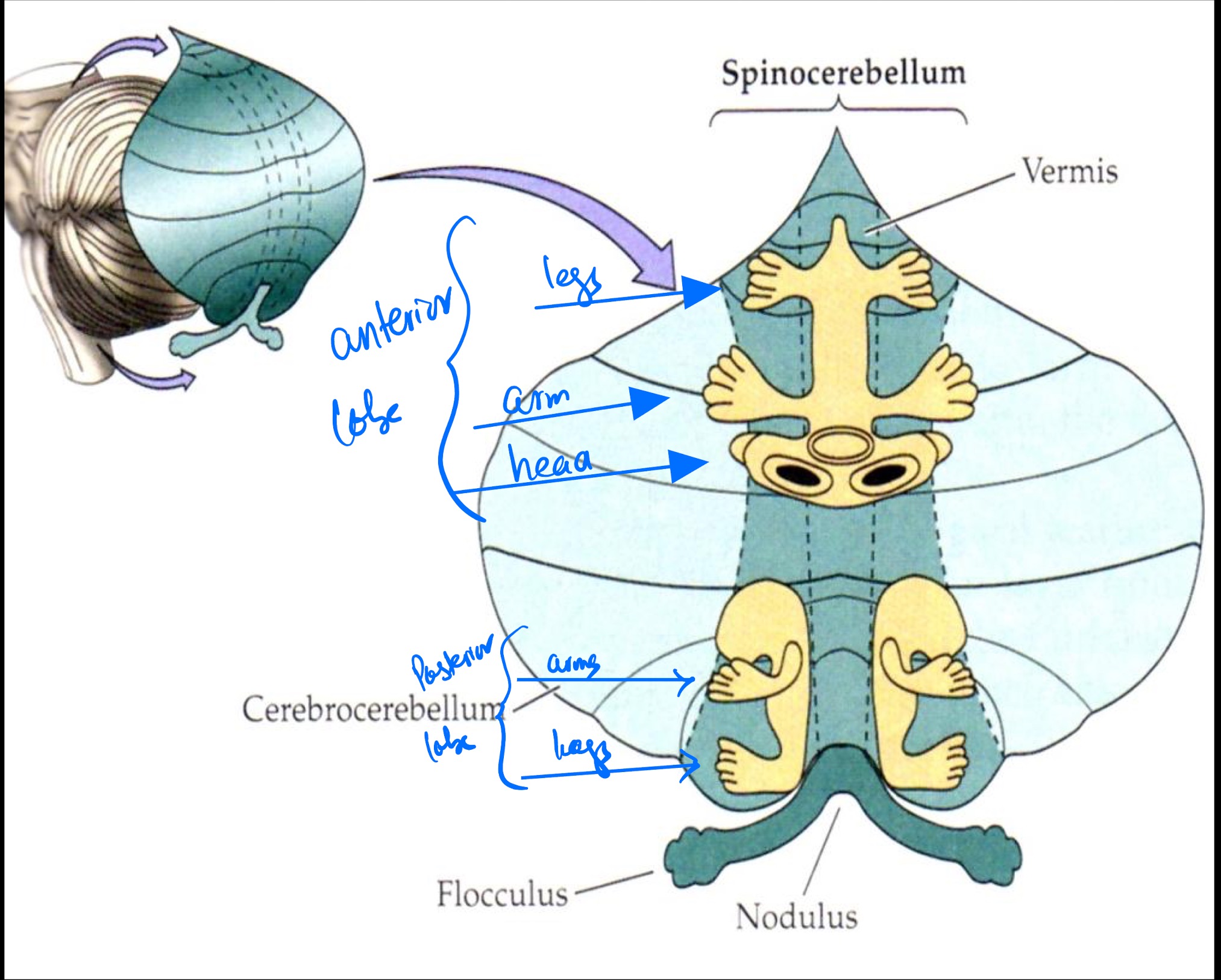
Somatotopic organization of the posterior lobe
NO trunk representation in vermis
Distal limbs (feet + arms) found in the intermediate zone

where are the distal limbs representation located on the cerebellum?
In all cases for the distal limbs are located in the intermediate zone of both the anterior and posterior zone
Where is the trunk representation located on the cerebellum?
The trunk is located in the vermis region of the anterior lobe ONLY.
it is not represented at the posterior lobe at all.
Describe the somatotopic representation of the lateral region of the cerebellum
No somatotopic organization of the lateral region.
Lateral region is involved in motor planning, it has no control over muscles
Function of the cerebellar pathways: Region → Primary role → Motor Pathways influenced
Lateral hemisphere
Lateral hemisphere → motor planning for extremities → LCST
Function of the cerebellar pathways: Region → Primary role → Motor Pathways influenced
Intermediate hemispheres
Intermediate hemispheres → Ongoing coordination of distal limbs and arms → LCST + Rubrospinal Tract
Function of the cerebellar pathways: Region → Primary role → Motor Pathways influenced
Vermis
Vermis → Proximal limb and trunk coordination in anterior lobe vermis region → ACST, Reticulospinal Tract, VST
Function of the cerebellar pathways: Region → Primary role → Motor Pathways influenced
Inferior Vermis + flocculonodular lobe
Inferior vermis + flocculonodular lobe → Balance and VOR → Medial Longitudinal Fasciculus (eye movement pathways)
Cerebellum acts as a comparator by detecting ____. It supplements and supports the ___ ___ cortex to perform ___, ___ movements
Cerebellum acts as a comparator by detecting errors. It supplements and supports the contrlateral motor cortex to perform smooth, coordinated movements
How is ongoing movement adjusted and modulated between cerebellum and cortex
Cerebellum receives:
Copy of efferent information regarding ongoing motor plans from the cortex via corollary discharge.
Lets cerebellum know what the intended movement is
At the same time, cerebellum receives ascending sensory information of what actually happened ipsilaterally via external feedback
Cerebellum then:
Compares the intended goal movement against the actual motor response
Projects information back to motor area to make corrections
Ongoing mvmt is adjusted and modulated this way between cerebellum and cortex