EXAM 2 - Review of CN, CSF, Blood Supply & Facial Muscles...Participatory exercise 2
1/118
There's no tags or description
Looks like no tags are added yet.
Name | Mastery | Learn | Test | Matching | Spaced | Call with Kai |
|---|
No analytics yet
Send a link to your students to track their progress
119 Terms
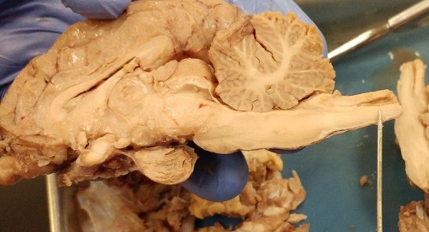
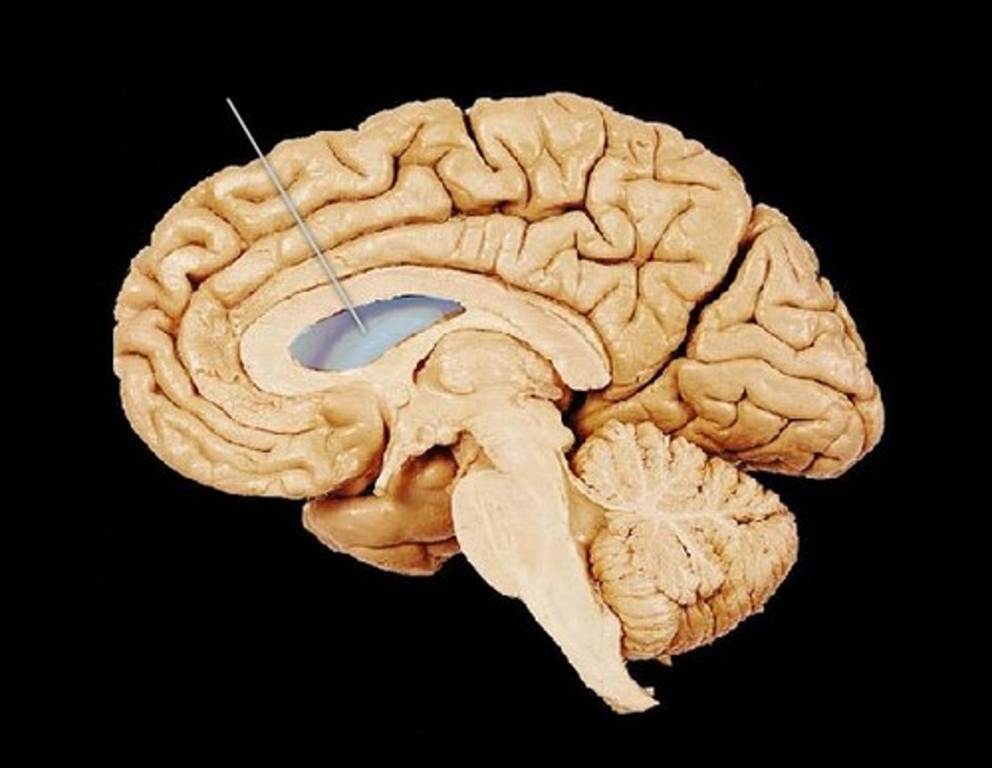
lateral ventricles of brain
ventricles found in each cerebral hemisphere

anterior horn of lateral ventricle
frontal lobe
inferior horn of lateral ventricle
temporal lobe
interventricular foramen brain
connects lateral ventricles to third ventricle
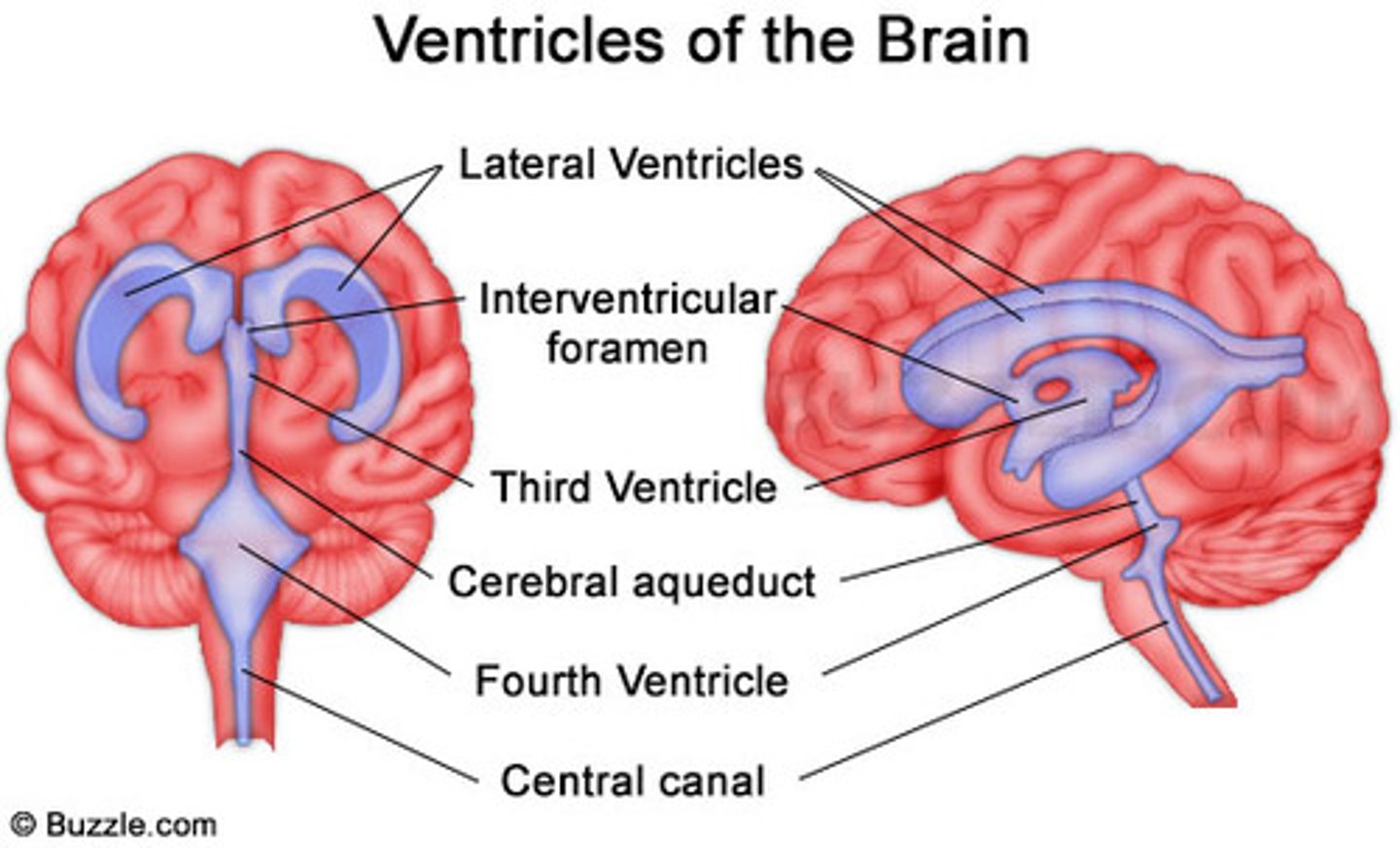
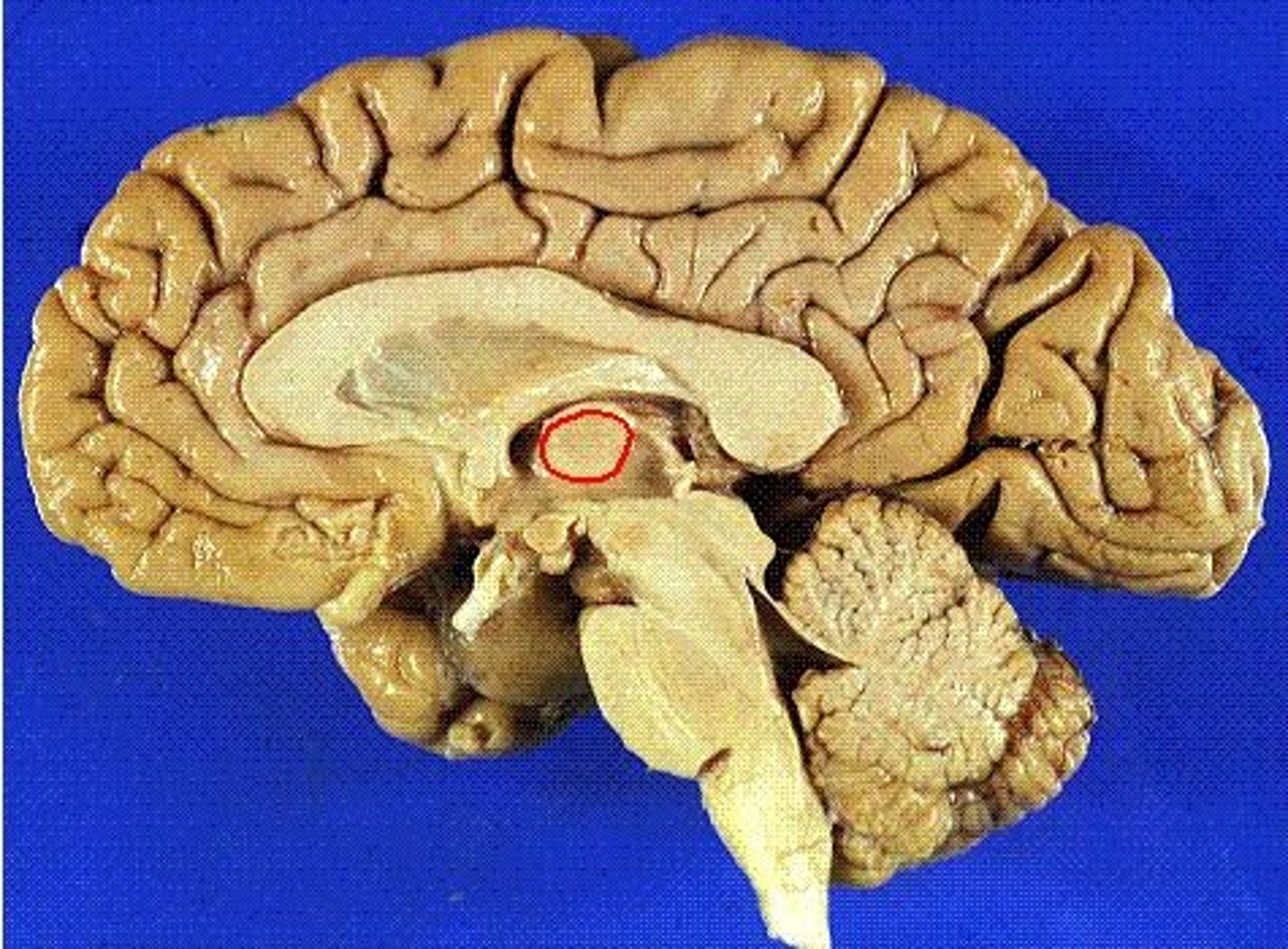
massa intermedia
A small, midline mass of gray matter that connects the right and left thalamic bodies
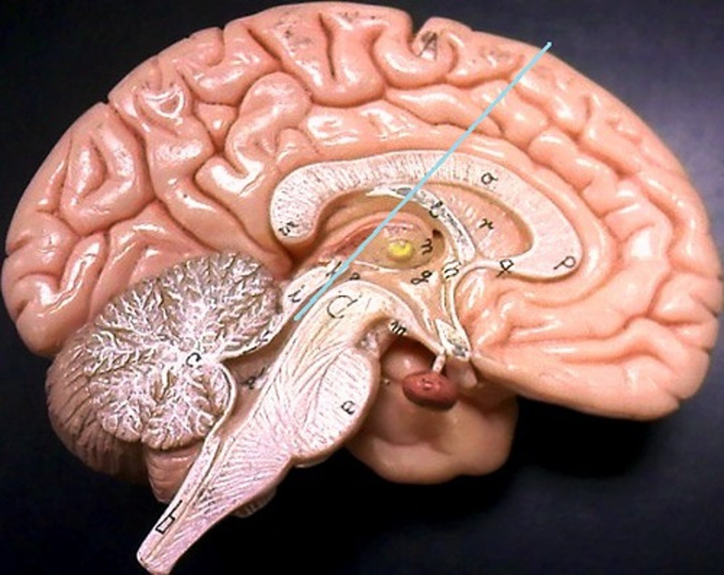
third ventricle
The midline ventricle that conducts cerebrospinal fluid from the lateral ventricles to the fourth ventricle.

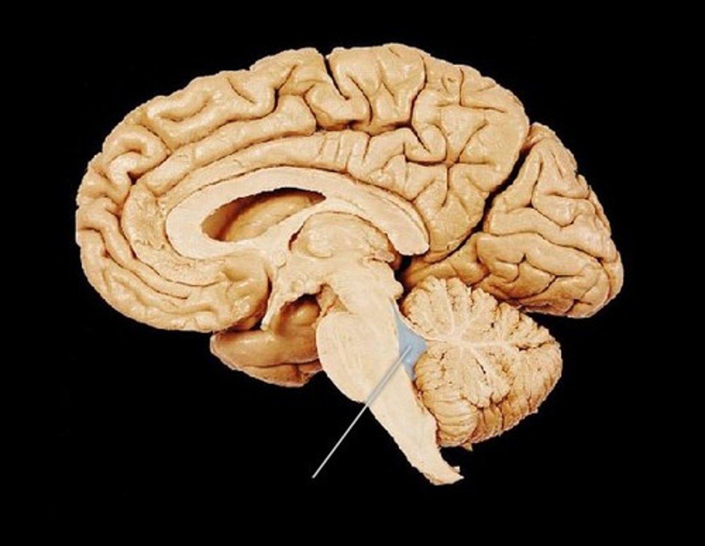
cerebral aqueduct
connects the third and fourth ventricles
fourth ventricle
the ventricle located between the cerebellum and the dorsal pons, in the center of the metencephalon
lateral aperture of fourth ventricle
Holes in 4th ventricle that allows CSF to pass through
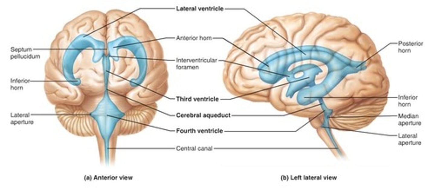
median aperture of fourth ventricle
An opening in the roof of the fourth ventricle that connects to the subarachnoid space

central canal of brain
- Hollow tube in spinal cord
- Continuous with ventricles
septum pellucidum
thin membrane that separates the anterior part of the lateral ventricles from each other.
choroid plexus
produces cerebrospinal fluid (CSF) within the ventricles

The flow of Cerebral Spinal Fluid (CSF) - starting in the lateral ventricle
i. Lateral ventricle
ii. Interventricular foramen (of Monro)
iii. Third ventricle
iv. Cerebral aqueduct
v. Fourth ventricle
vi. From the fourth ventricle, CSF can escape via the lateral apertures, medial aperture, or central canal.
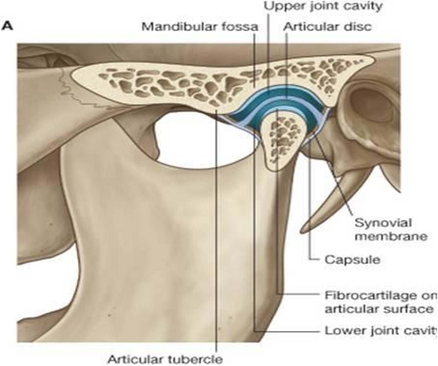
2 articular surfaces of temporomandibular joint
2 articular surfaces and their hyaline cartilage:
superior: articular tubercle and mandibular fossa of squamous portion of temporal bone
inferior: head of condyle of mandible
other structures in TMJ
articular/joint capsule
articular disc
synovial membrane
fibrous capsule
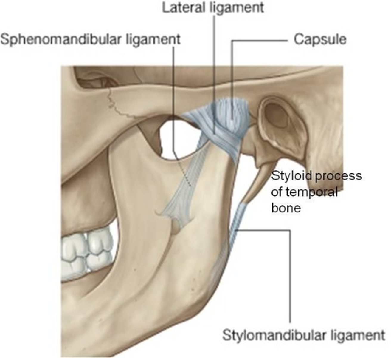
3 ligaments
Articular Disc in TMJ
fibrocartilage that is more firmly attached/moves with the mandible
separates the synovial cavity into superior and inferior compartments
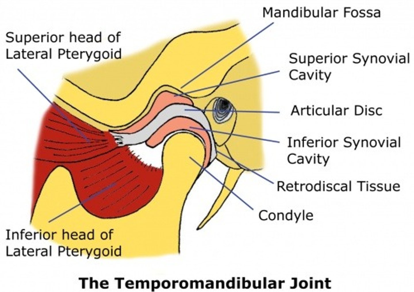
synovial membrane of TMJ
gradually wears away as we age
Lateral (TMJ) ligament
lateral thickening fibrous capsule
zygomatic process of temporal bone and the articular tubercle to the neck of the mandible

stylomandibular ligament
mandible to cranium; weak reinforcement of TMJ; styloid process of temporal bone to the angle of the mandible
sphenomandibular ligament
mandible to cranium; weak reinforcement of TMJ; spine of sphenoid bone to the lingula of the mandible

When the mouth opens (mandibular depression) what happens TMJ
the head of the mandible with the articular disc slides anteriorly until the head lies inferior to the articular tubercle. There is also rotation of the mandibular head on the inferior surface of the articular disc.
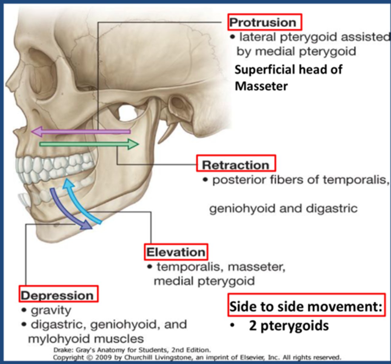
When the protrusion and retrusion of mandible, what happens TMJ
the heads of the mandible (with their articular discs) slide anteriorly (protrusion) and posteriorly (retrusion) on the articular surface of the temporal bone
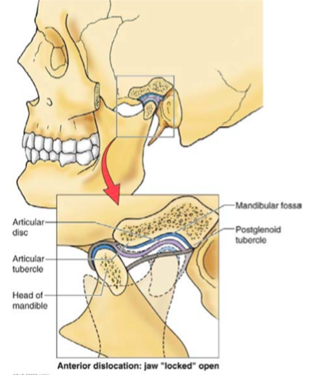
Dislocation of TMJ
refers to the case where the heads of the mandible and their articular discs slide anteriorly past the articular tubercle of the temporal bone
Elevation of mandible/closing mouth TMJ muscles
temporalis
masseter
medial pterygoid muscles
mandibular protrusion TMJ muscles
lateral pterygoids aided by medial pterygoids
mandibular retrusion TMJ muscles
temporalis muscle (posterior fibers)
Trigeminal nerve V1
opthalmic
general sensory for the forehead
Trigeminal nerve V2
general sensory for the region below the eyes
trigeminal nerve V3
general sensory for the lower jaw and tongue and motor to the muscles of mastication
dermatomes
areas of the body supplied by sensory fibers associated with a particular dorsal nerve root of a spinal nerve
Regions of the face can be divided into ______ , however
dermatomes
it is not the spinal roots that provide sensory innervation for the majority of these regions, but the branches of the trigeminal nerve
The posterior regions of the head receive sensory input from
the dorsal rami of the cervical spinal nerves
-primarily C2 while the region between receives supply from C3 (branches from cervical plexus)
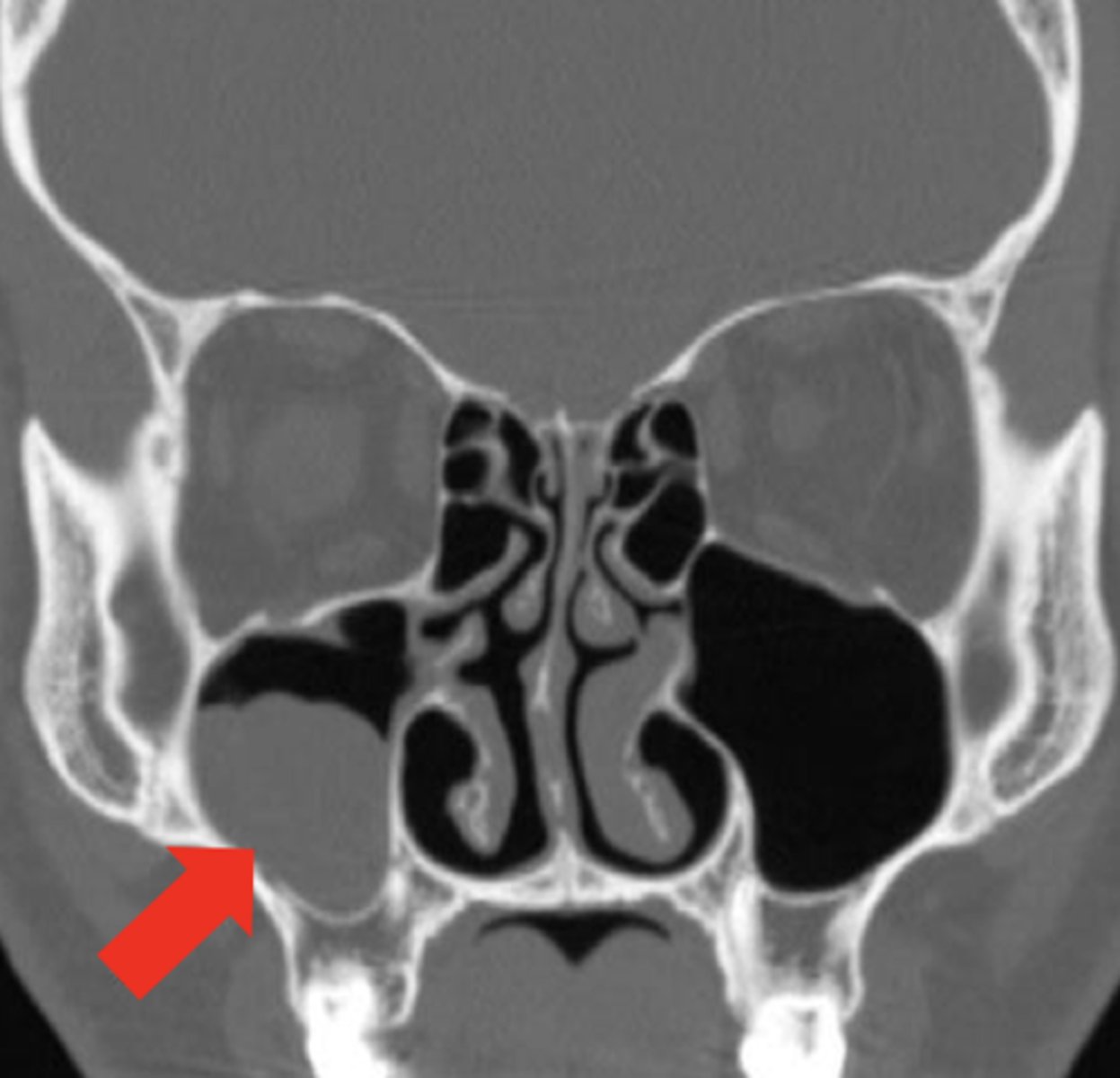
maxillary sinusitis
most common of all the sinuses to be infected, infection can spread from sinus to upper teeth...s/s of sinus infection plus tooth pain (because the V2 branch also innervates teeth via superior alveolar nerve)
Facial nerve
special motor to the muscles of facial expression and special sensory for taste from the anterior 2/3's of the tongue
5 main branches of facial nerve
Temporal
zygomatic
buccal
mandibular
cervical
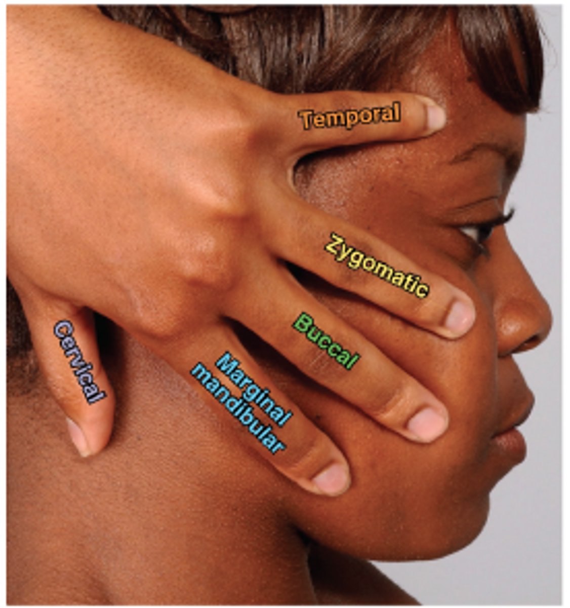
clinical application of facial nerve
bell's palsey
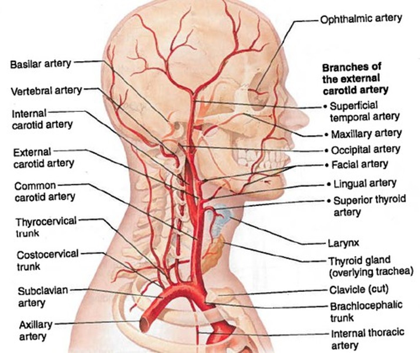
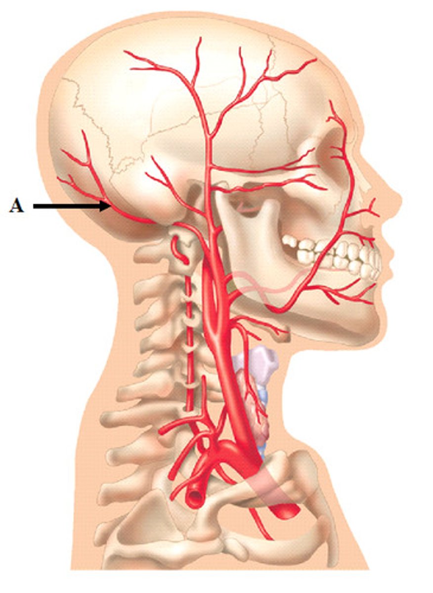
main arteries of the head
supraoribtal arteries
infraorbital arteries
facial arteries (palpate pulse)
maxillary arteries
lingual arteries
superficial temporal arteries (palpate pulse)
occipital arteries
supraoribital arteries

infraorbital arteries
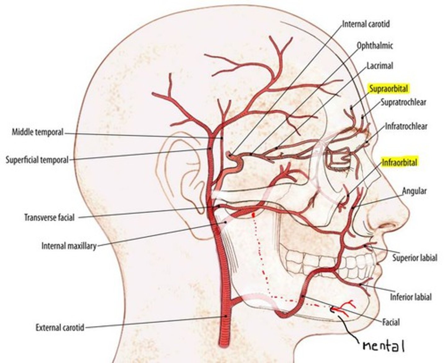
facial arteries
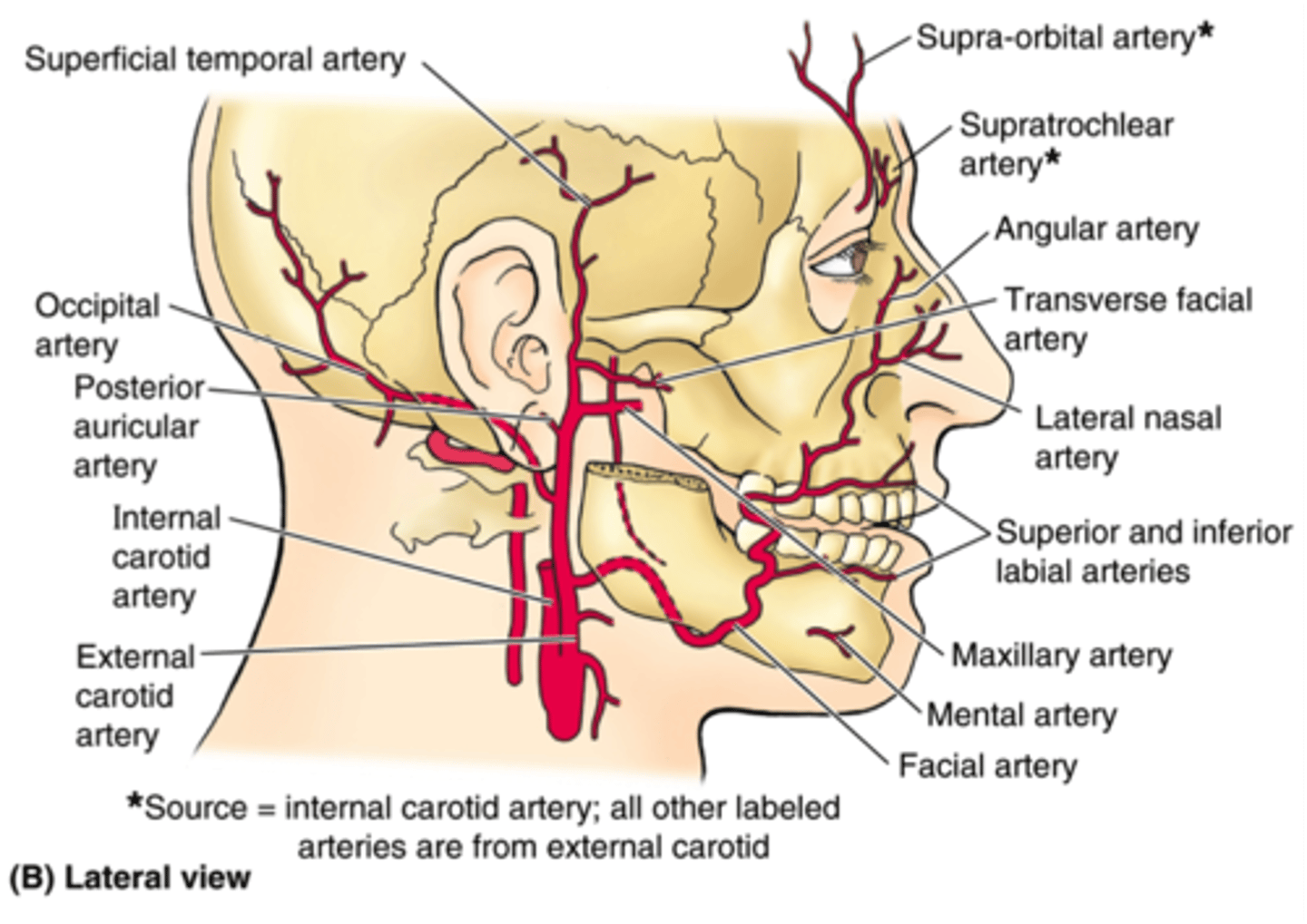
maxillary arteries
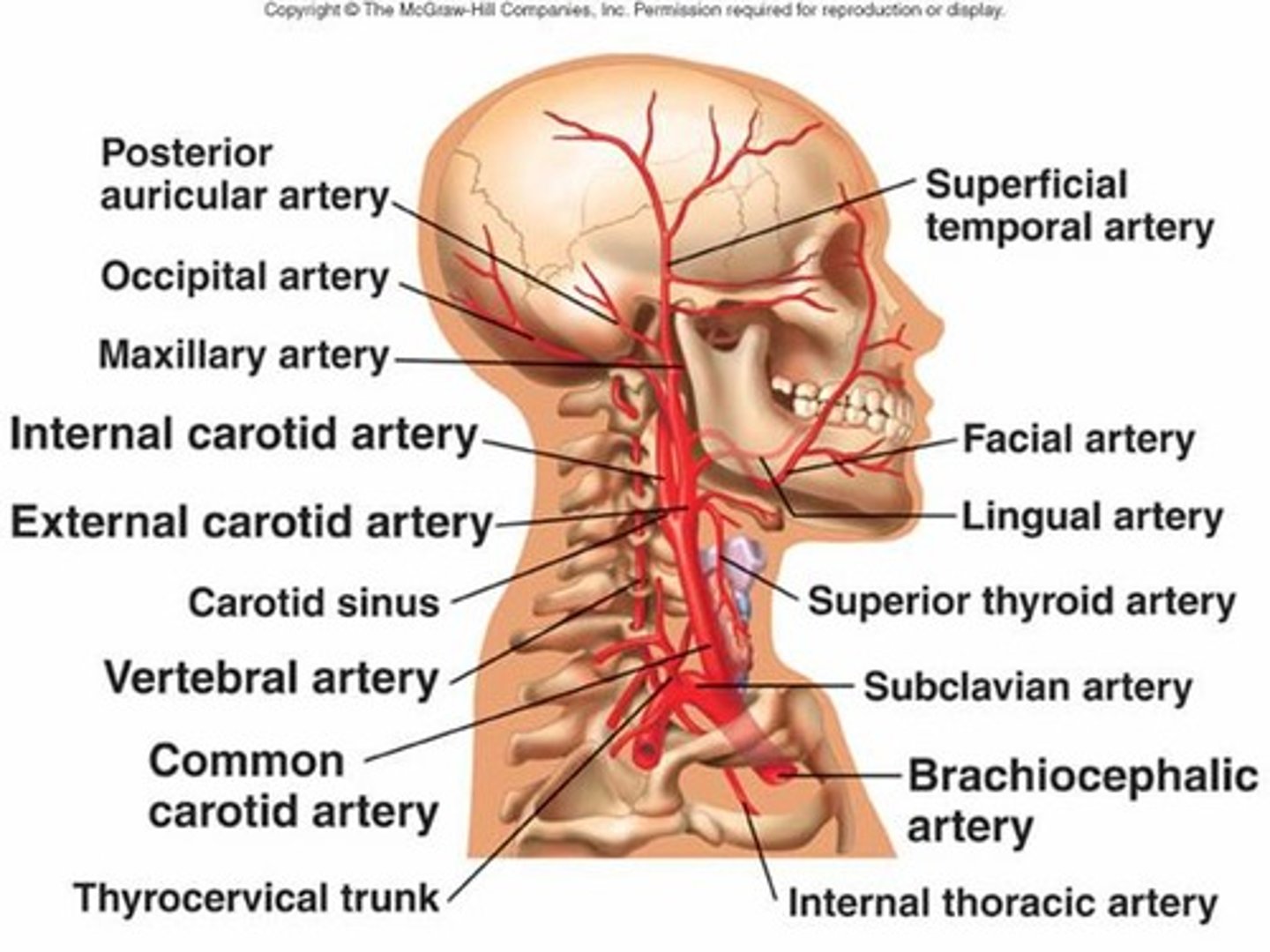
lingual arteries
superficial temporal artery
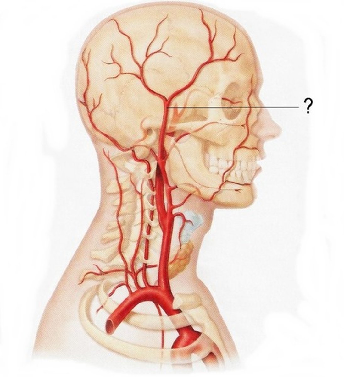
occipital arteries
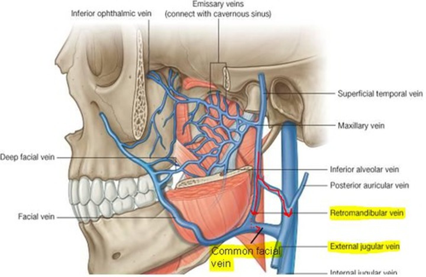
Primary venous drainage for the face
facial vein and this vessel joins with the retromandibular vein and they both drain into internal jugular vein in neck
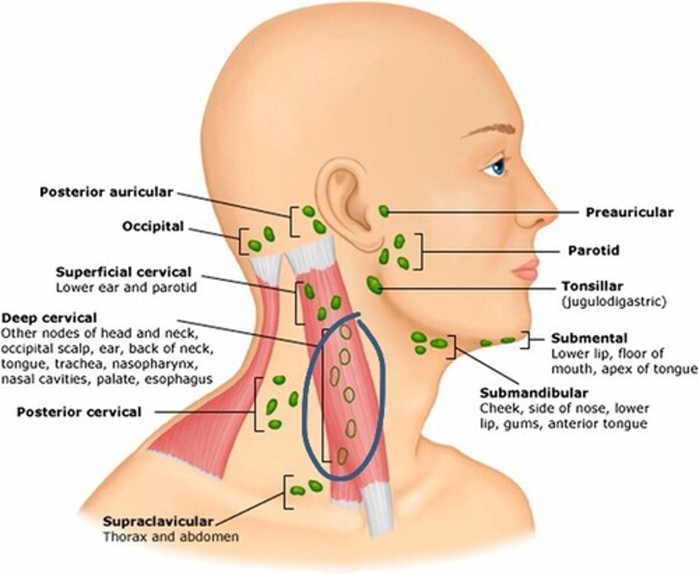
lymphatics superficial group
occipital
retroauricular (mastoid)
parotid
submandibular
superficial cervical
deep cervical
occipital node
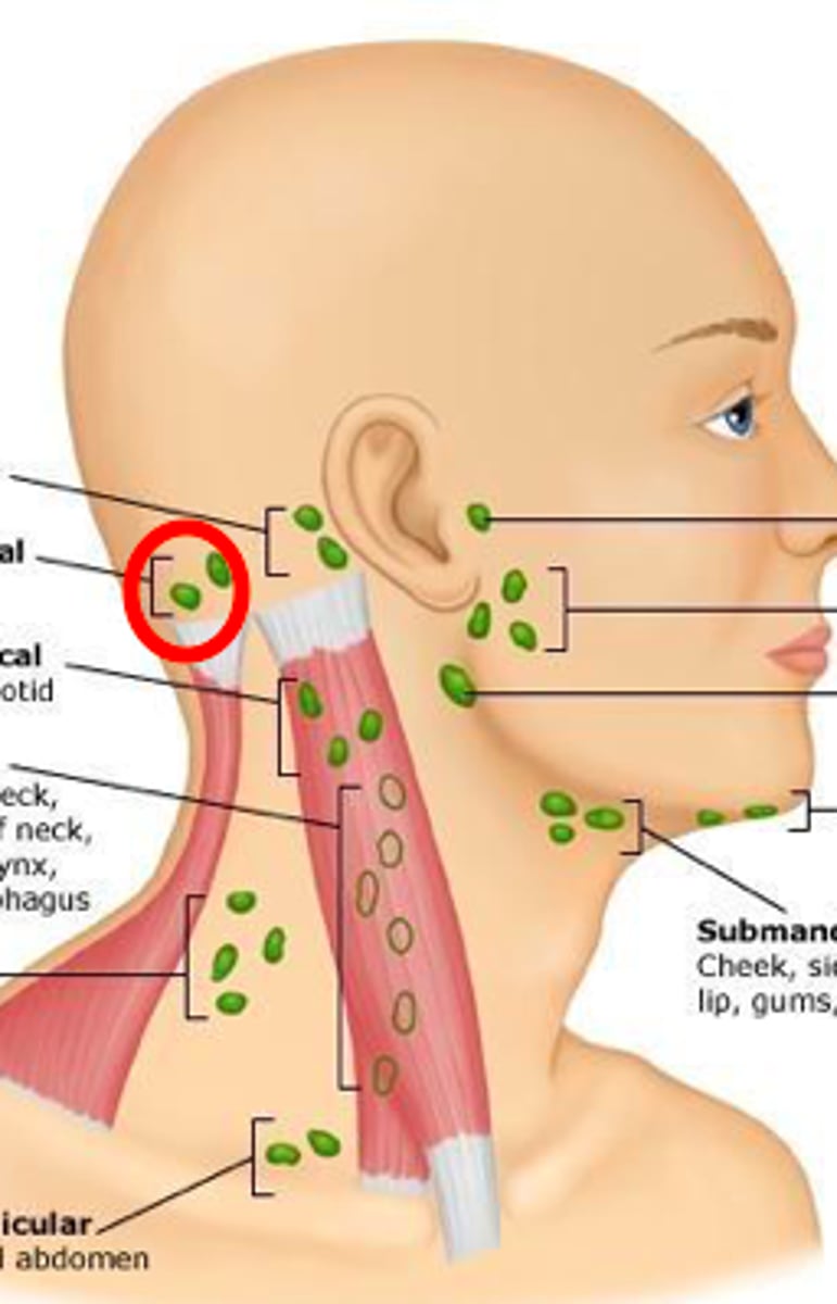
retroauricular node
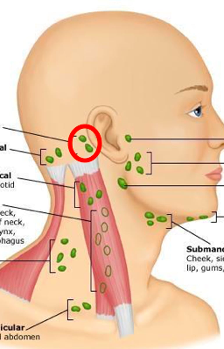
parotid node

submandibular node
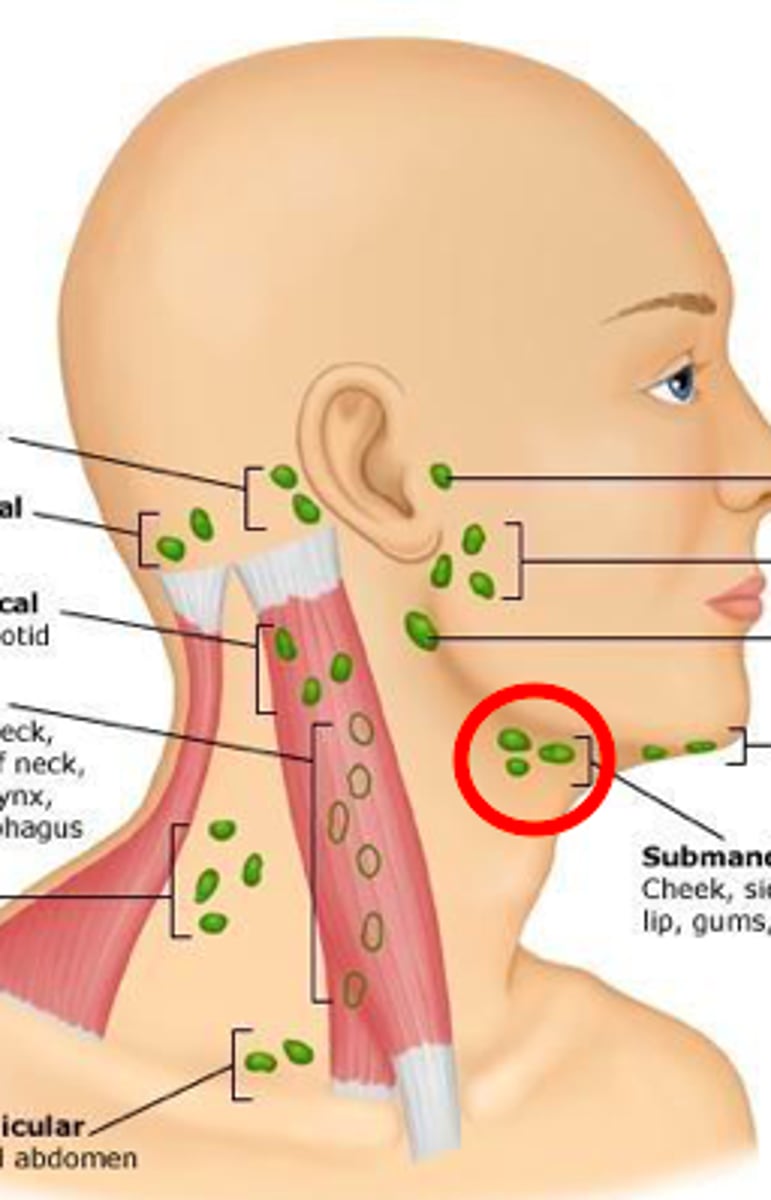
superficial cervical node
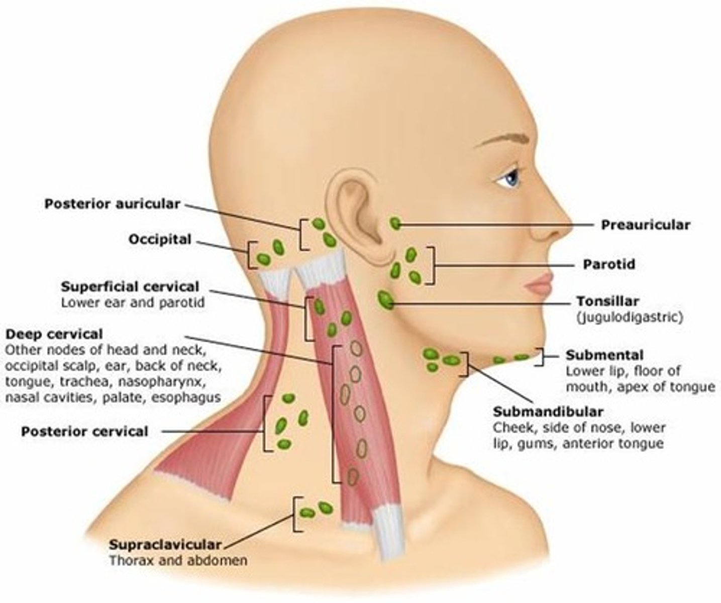
deep cervical node
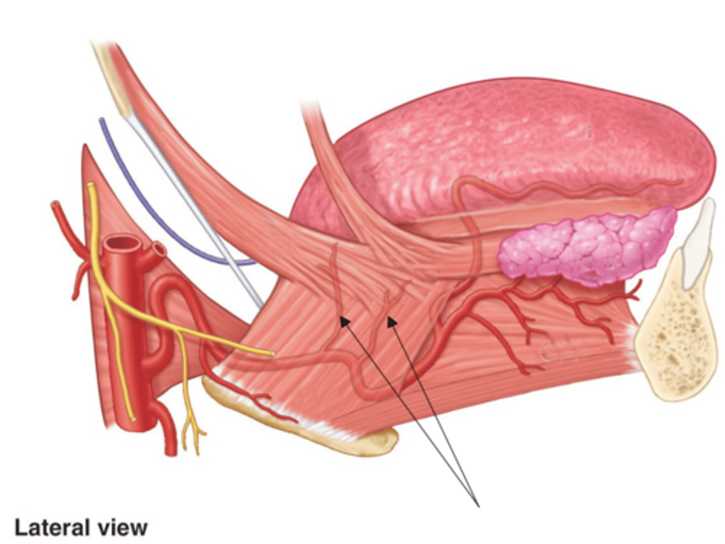
sublingual gland
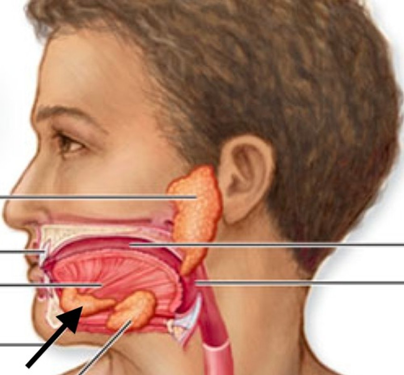
submandibular gland
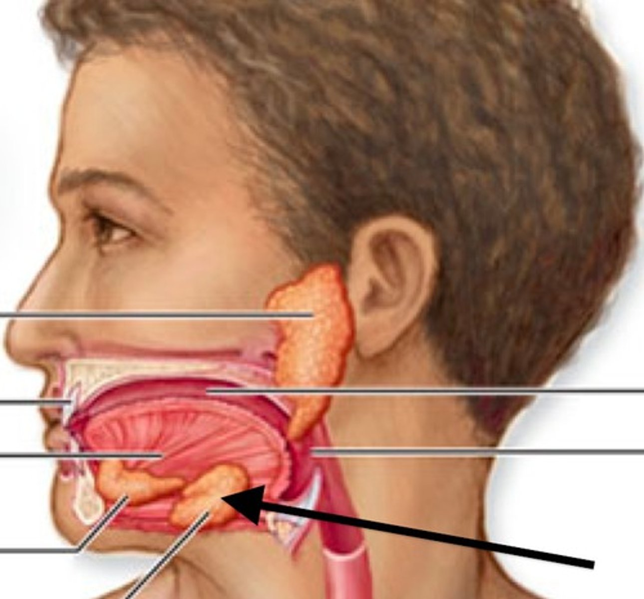
Parotid salivary gland
many structures are related to or pass through
located inferior and just anterior to each external auditory meatus and between each ramus of the mandible and each mastoid process of the occipital bone
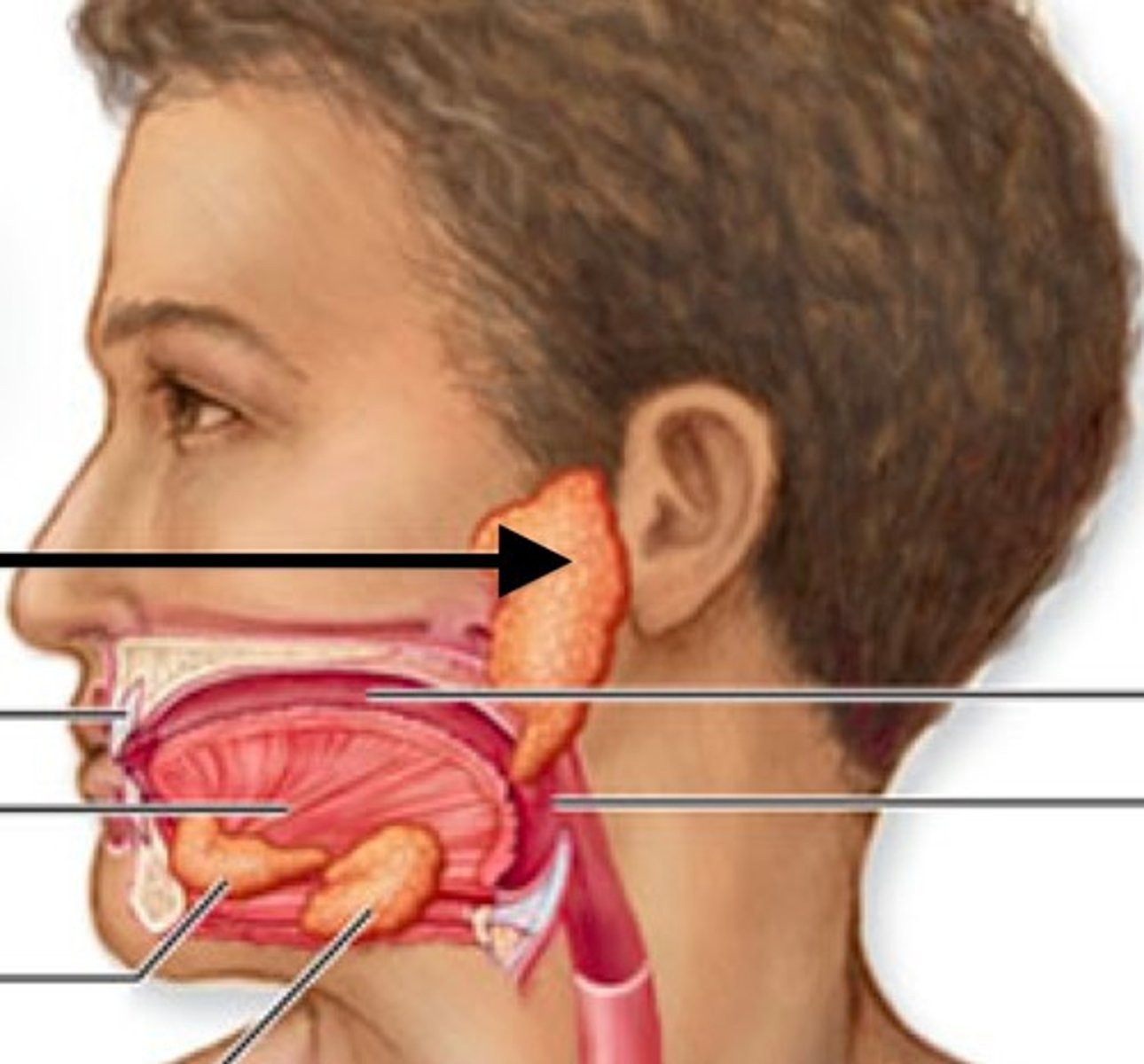
important structures within the parotids
facial nerve and branches
retromandibular vein
external carotid artery
parotid lymph nodes (superficial; drain into superficial and deep cervical nodes)
Clinically important structures related to the parotids
CN V2 (auricotemporal nerve)
great auricular nerve (C2/C3)
glossopharyngeal nerve
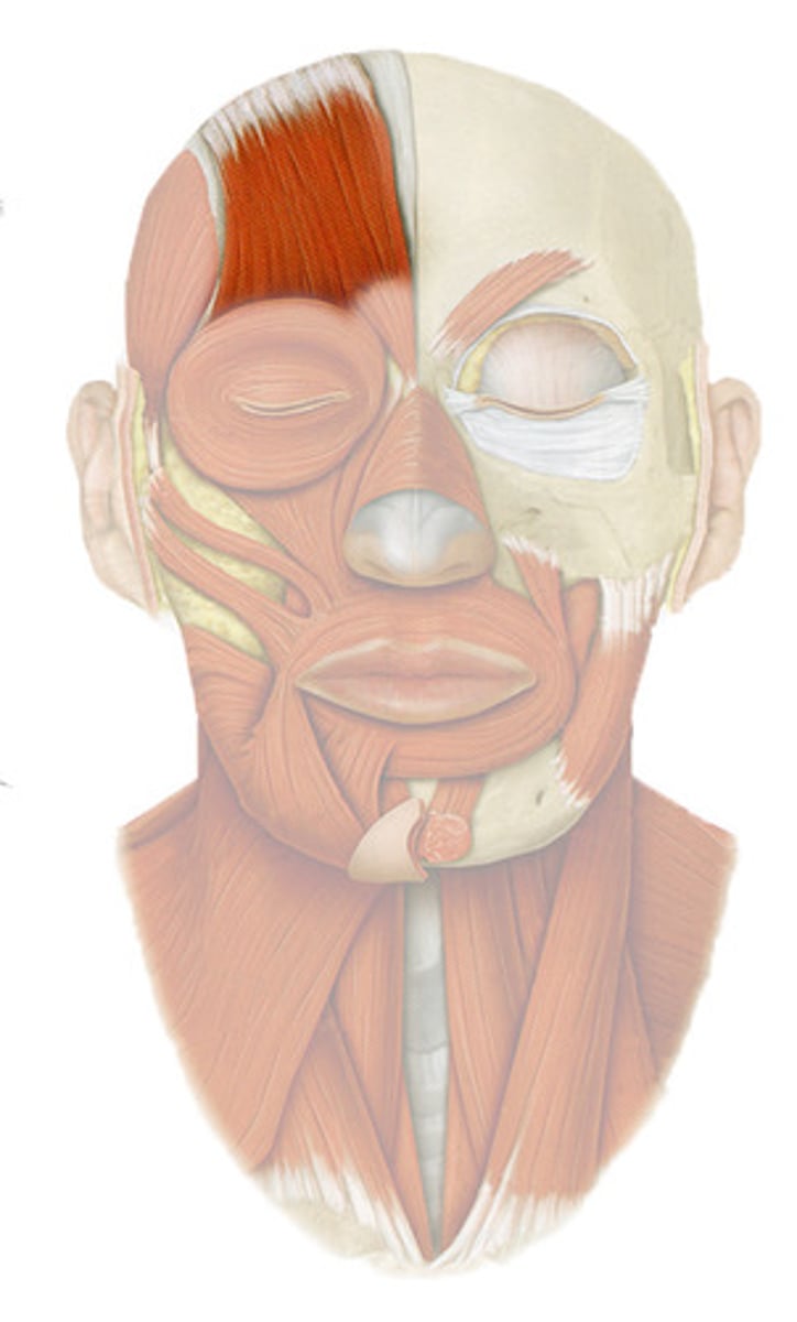
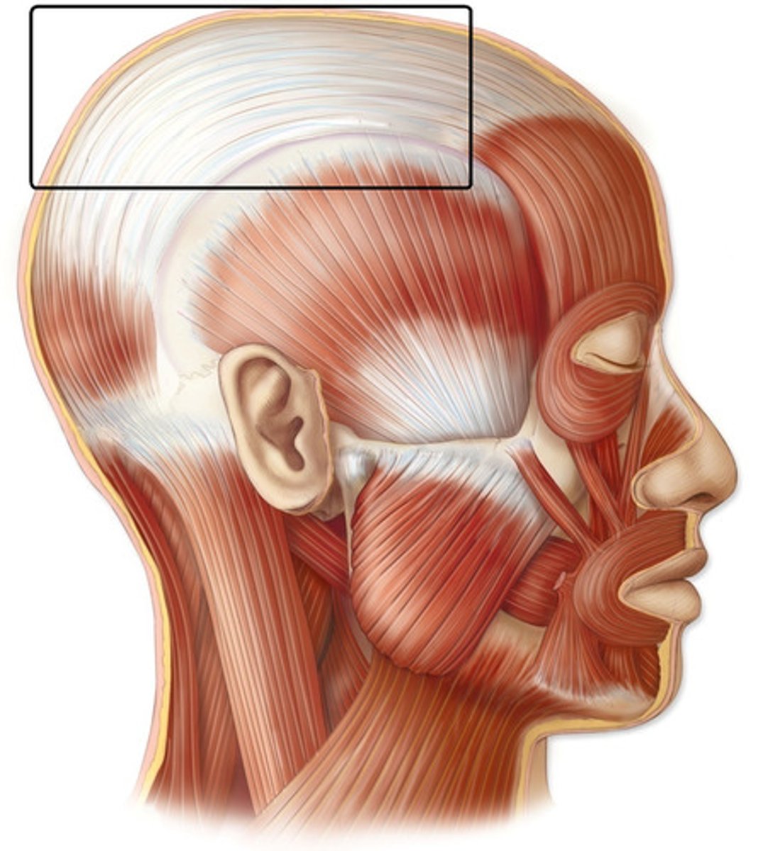
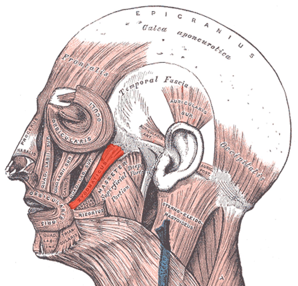
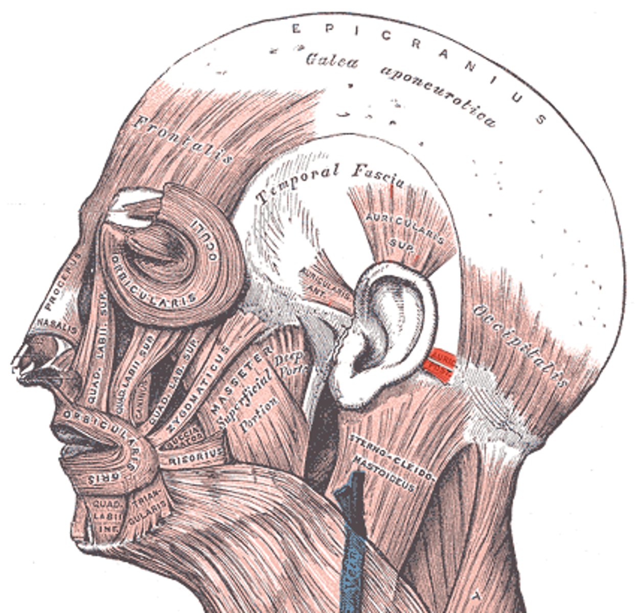
Occipitofrontalis (frontal belly) action
innervation
Frontal belly covers forehead;
Elevates eyebrows & wrinkles forehead horizontally
EMOTION: Surprise, Curiosity
N: facial
Occipitofrontalis (occipital belly)
action and nerve
occipital belly covers posterior skull; moves scalp posteriorly
EMOTION: Surprise, Curiosity
N: facial
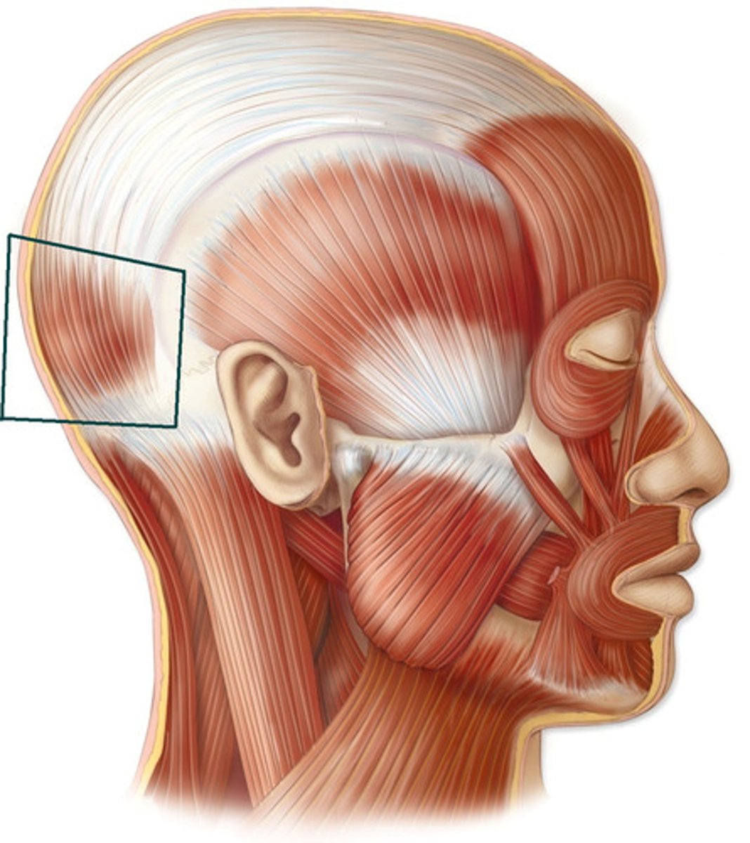
Occipitofrontalis: Epicranial aponeurosis
connects the frontal and occipital bellies
Temporoparietalis muscle
action and nerve
tightens scalp
N: facial nerve
emotion:
24
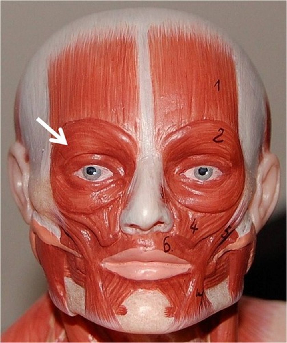
Orbicularis oculi muscle
action and nerve
Closes eyelids
EMOTION: Winking, blinking; squinting
N: facial nerve
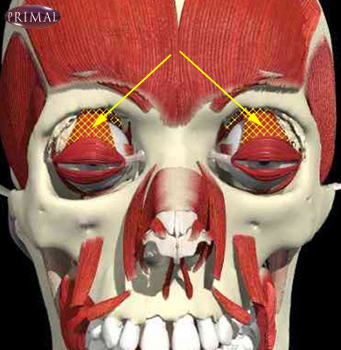
Corrugator supercilii muscle action and innervation
Draw eyebrows inferomedially creating vertical wrinkles above the nose
EMOTION: Concern, skepticism
N: facial nerve
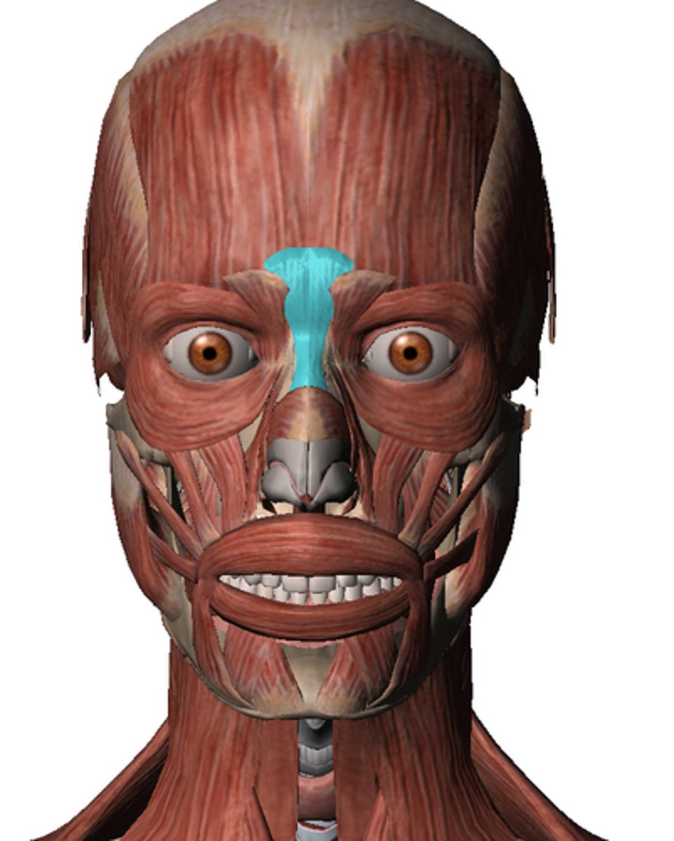
Nasalis muscle
action and innervation
transverse part: compresses nostrils
alar part: dilates nostrils
EMOTION: Flares nostrils, as with anger
N: facial nerve
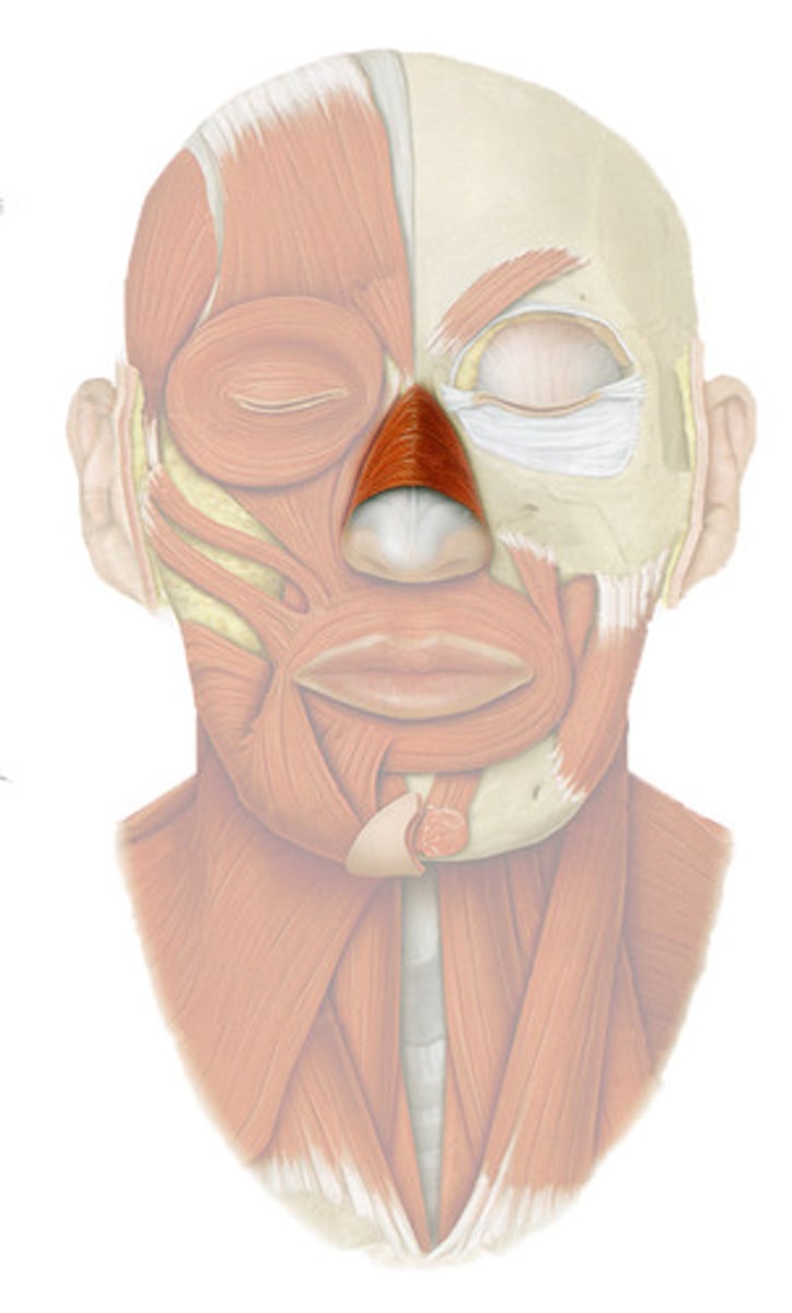
Procerus action and innervation
Depresses medial eyebrow, transverse wrinkles over the nose
EMOTION:Dislike or distain
N: facial nerve
Zygomaticus major action and innervation
elevates angle of mouth
EMOTION: smile
N: facial nerve
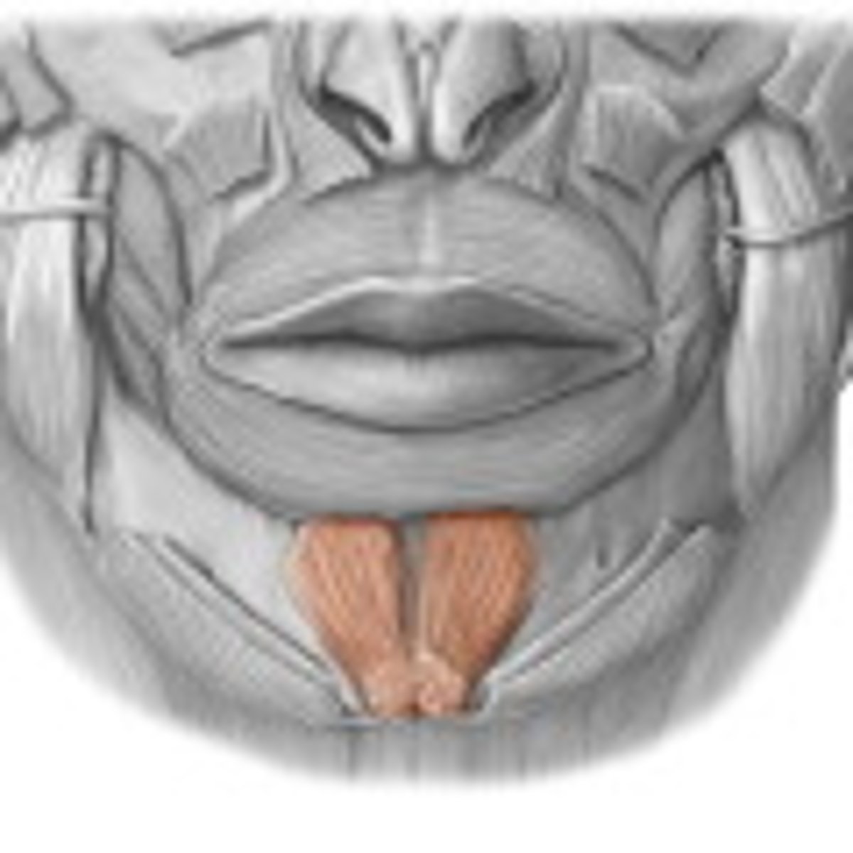
Mentalis action and innervation
Elevates and protrudes lower lip
EMOTION: pouting
N: facial nerve
Depressor labii inferioris action and innervation
Depresses the lower lip
EMOTION: Frown
N: facial nerve
Depressor anguli oris action and innervation
depresses angle of mouth
EMOTION: frown
N: facial nerve
Risorius
pulls angle of mouth laterally
EMOTION: allows for smiling
N: facial nerve
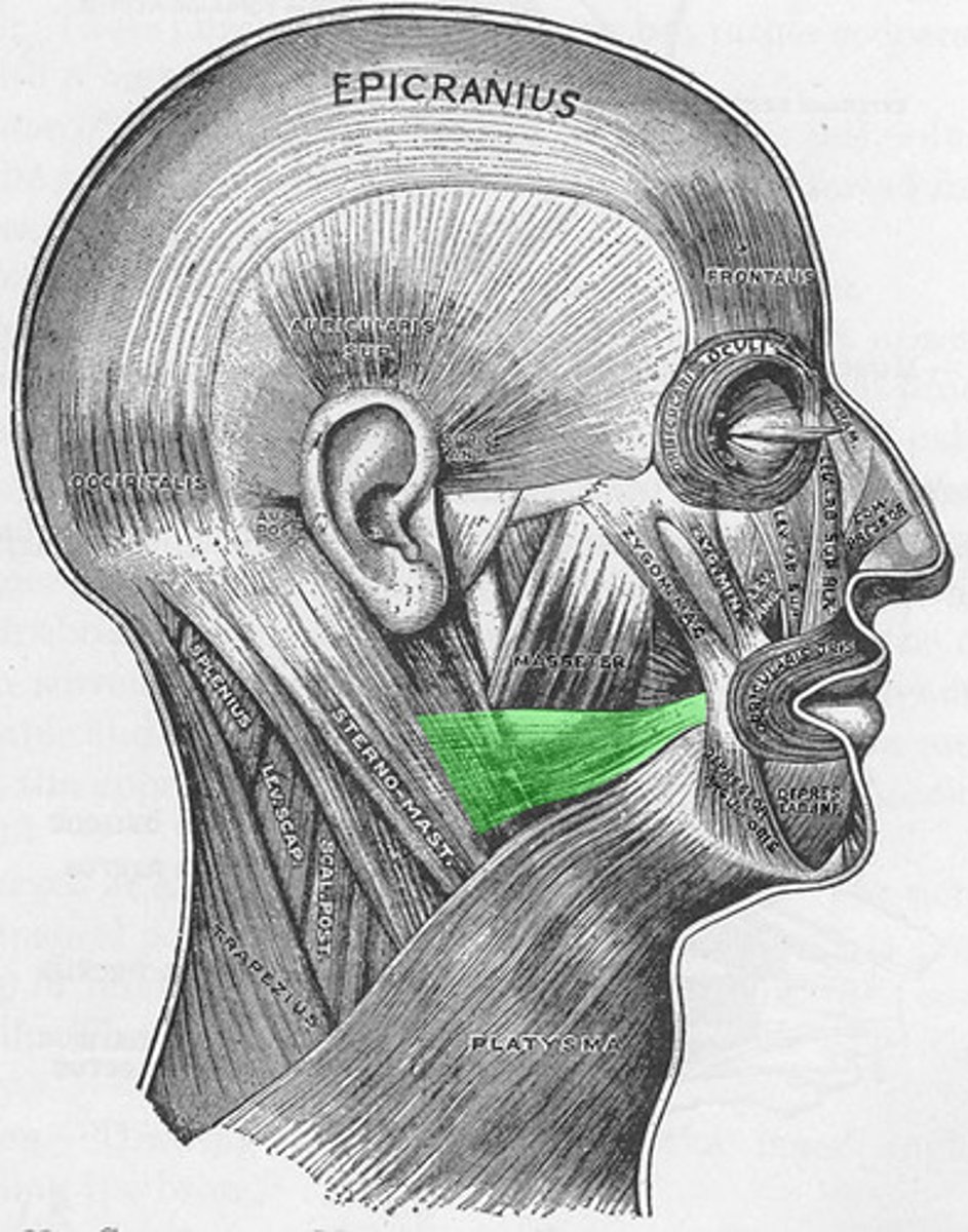
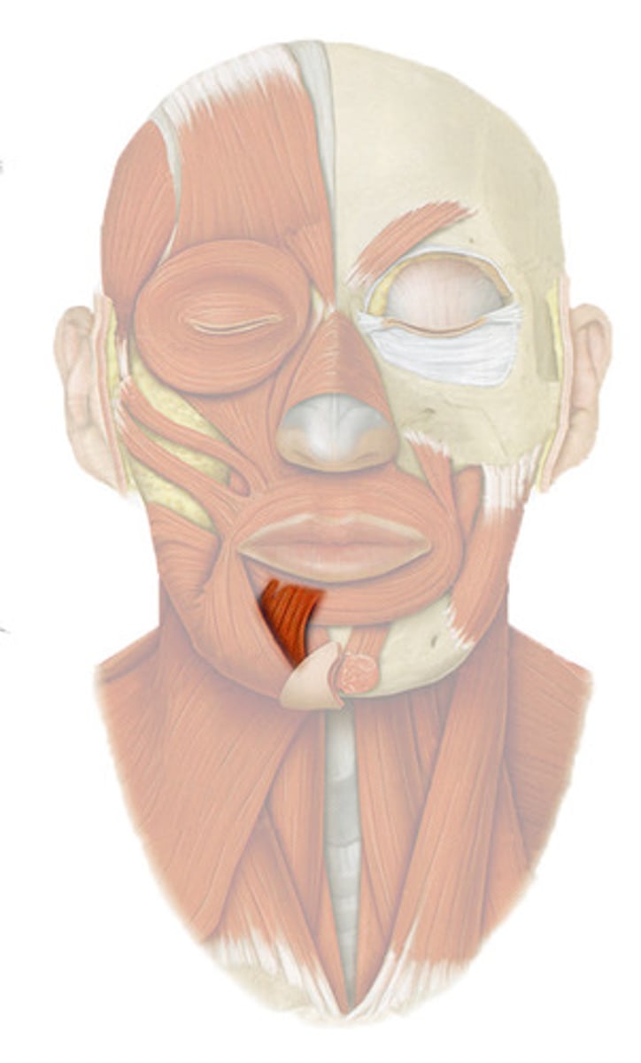
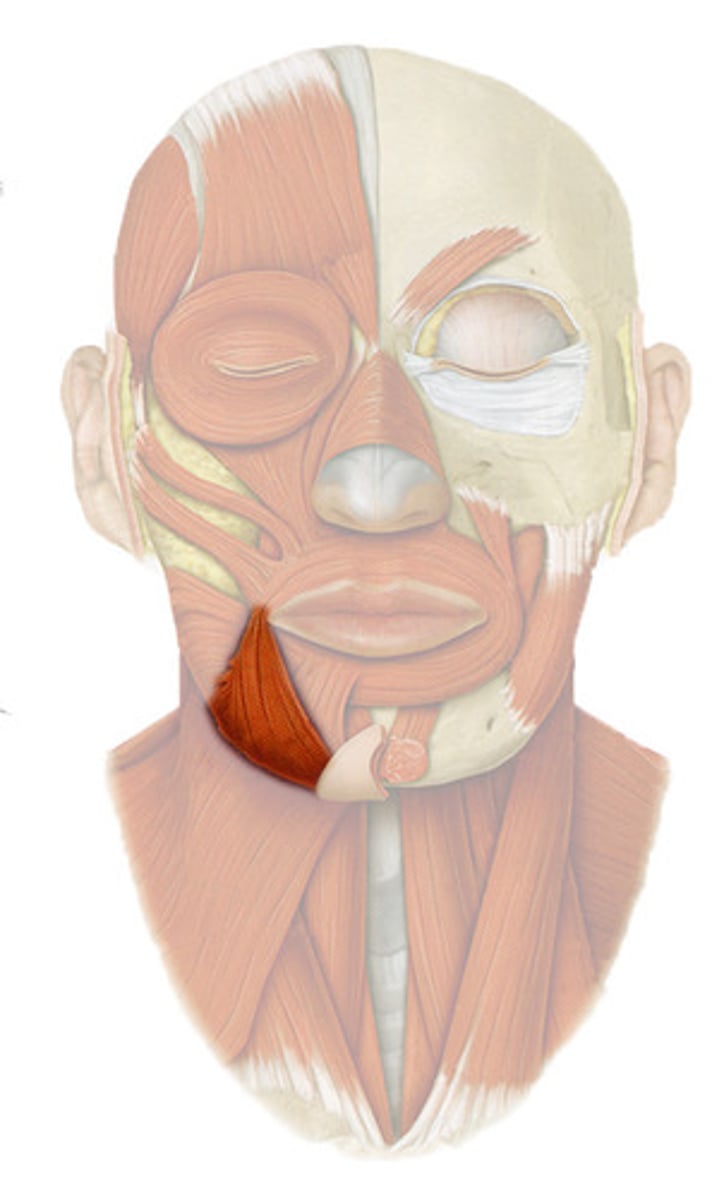
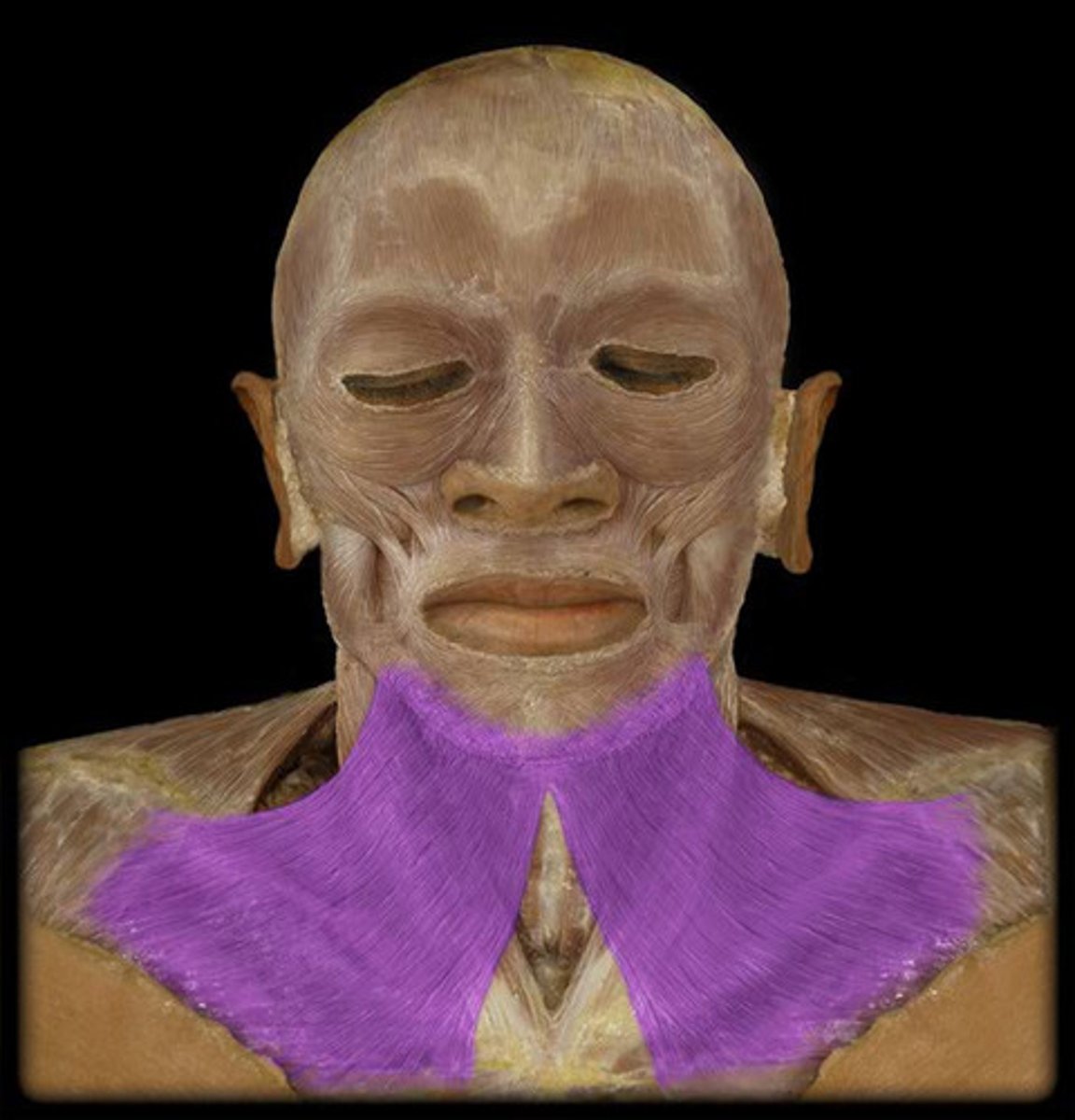
Platysma
Tenses the skin of the neck, depresses mandible
expression: eek
N: facial nerve
Auricularis
ant: elevates and draws auricle forward
post: elevate and retract auricle
sup: retracts and elevates auricle
N: facial nerve
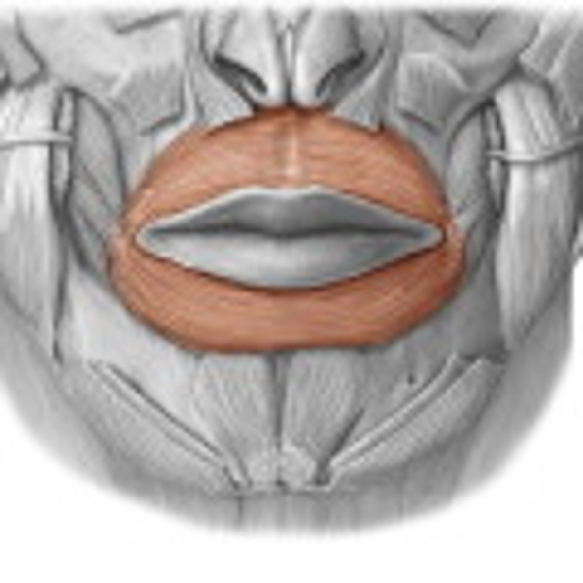
Orbicularis oris
compresses and protrudes lips
EMOTION: kissing, whistling
N: facial nerve
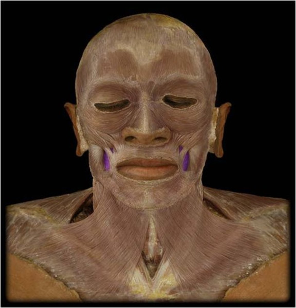
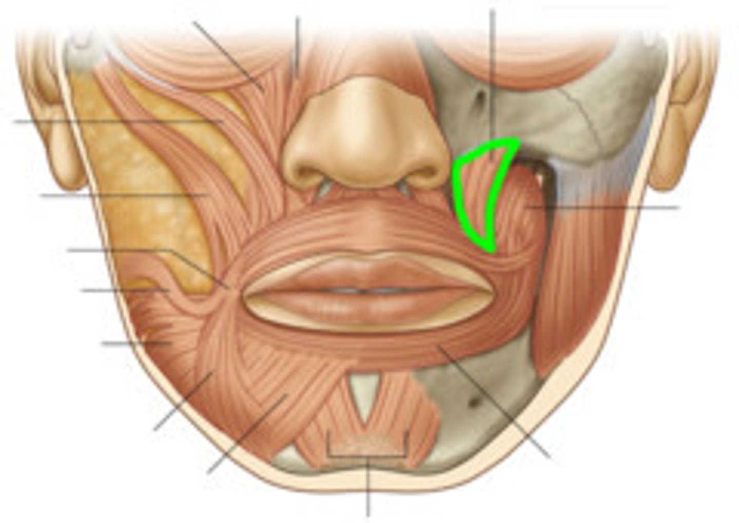
Buccinator
deep to the masseter; Compresses cheek; aid in mastication
EMOTION: Whistling, sucking
N: facial nerve
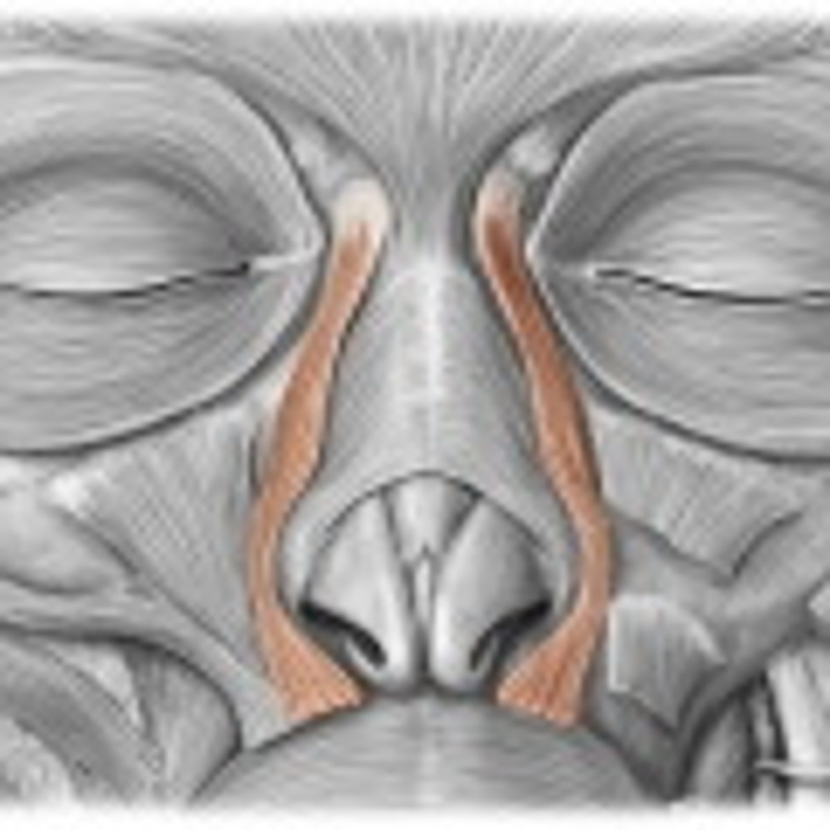
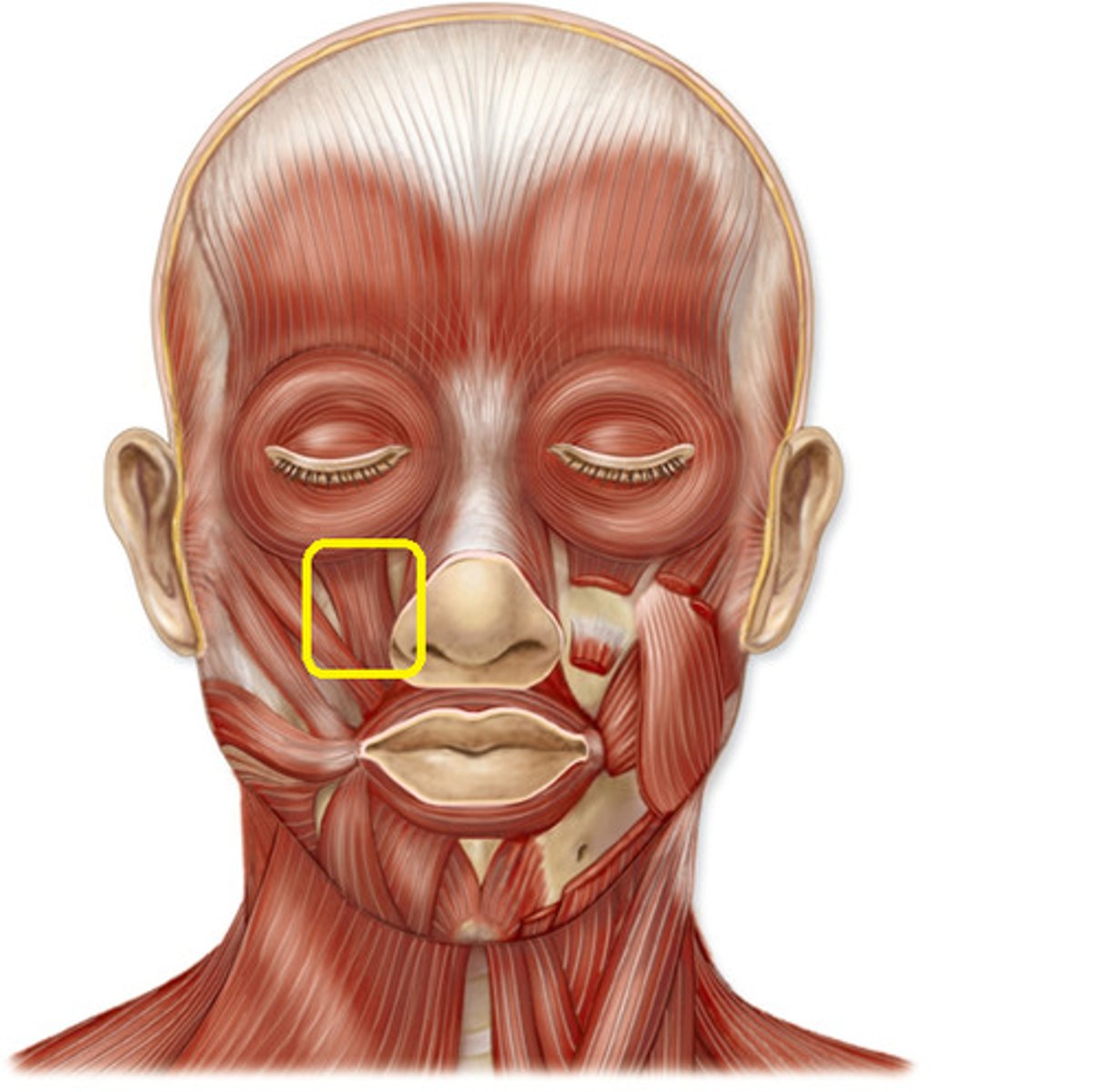
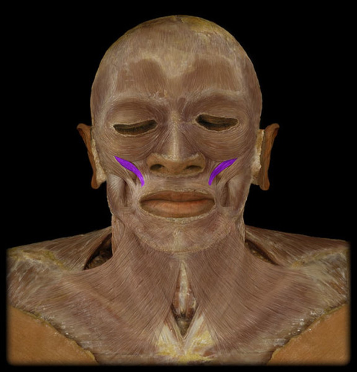
Levator labii superioris alequae nasi
(LLSAN)
Elevates the upper lip, dilates the nostrils
EMOTION: "elvis" snarl
N: facial nerve
Levator labii superioris
Elevates and everts upper lip
EMOTION: dilator of mouth look...teeth coming together
N: facial nerve
Zygomaticus minor
elevates upper lip
EMOTION: smile
N: facial nerve
Levator anguli oris
elevates angle of mouth
EMOTION: smiling
N: facial nerve
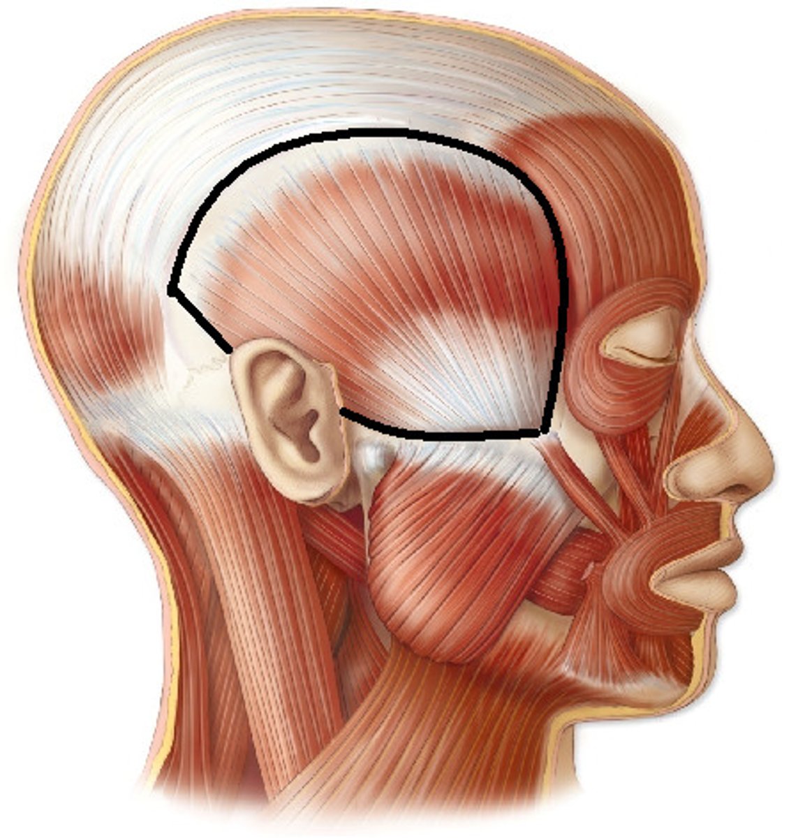
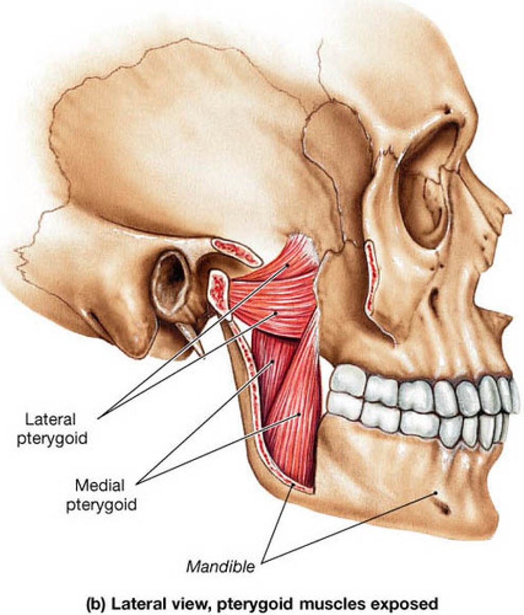
muscles of mastication
masseter
temporalis
medial pterygoid
lateral pterygoid
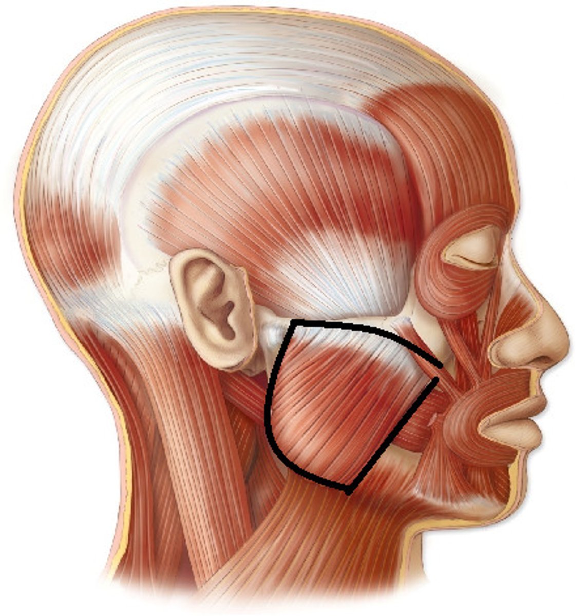
masseter
OIAN
Origin: zygomatic arch
Insertion: mandible
action: elevate, protract, and retract mandible
innervation: trigeminal, V3
temporalis OIAN
origin: temporal fossa
insertion: mandible
action: elevate and retract mandible
innervation: All trigeminal, V3
medial pterygoid OIAN
origin: pterygoid plate insertion: mandible action: elevate and protract mandible, and lateral movement of opposite side of mandible
innervation: All trigeminal, V3
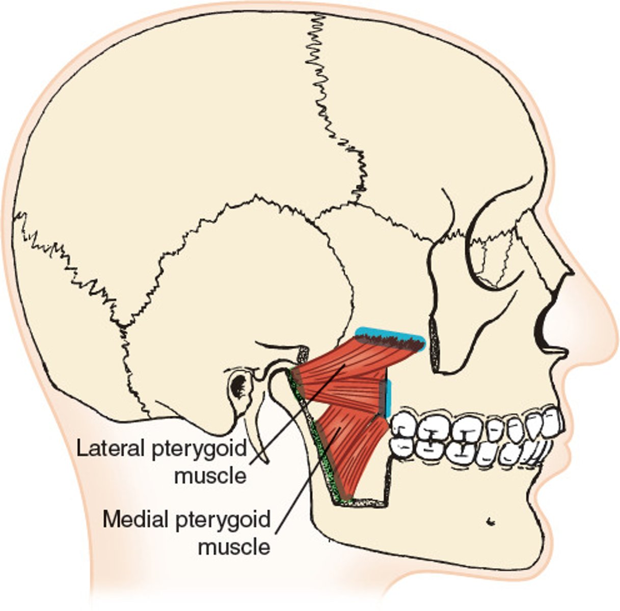
lateral pterygoid OIAN
origin: pterygoid plate
insertion: mandible
action: depress and protract mandible, and lateral movement of opposite side of mandible
innervation: All trigeminal, V3
muscles of the tongue
genioglossus, styloglossal muscle, hyoglossal muscle, palato(chondro)glossal muscle
genioglossus action and innervation
action: depresses and protrudes tongueinnervation: CN XII (hypoglossal)
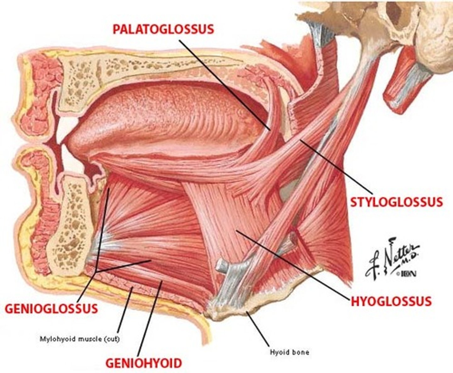
hyoglossal muscle action and innervation
depresses tongue innervation: CN XII (hypoglossal)
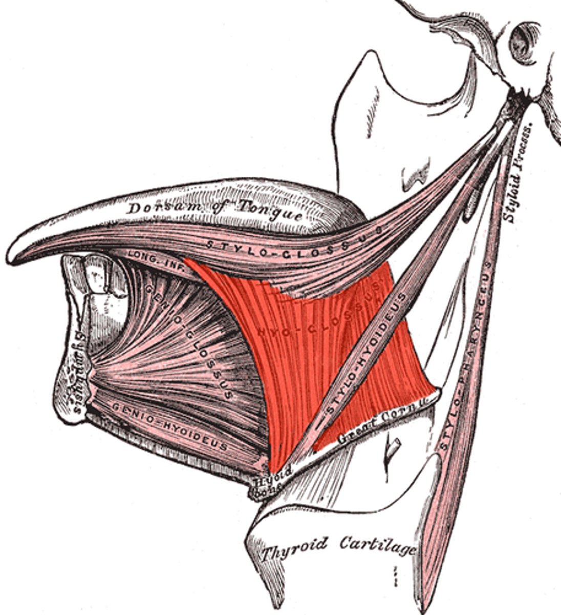
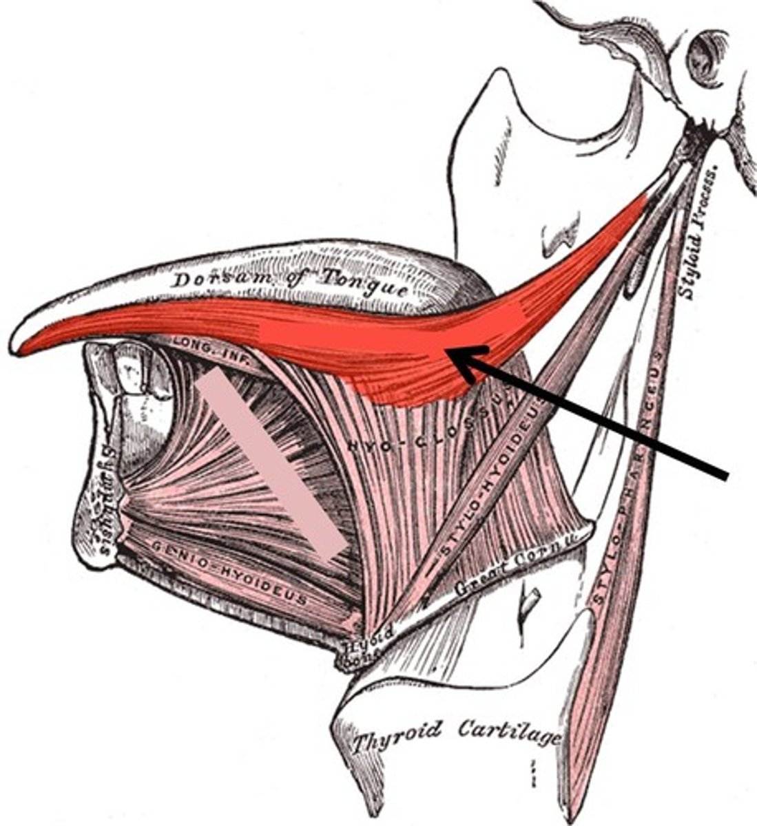
styloglossal muscle action and innervation
retrude and curls the tongue
innervation: CN XII (hypoglossal)
palato(chondro)glossal action and innervation
elevates posterior tongue to palateinnervation: CN X (Vagus)
CN I
Olfactory Nerve;
Sensory
Smell

extraocular muscles
inferior rectus
medial rectus
lateral rectus
superior rectus
superior oblique
inferior oblique
levator palpebrae superioris
inferior rectus action and innervation
depresses eye
III (oculomotor)
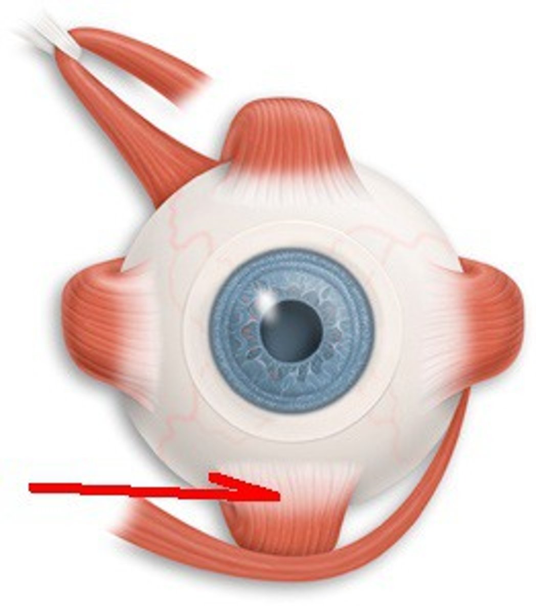
medial rectus action and innervation
moves eye medially (III oculomotor)
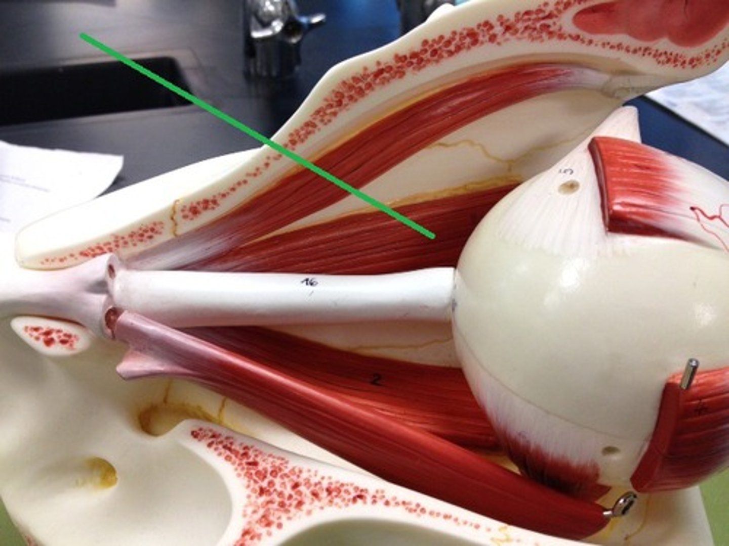
lateral rectus action and innervation
moves eye laterally (VI abducens)
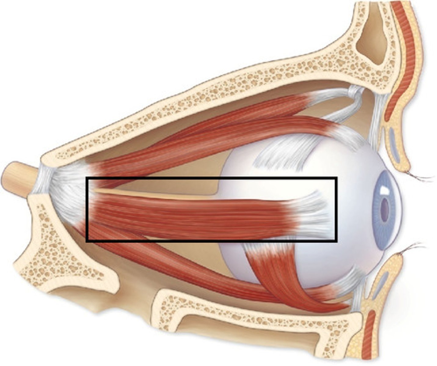
superior rectus action and innervation
elevates eye;
III oculomotor
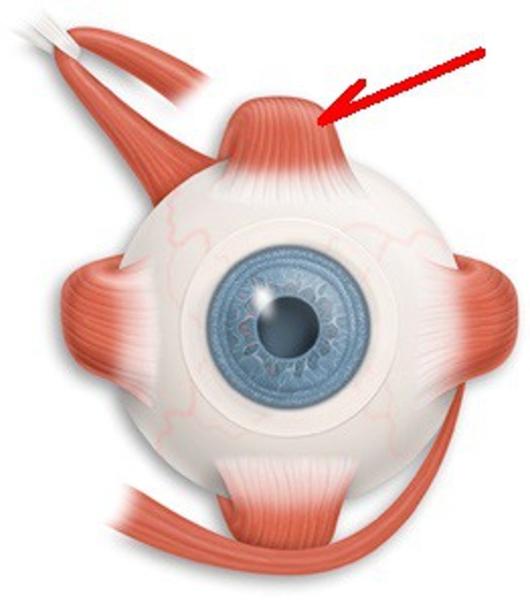
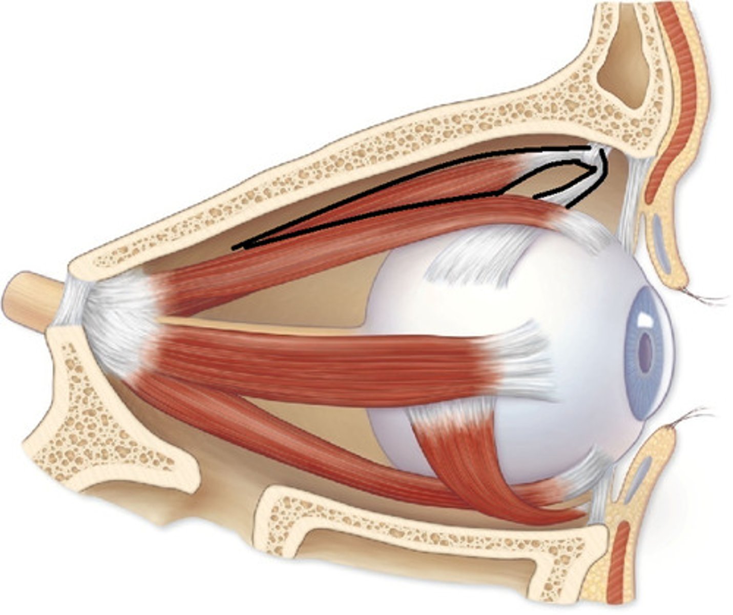
superior oblique action and innervation
Depresses eye and turns it laterally
CN IV (trochlear)
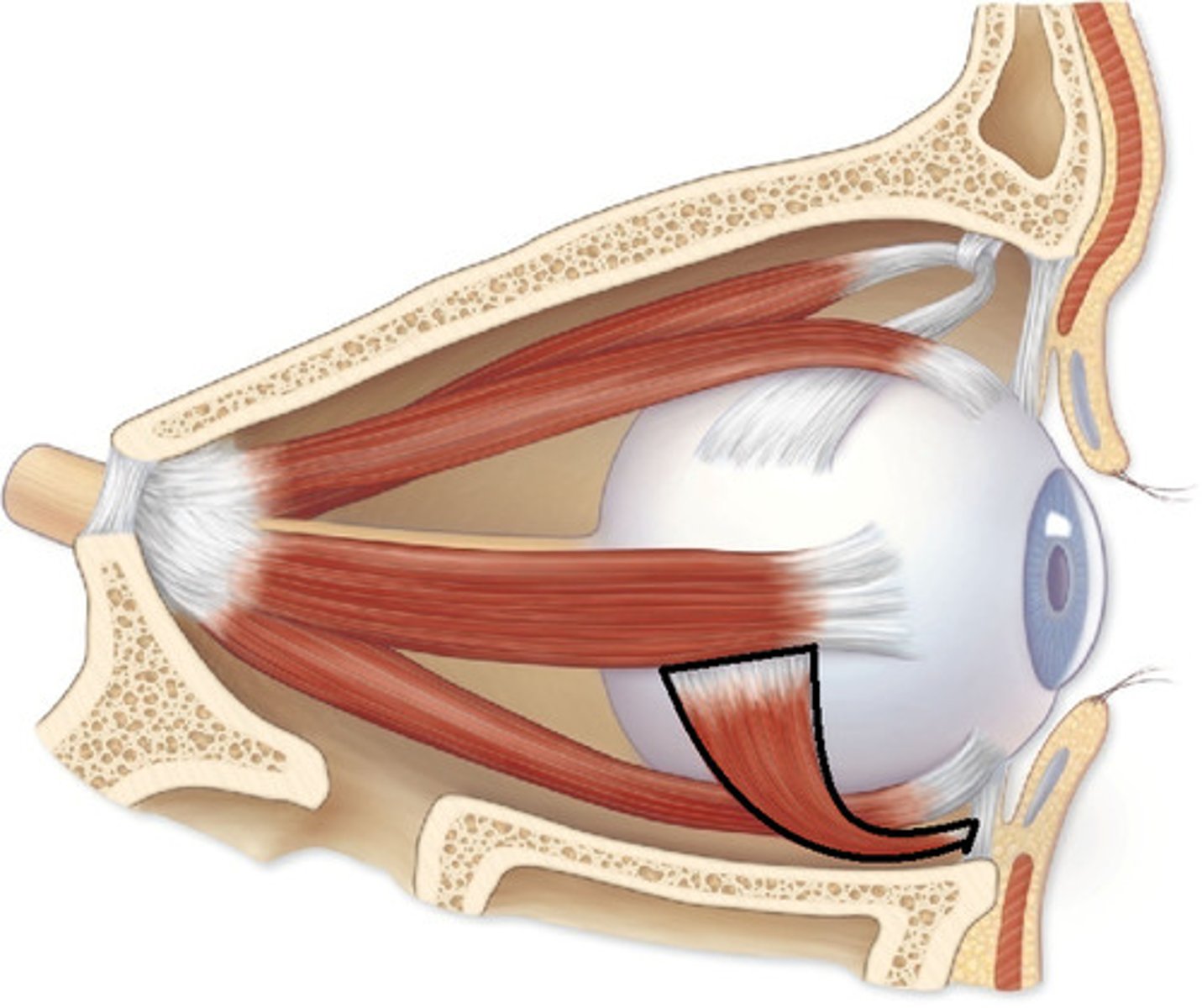
inferior oblique action and innervation
Elevates eye and turns it laterally
III (oculomotor)
levator palpebrae superioris action and innervation
action: elevates upper eyelid
innervation: III (oculomotor)
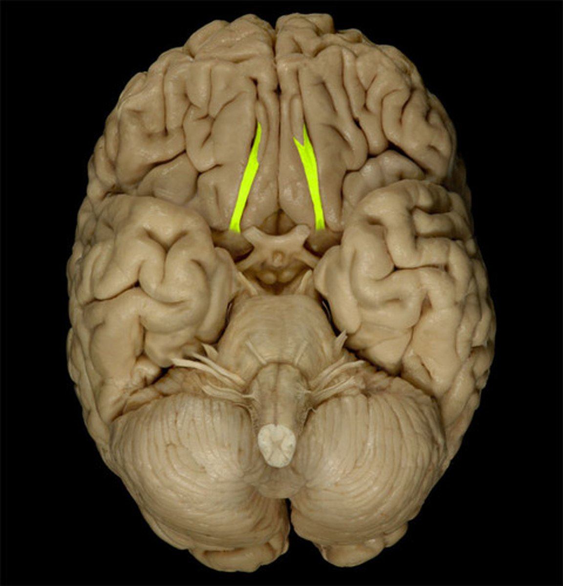
olfactory bulb
where the olfactory nerves synapse, on the inferior aspect of the frontal lobe
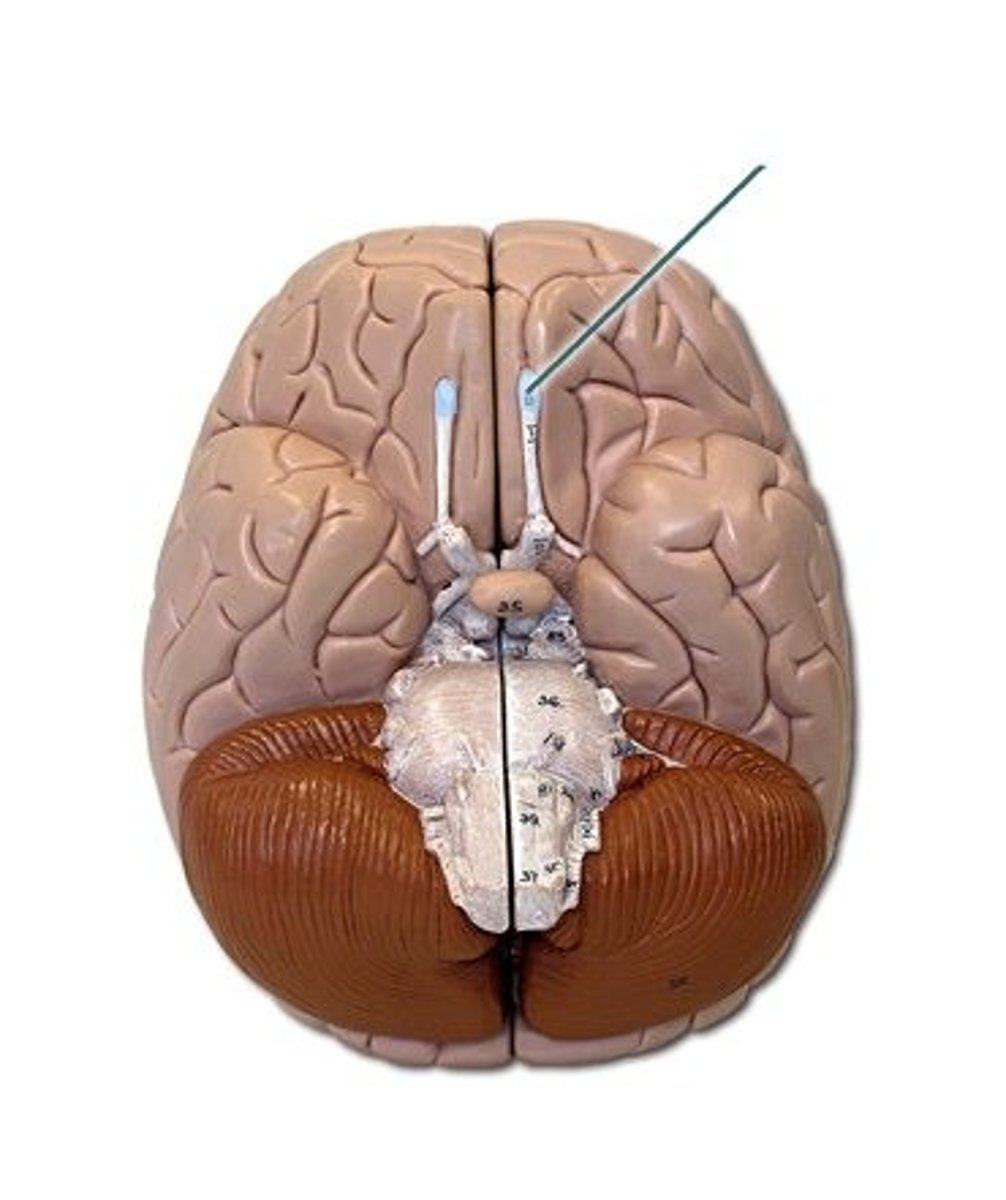
olfactory tract
originates in the olfactory bulb and runs caudally on the ventral aspect of the frontal lobe.