identification final pathology
1/40
There's no tags or description
Looks like no tags are added yet.
Name | Mastery | Learn | Test | Matching | Spaced | Call with Kai |
|---|
No analytics yet
Send a link to your students to track their progress
41 Terms
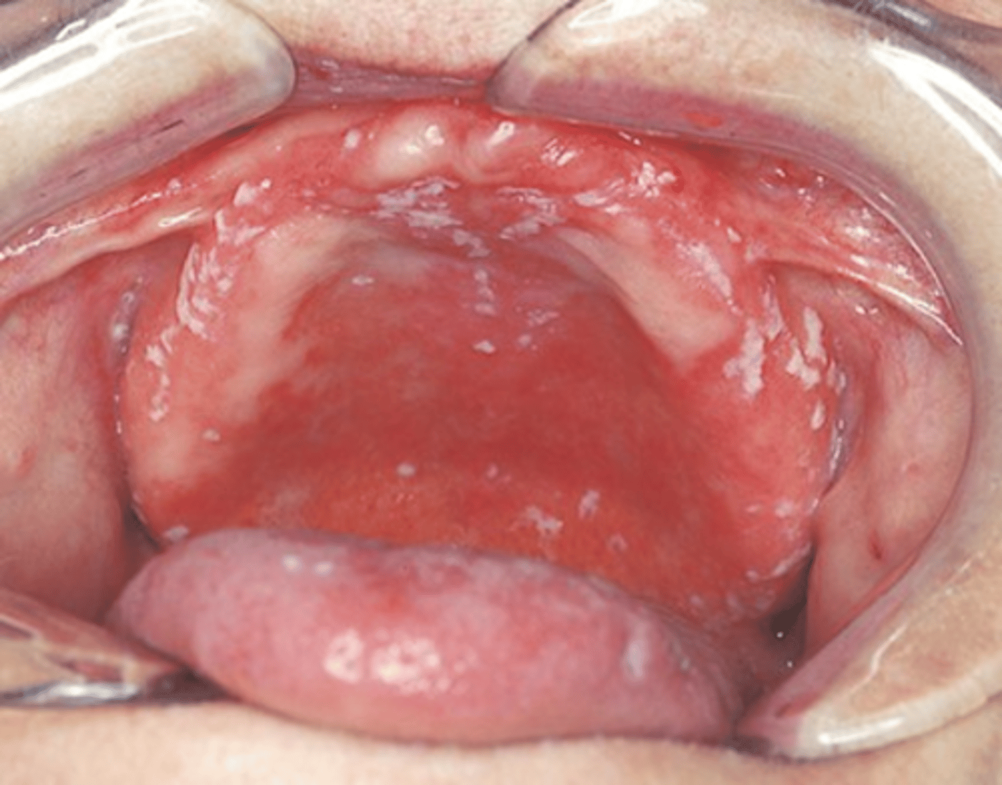
pseudomembranous candidiasis
-white curdlike material that can be wiped off
-on occasion a burning sensation is felt and the patient may complain of a metallic taste
Calcifying Odontogenic Cyst
-composed of odontogenic epithelium that contains "ghost cells"
-also referred to as gorlin cyst
-most commonly seen individuals younger than 40 years
-lesions occur equally in the maxilla and mandible, most often in the incisor and cuspid area
-present as well defined unilocular or multilocular radiolucency
dens in dente
-also called dens invaginatus
-tooth within a tooth
Trisomy 13 (Patau Syndrome)
-abnormalities in multiple organs
turner syndrome
-webbing of the neck
-caused by patient has only one X chromosome or monosomy X
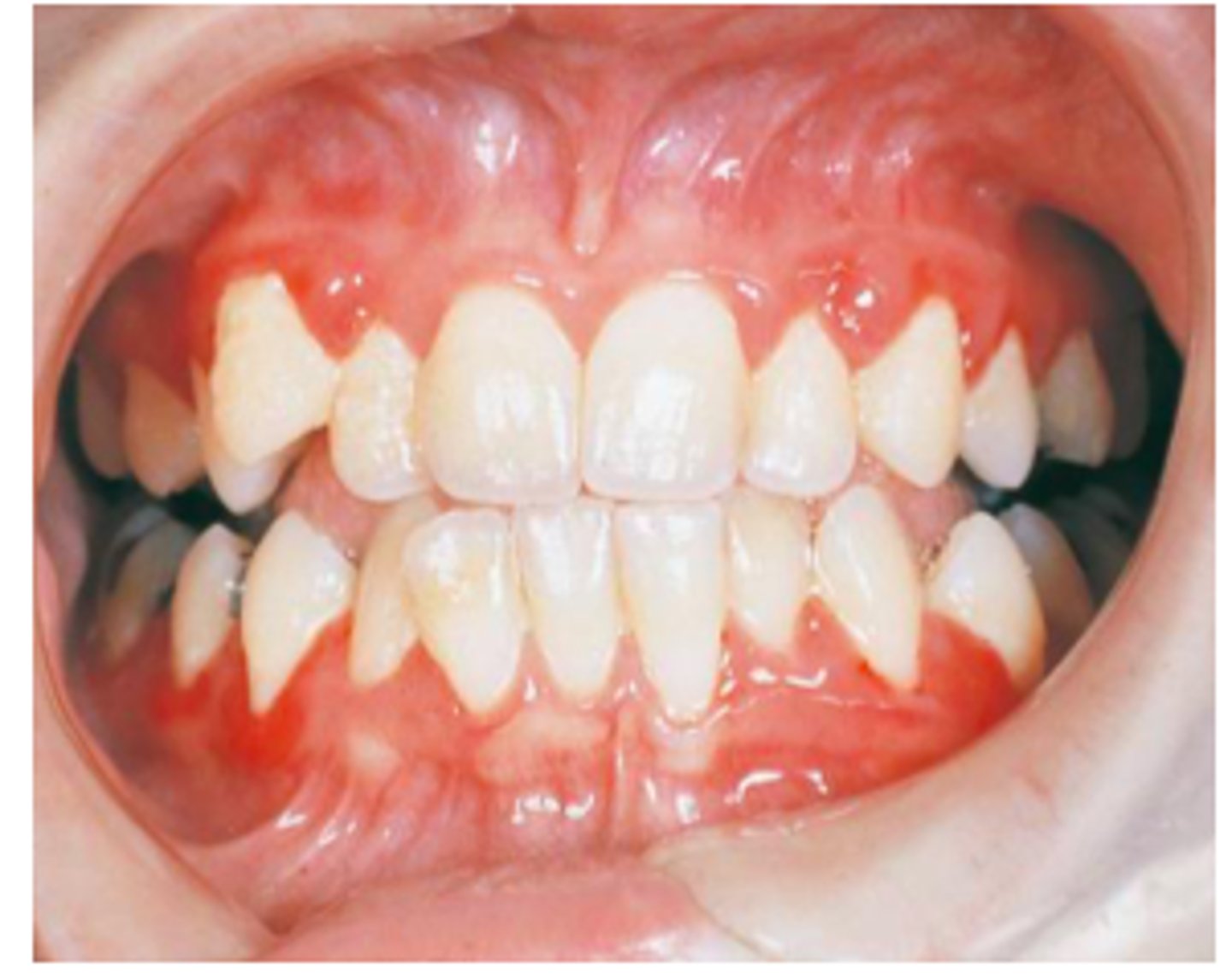
Cyclic Neutropenia
-by decrease in the number of circulating neutrophilic leukocytes, white blood cells help fight infection and remodel the body.
-autosomal dominant
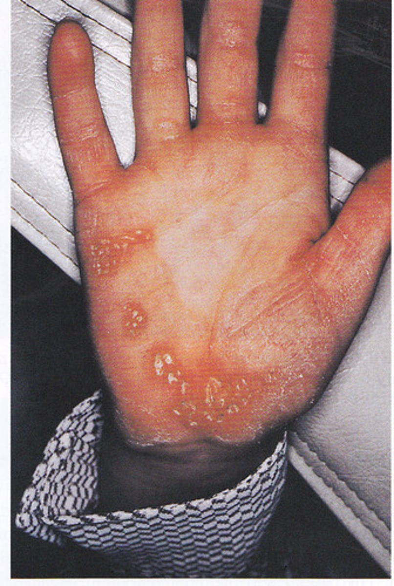
Papillon-Lefevre Syndrome
-lesion of the hand remain reddish white scaly, thick areas of hyperkeratinization
-autosomal-recessive inheritance
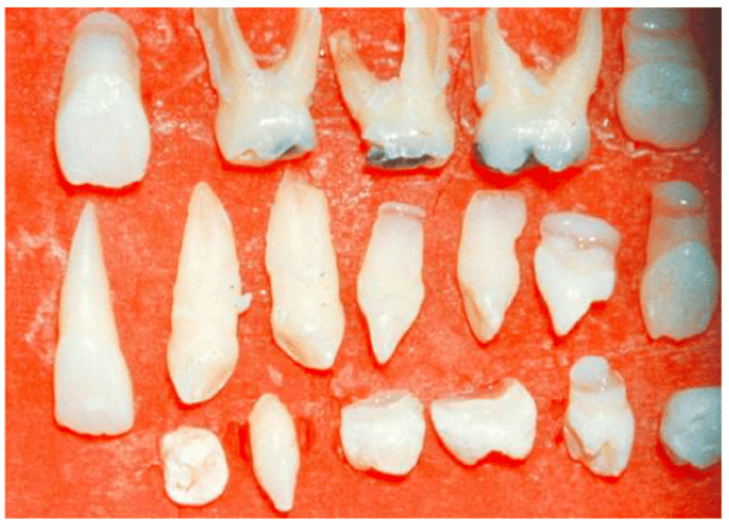
cleidocranial dysplasia
-multiple extracted supernumerary teeth rom the patient with cleidocranial dysplasia
-results in cranium developing into a mushroom shape because the fontanelles remain open
-the premaxilla is generally underdeveloped
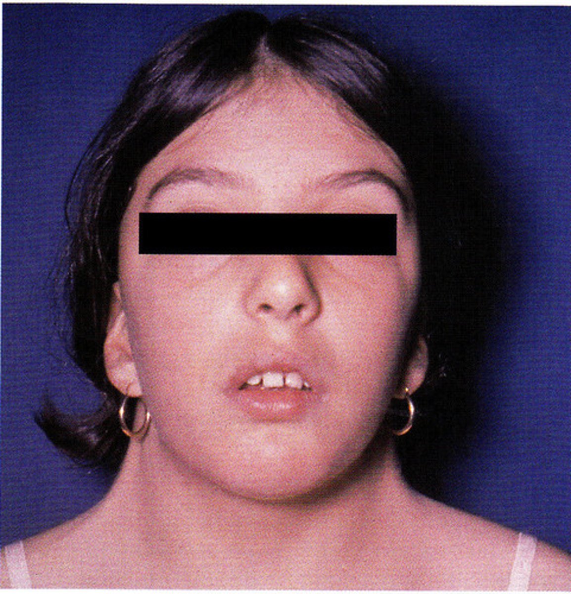
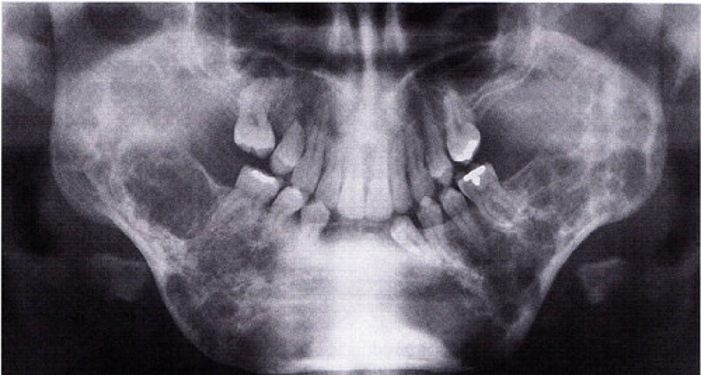
cherubism
-bilateral facial swelling that appears when the patient is between 1.5 and 4 years of age
-soap bubble appearance
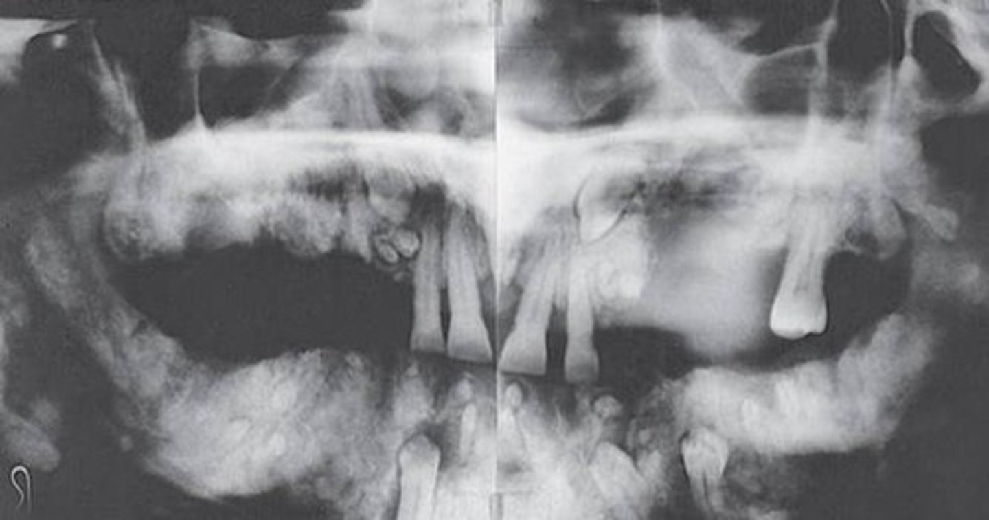
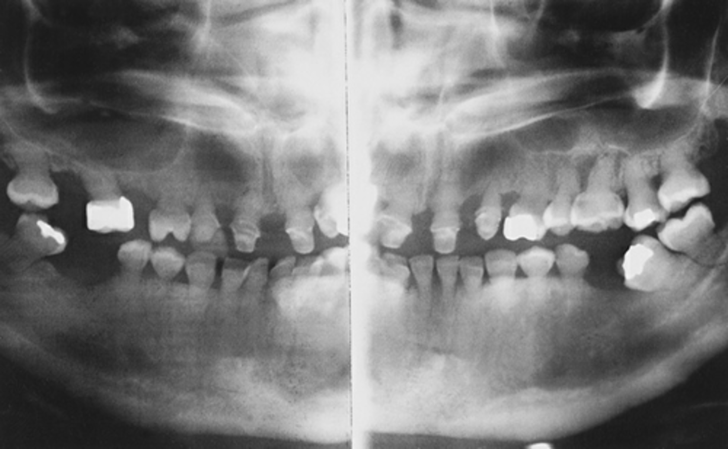
Gardner syndrome
-panoramic radiograph of patient with gardner syndrome shows multiple osteomas and odontomas
-also known as familial adenomatous polyposis
-benign bone growths
-teeth can exhibit hypercementosis and fail to erupt
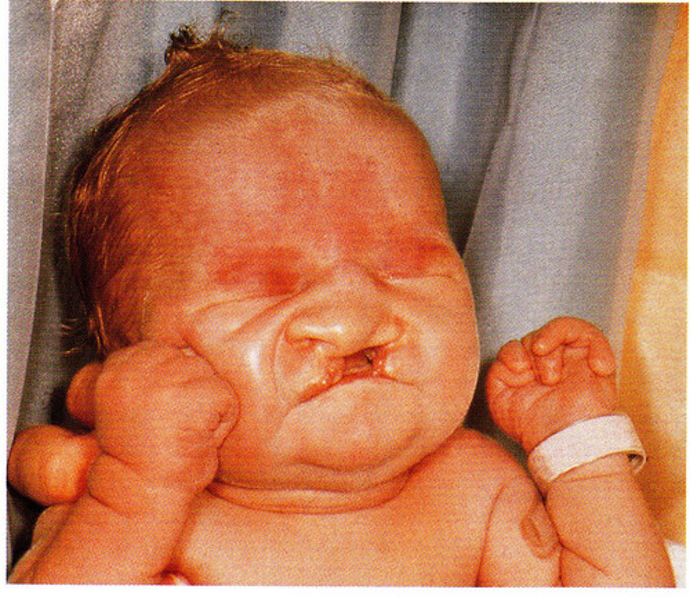
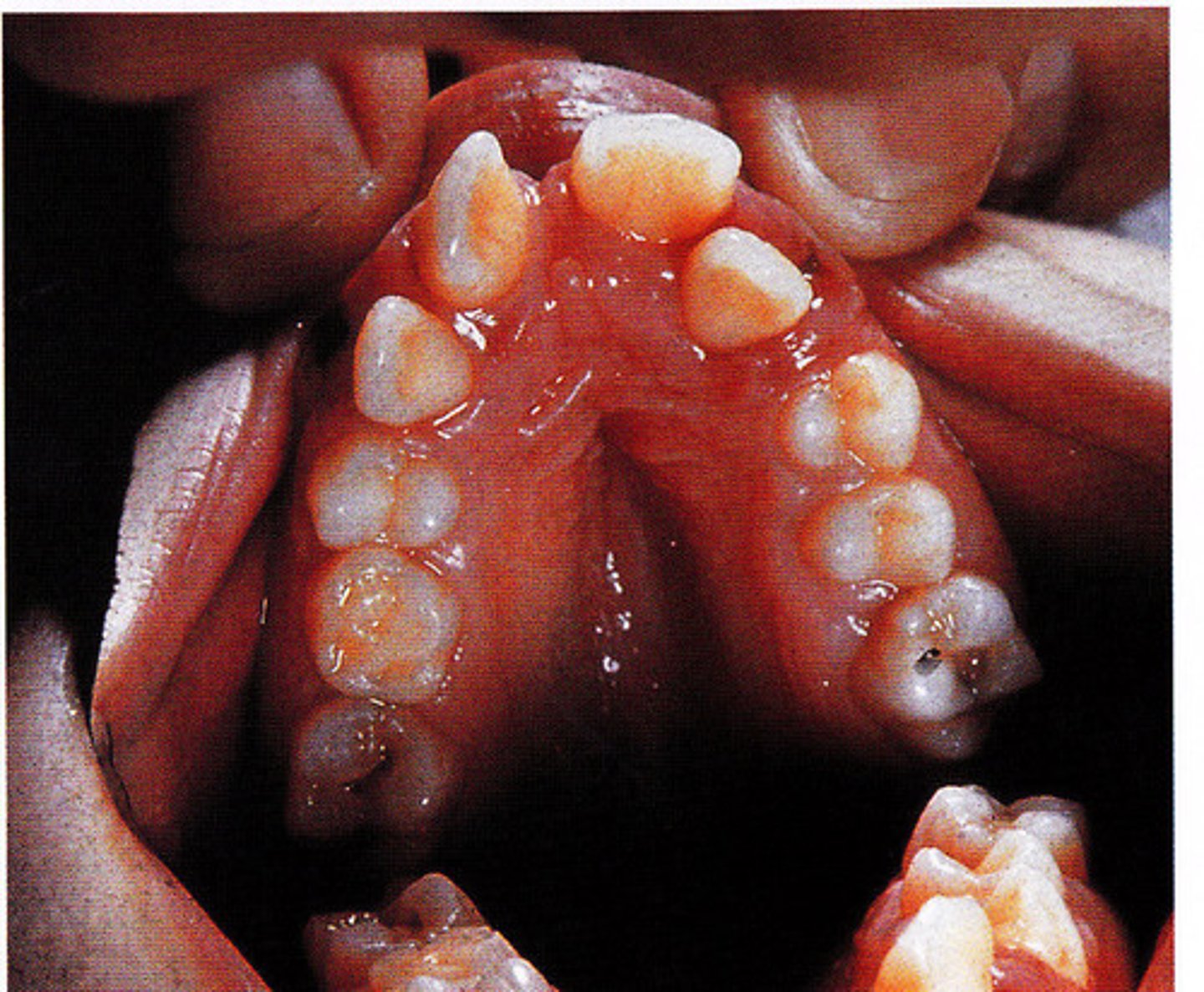
Mandibulofacial Dysostosis
-markedly high-arched palate and malpositioned teeth in a patient with mandibulofacial dysostosis
-also known as treacher collins syndrome
-results in a dysmorphic face, including downward sloping of the palpebral fissures.
-mouth appears fishlike
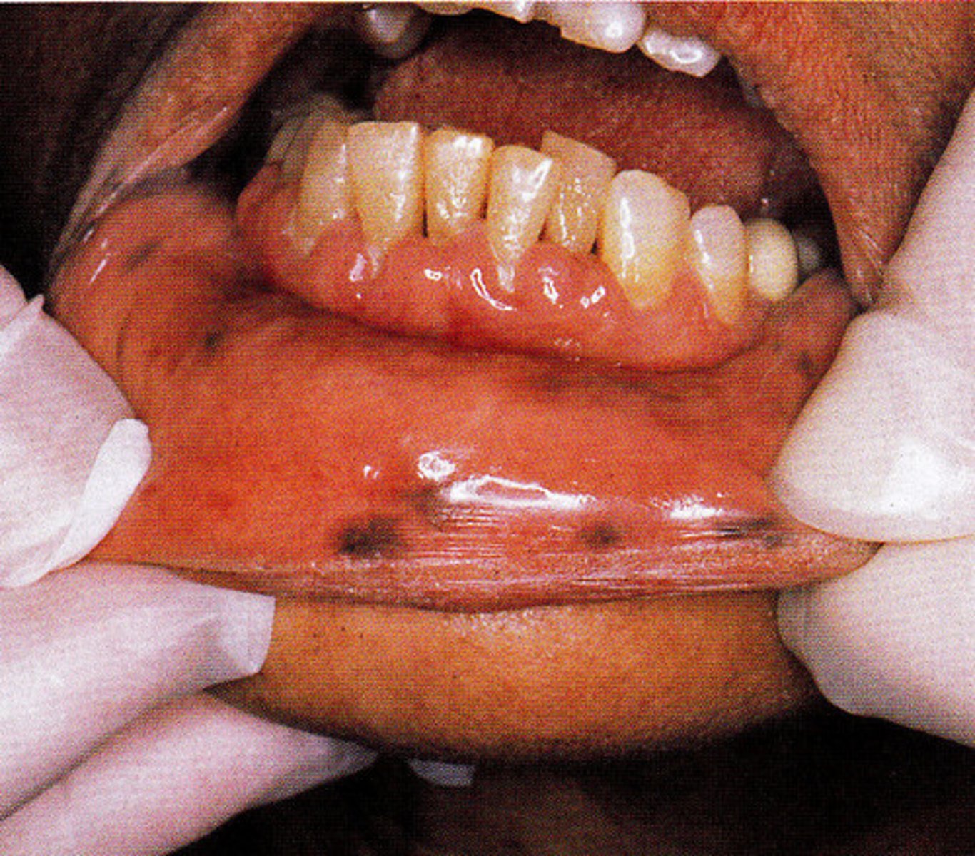
Peutz-Jeghers syndrome
-multiple small to medium sized pigmented macules on the labial mucosa of a patient with peutz-jeghers syndrome
-also known as hereditary intestinal polyposis syndrome
-pigmentation occurs around the eyes, nose and mouth
-associated with gastrointestinal polyposis
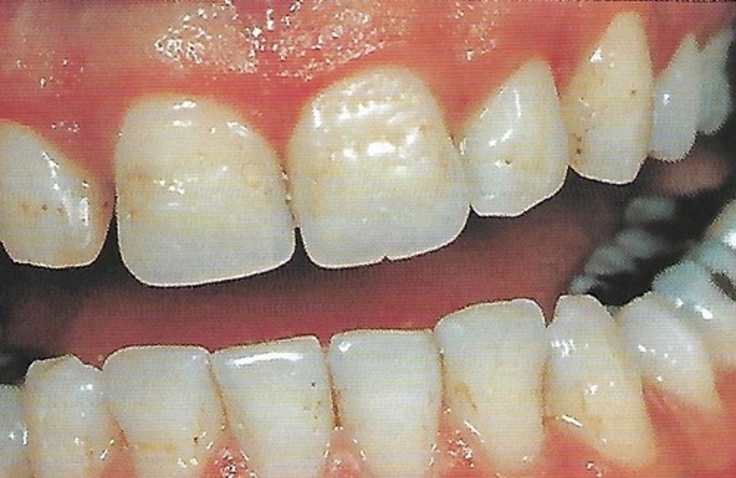
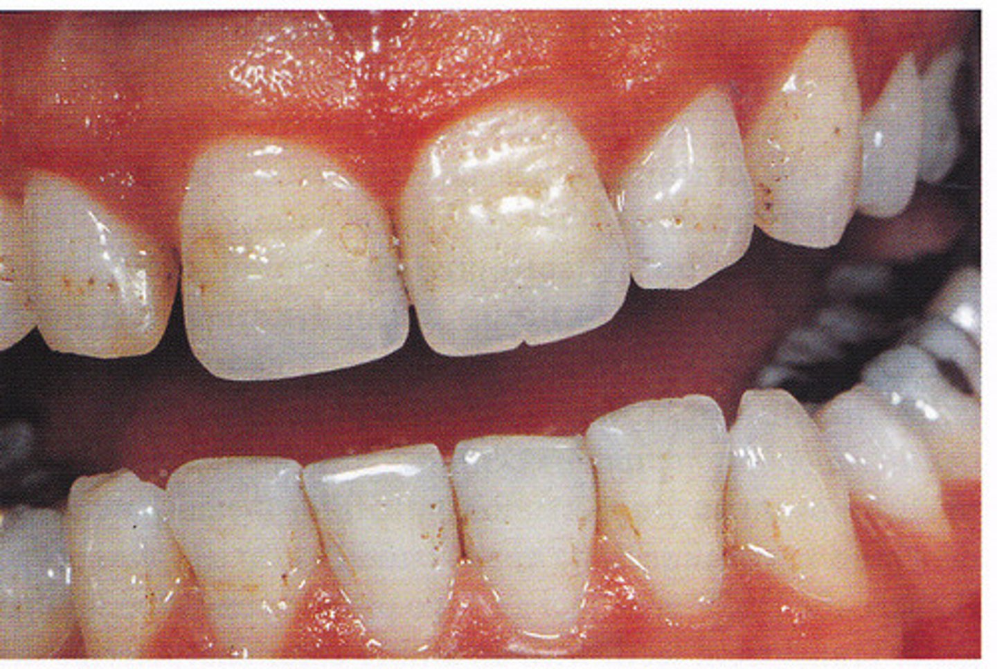
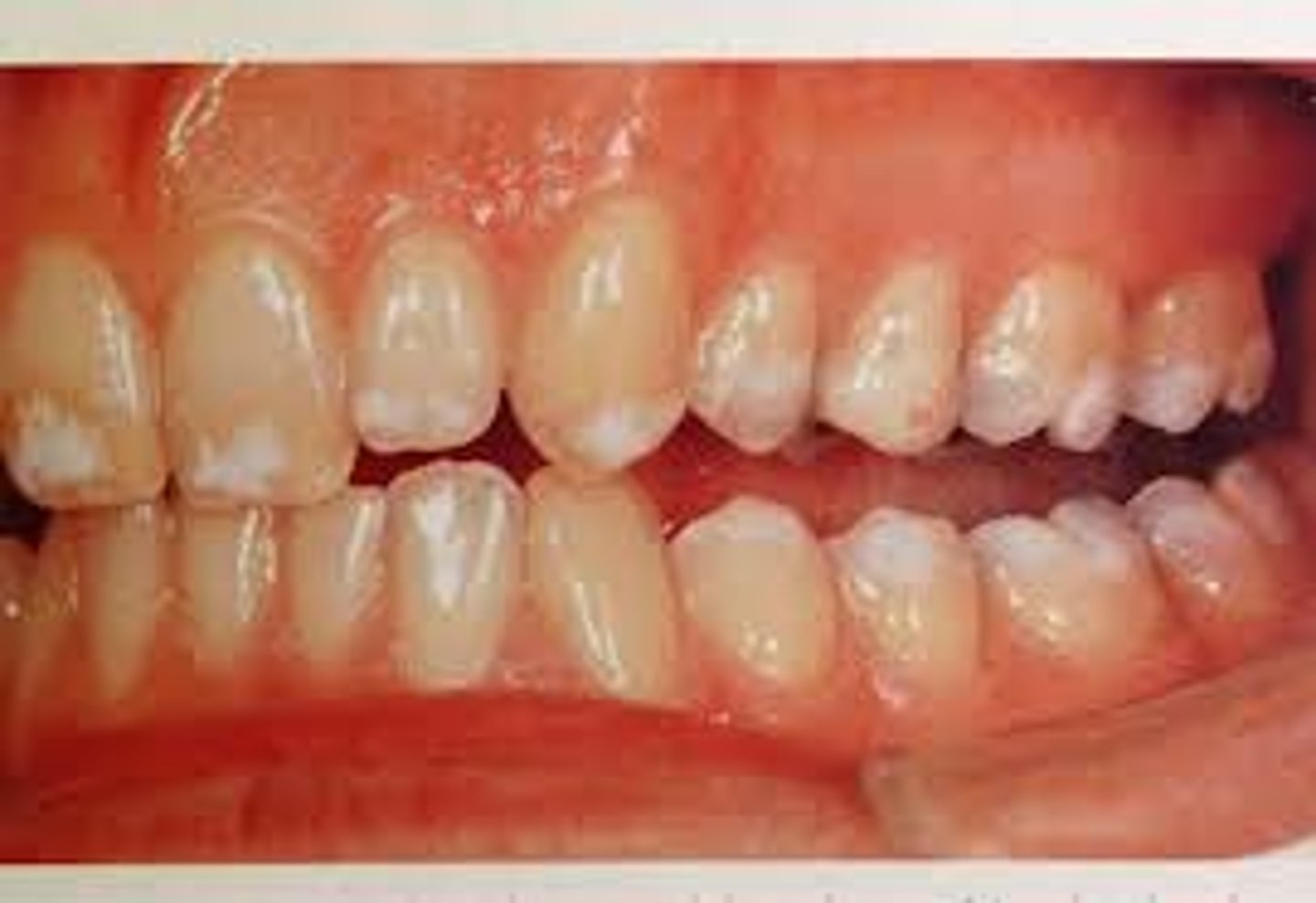
Pitted autosomal dominant Amelogenesis imperfecta
-multiple pits on the labial surface of the teeth
-4 types
Type 1 hypoplastic amelogenesis imperfecta
-characterized by tooth enamel that does not develop to a normal thickness because of failure of the ameloblasts to lay down enamel matrix properly
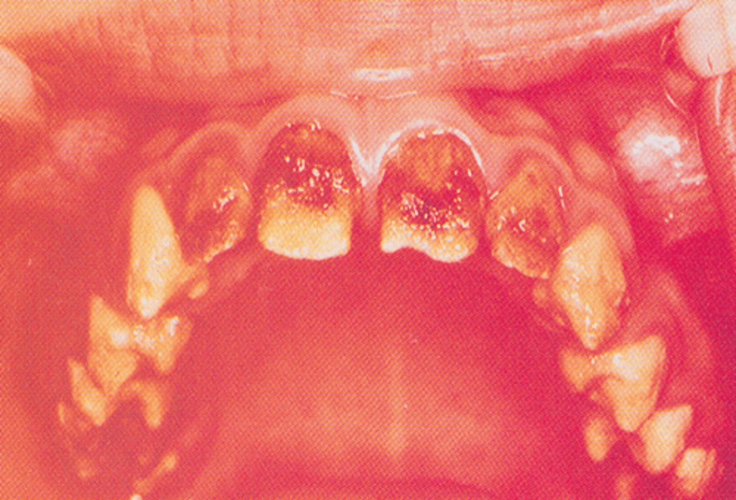
Type 2: Hypocalcified Amelogenesis Imperfecta
-loss of enamel is exhibited in the teeth
-enamel of normal thickness that is poorly calcified
-yellow to orange enamel
Type 3: Hypomaturation Amelogenesis Imperfecta
-an enamel of mottled appearance but normal thickness
-snowcapped amelogenesis imperfecta is a special type
Type 4: Hypoplastic-Hypomaturation Amelogenesis Imperfecta
-thin enamel yellow to brown and pitted
-on radiographs the enamel has a radiodensity similar to dentin and single rooted teeth have large pulp chambers and molar teeth appear as taurodonts.
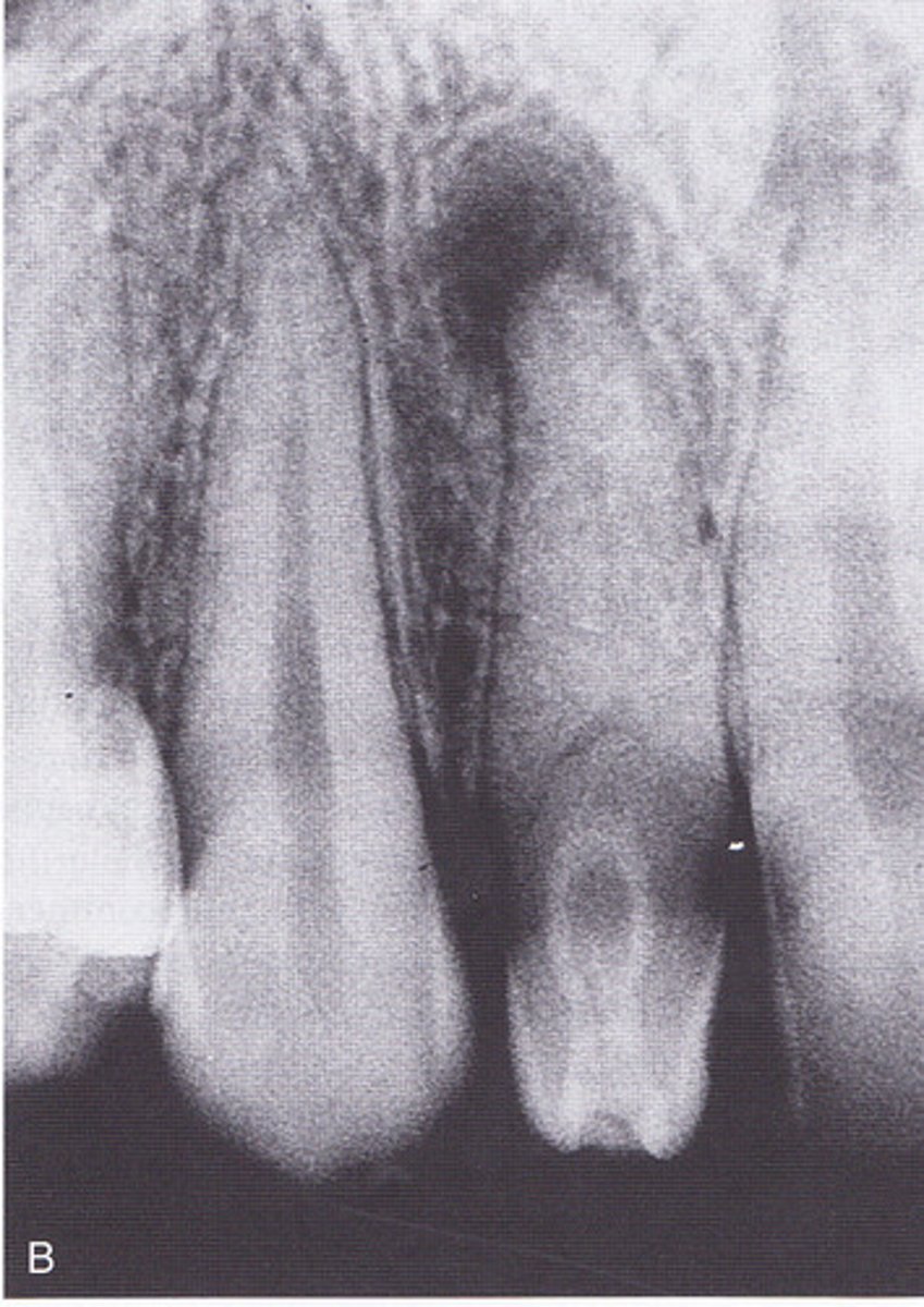
dentinogenesis imperfecta
-shorts roots almost complete lack of pulp chamber
-is marked by bulbous crowns that are colored from opalescents brown to brownish blue
Radicular dentin dysplasia
-shows blunted and short tooth roots, the few remaining pulp chambers have a half moon appearance
-teeth with normal crowns and abnormal roots
-the basic defects seems to lie in a disturbance in the hertwig epithelial root sheath
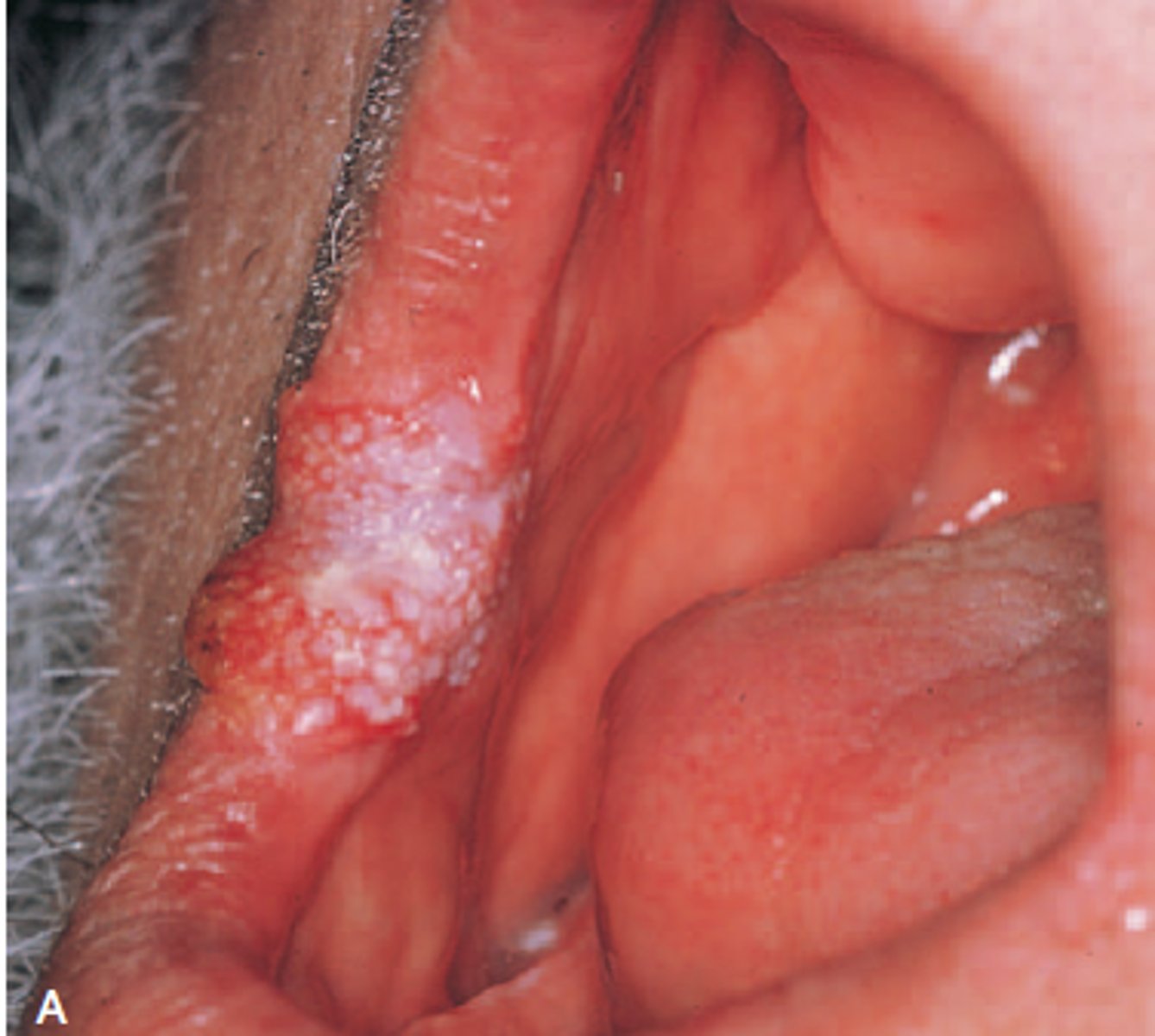
papilloma Figure 7.2 A
-papilloma at the junction of the hard and soft palate shows a cauliflower like appearance and rough surface from fingerlike projections
-benign tumor of squamous epithelium that arise as a small, exophytic, pedunculated or sessile growth
-other oral lesions may resemble a papilloma clincily are verruca vulgaris (common wart)
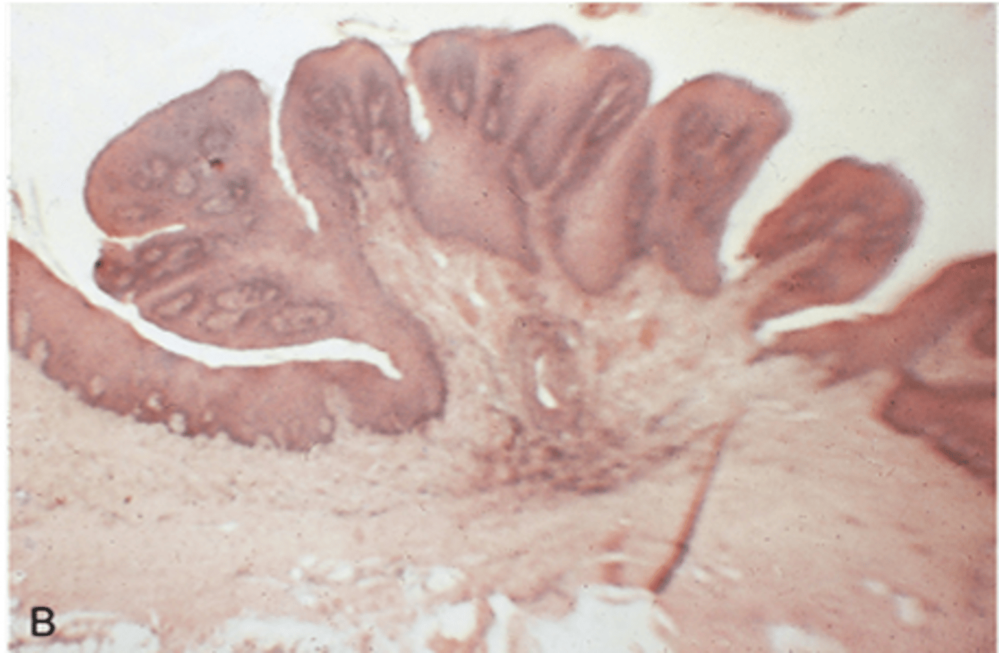
Papilloma Microscopic
-microscopic appearance of a papiloma shows fingerlike projections surfaced by squamous epithelium and supported by thin cores of fibrous connective tissues
-a central core of vascular fibrous connective tissue supports each papillary projection
Verrucous Carcinoma
-commissure and anterior buccal mucosa
-is a specific type of squamous cell carcinoma that is separated from other squamous cell carcinoma because it has a much better prognosis
-appears as a slow growing exophytic tumor with a pebbly white and red surface
-most cases occurs in men older than 55 years of age and involves the vestibule and buccal mucosa
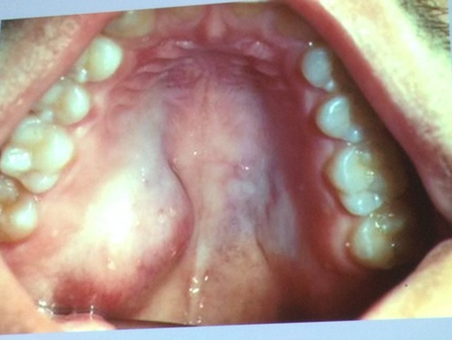
Benign salivary gland tumor
-arises in either the major or the minor salivary gland
-tumors of minor salivary glands origin are much more common in the upper lip than the lower lip
-benign tumors of salivary gland origin are called adenomas
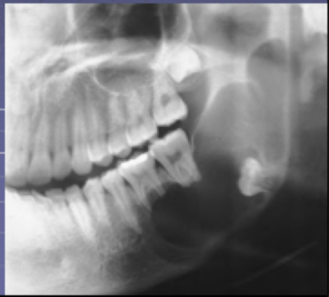
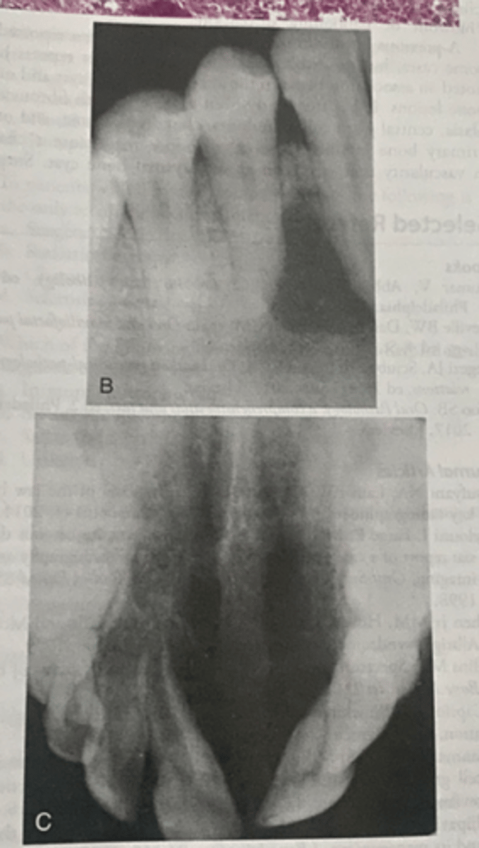
ameloblastomas B
-show multilocular radiolucencies in the molar areas if the mandible
-is benign, slow growing but locally aggressive epithelial odontogenic tumor that may arise in either the maxilla or the mandible
-it is a non encapsulated tumor that infiltrates into surrounding tissue and can cause extensive destruction
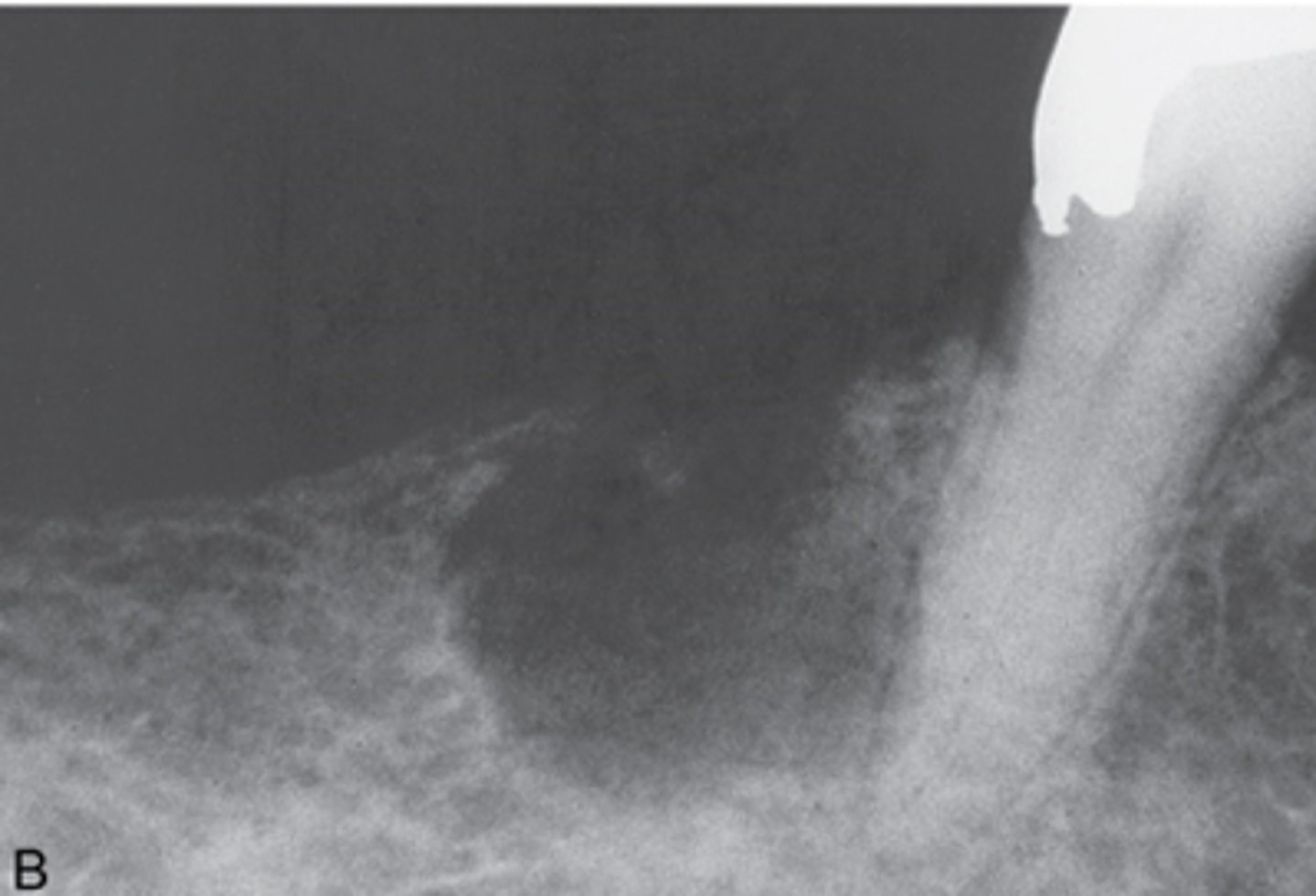
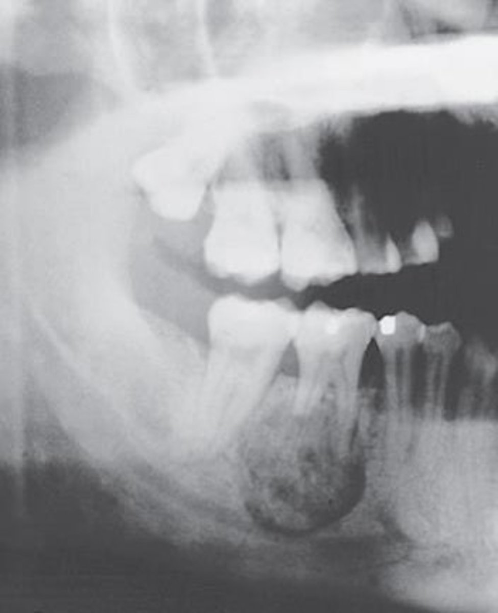
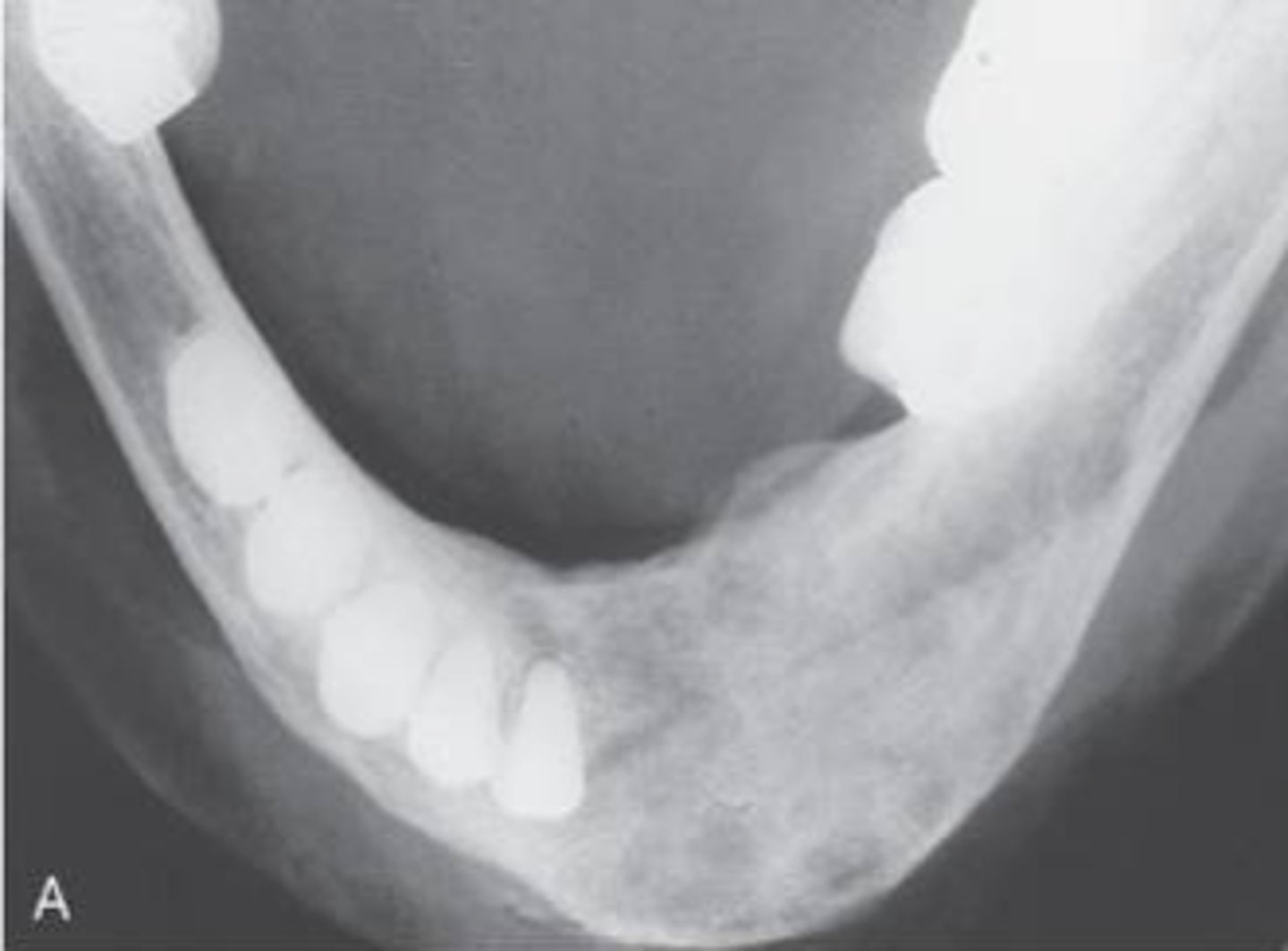
Benign Cementoblastoma
-shows a well circumscribed radiopaque mass surrounded by a radiolucent halo and attached to the root of a mandibular first molar
-pain is a frequent symptom
-occurs in young adults and most occur in patients under 30
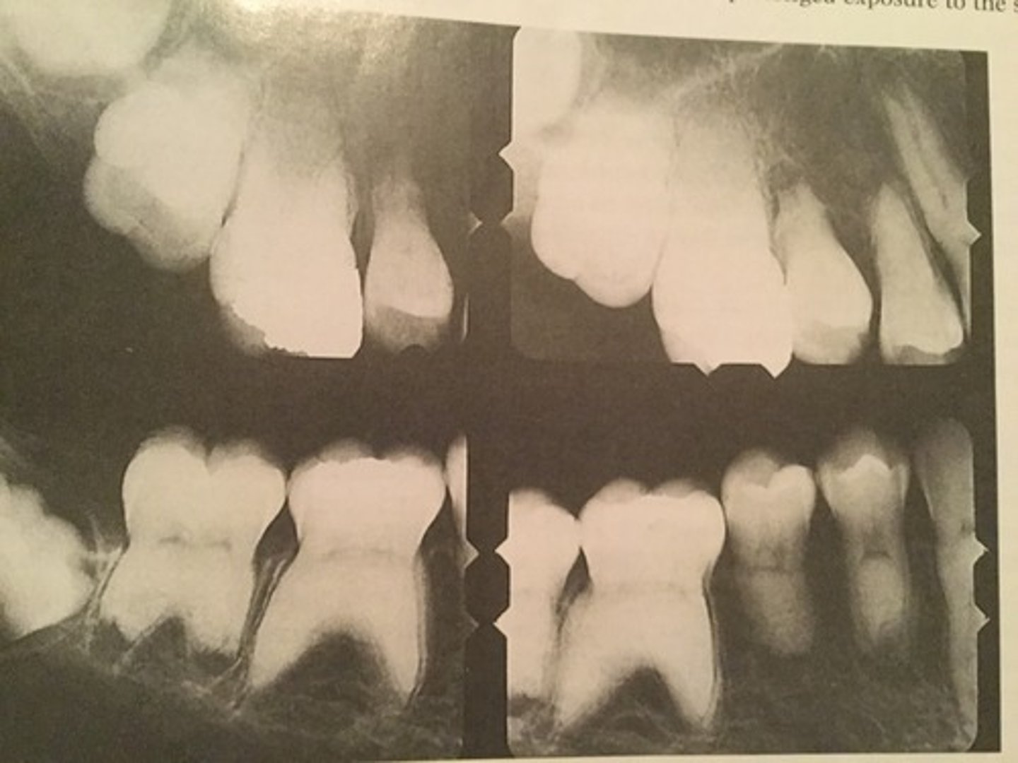
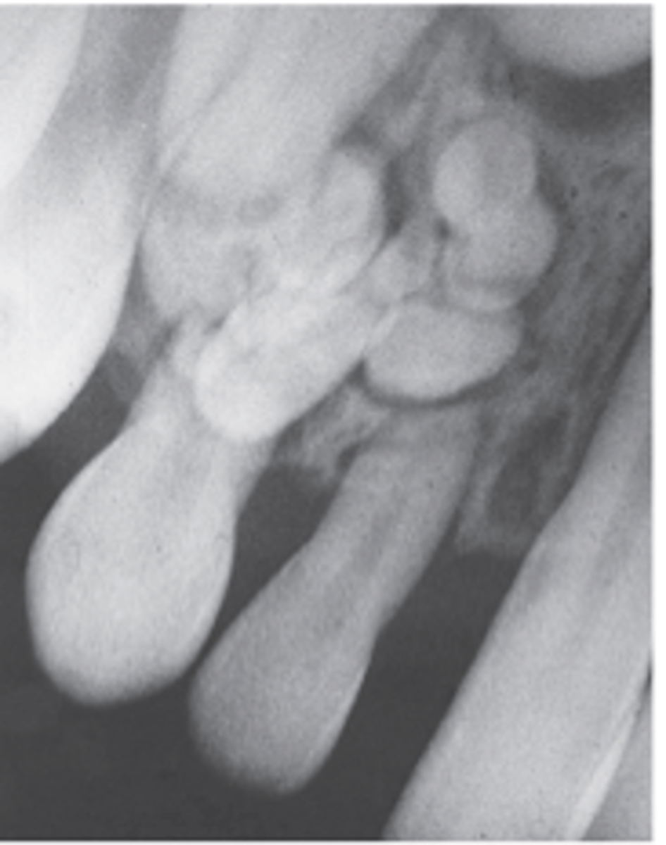
Compound odontoma
-shows a collection of numerous small toothlike radiopacities surround by a radiolucent halo
-do not exhibit unlimited growth potential
-described as a developmental lesion (hamartomas) rather than true tumors
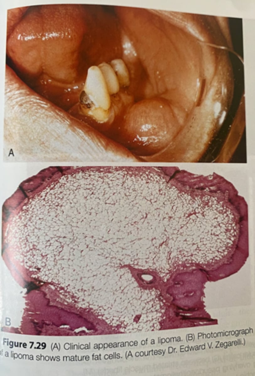
lipoma
-benign tumor of adipose tissue
-clinically appears as a yellowish mass that is surfaced by a thin layer of epithelium
-mature fat cells
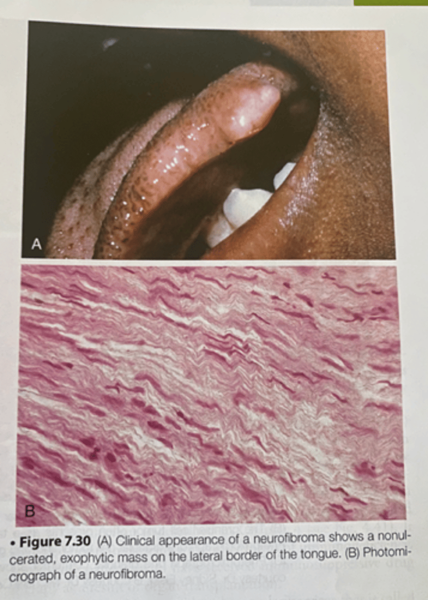
neurofibroma
-a nonulcerated ,exophytic mass on the lateral border of the tongue
-benign tumor derived from nerve tissue
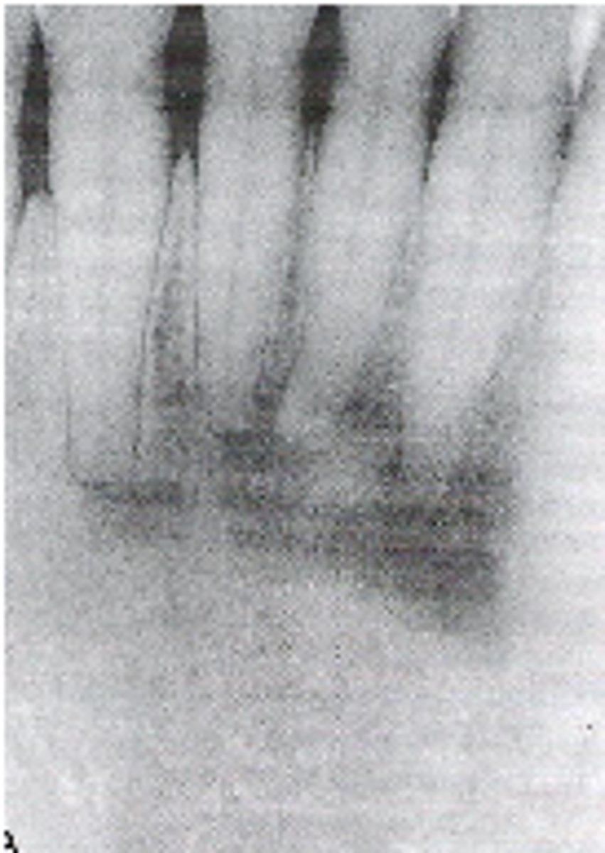
Periapical cemento-osseous dysplasia
-lesion is asymptomatic and is discovered on routine radiographic examination
-occurs most commonly in the anterior mandible of patients older than 30 years of age
-more common in women than men, predilection for this disease in black women
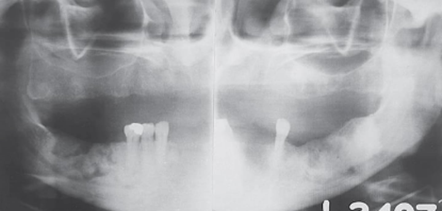
florid cemento osseous dysplasia
-shows irregular radiopaque masses in both the left and right posterior mandible
-disordered cementum and bone deposition
-involves multiple quadrants in the posterior maxilla and mandible
-most often occurs in black women older than 40 years of age
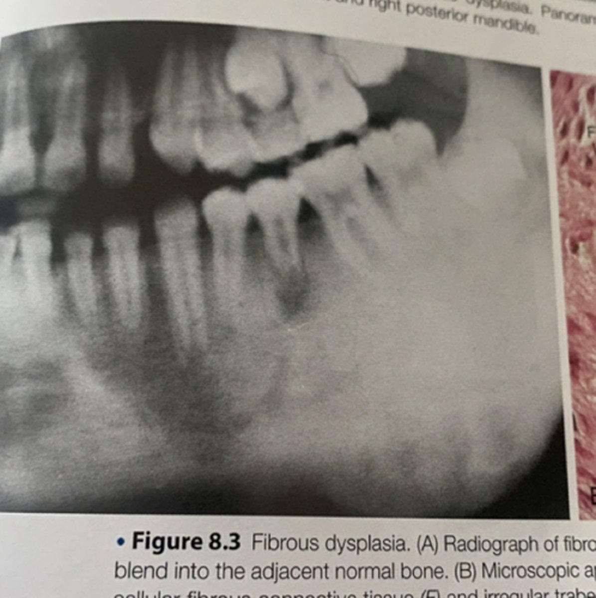
fibrous dysplasia
-is a developmental disease that is charcterized by the replacement of bone with abnormal fibrous connective tissue
-a genetic mutation has been identified as the underlying cause of this disorder
-
paget disease
-enlargement of the maxilla with spaces between the teeth
-radiograph demonstrating irregular opacification that is also referred to as a cotton wool appearance
Central Giant Cell Granuloma
-may occur on the gingiva or alveolar ridge or in the maxilla or mandible
-divergence of the root of adjacent teeth is a common feature
acromegaly
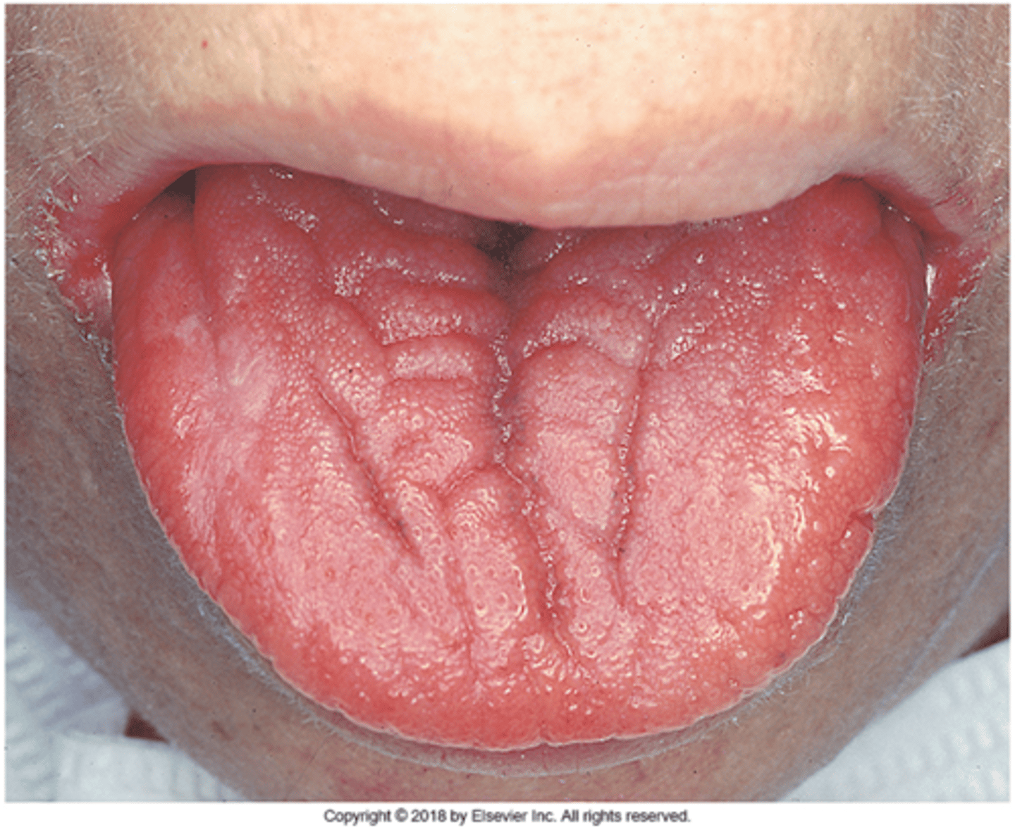
-enlarged tongue
-rare condition that results when the hypersecretion of growth hormones occurs in adult like after closure of epiphyseal plates of the long bones

hyperparathyroidism
-results from excessive secretion of parathyroid hormone (parathormone)
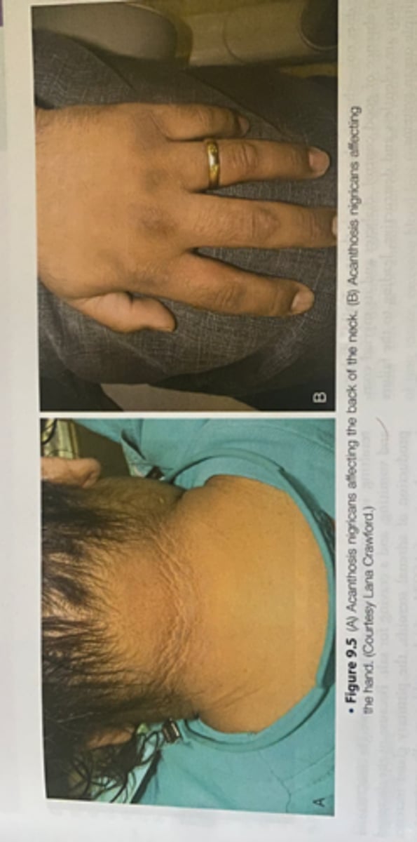
Acanthosis nigricans
-skin disorder
-has been reported to be associated with type 2 diabetes
iron deficiency anemia
-tongue is devoid of filiform papillae
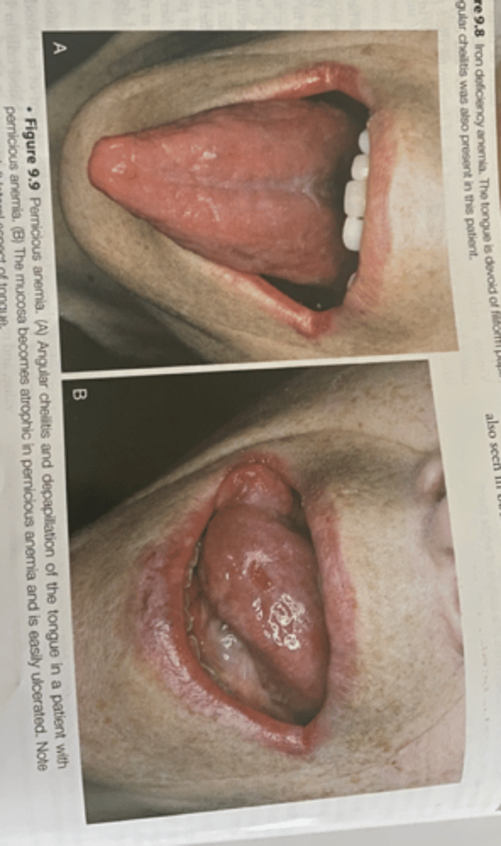
pernicious anemia
-Vitamin B12 deficiency that is caused by a deficiency of intrinsic factors a substance secreted by the parietal cells of the stomach
-
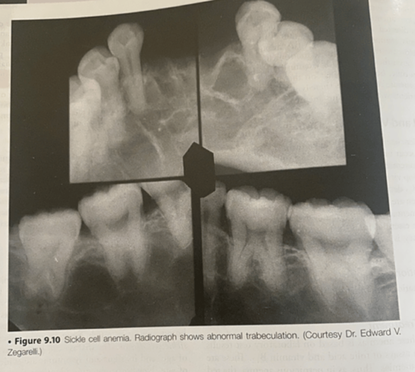
sickle cell anemia
-abnormal trabeculation
-most common inherited disorder of red blood cells
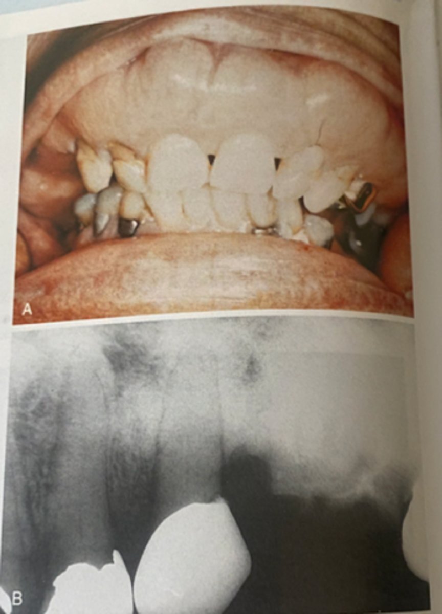
osteonecrosis
-staged based on size, location and symptoms. -bisphosphonate therapy
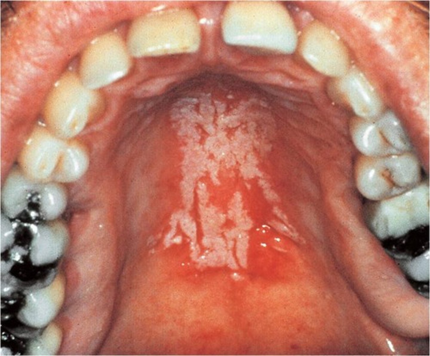
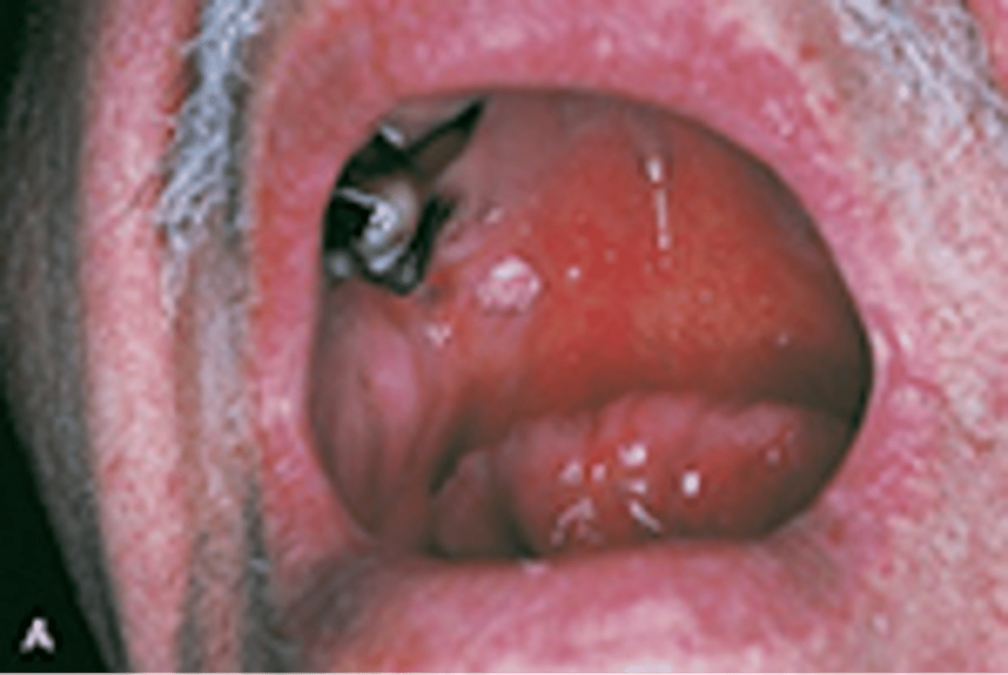
candidiasis in a patient taking prednisone
-