ch 22 respiratory system - label / fill in blank
1/36
There's no tags or description
Looks like no tags are added yet.
Name | Mastery | Learn | Test | Matching | Spaced | Call with Kai |
|---|
No analytics yet
Send a link to your students to track their progress
37 Terms
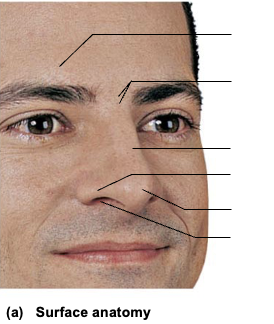
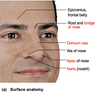

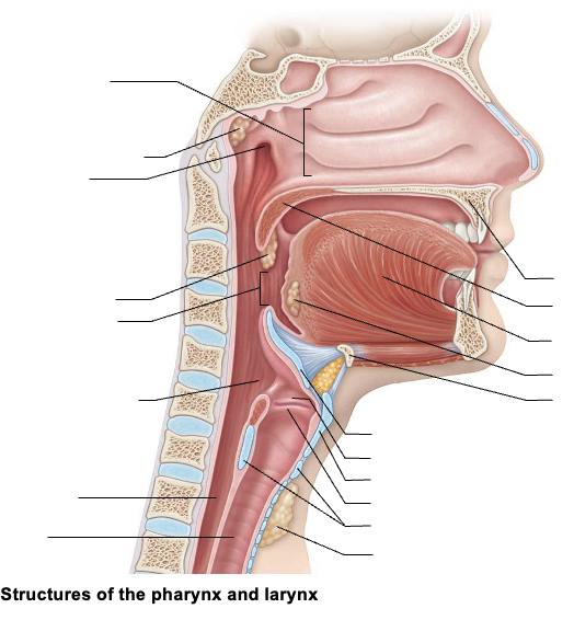

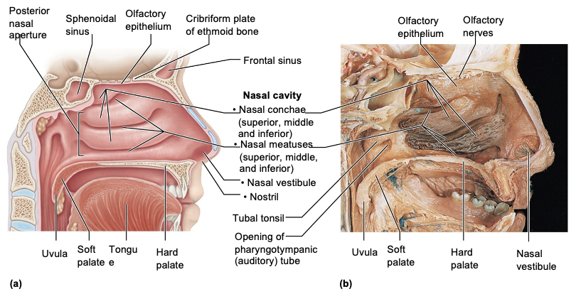


On a sagittal head model, a structure is a small chamber just inside the nostril with hairs. What is it?
Vestibule

On a nasal cavity image, three curved bony projections are visible. What are they?
Nasal conchae (turbinates)
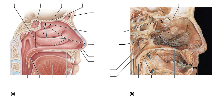
On a nasal cavity model, the spaces beneath the conchae are labeled. What are they?
Meatuses

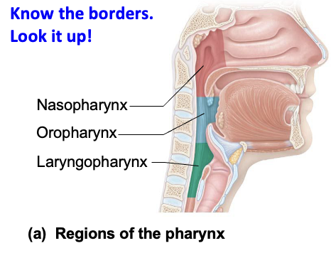
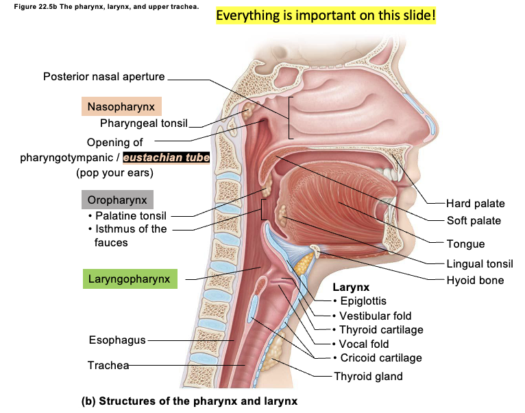
On a sagittal model, the region posterior to the nasal cavity and above the soft palate is labeled. What is it?
Nasopharynx
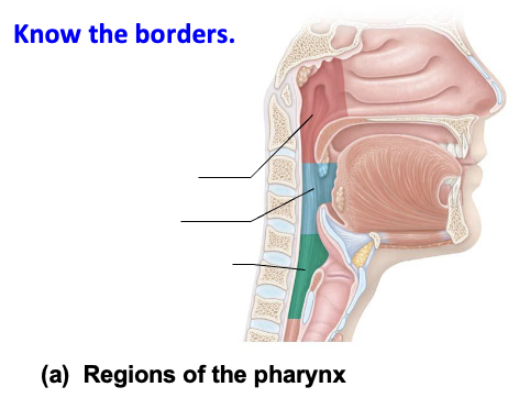
On a model, a region between the soft palate and epiglottis is shown. Identify it.
Oropharynx

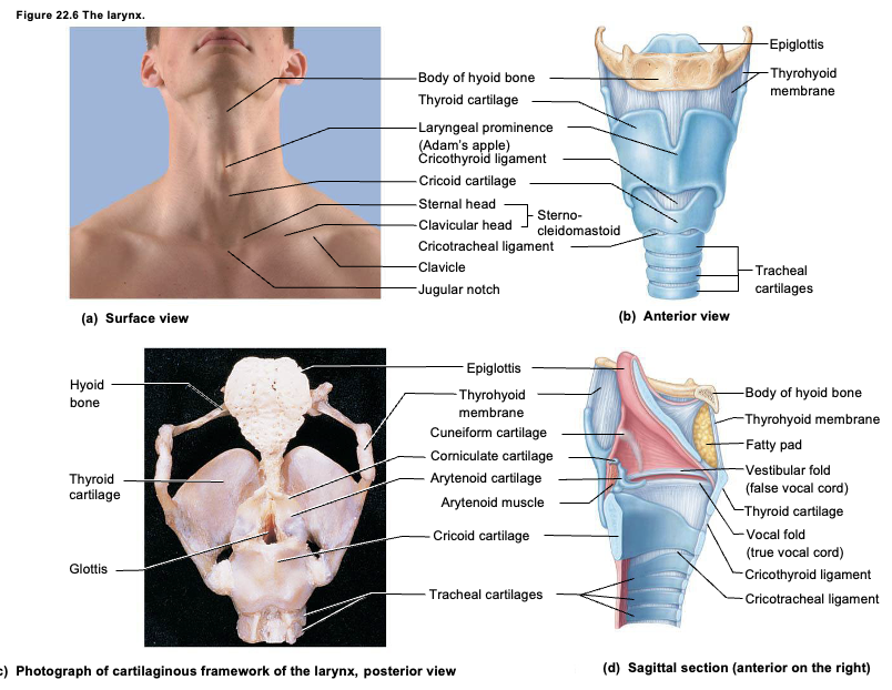
On a larynx model, a leaf-shaped flap above the glottis is shown. What is it?
Epiglottis
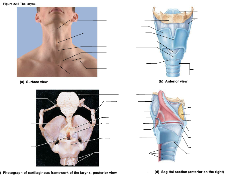
On a larynx model, the large anterior shield-shaped cartilage is labeled.
Thyroid cartilage

On a larynx image, the inferior ring-shaped cartilage is identified.
Cricoid cartilage
On an internal larynx view, the superior folds that do NOT produce sound are labeled.
Vestibular folds
On the same image, the inferior folds responsible for sound are labeled.
Vocal folds
On a histology slide, you see ciliated pseudostratified columnar epithelium with goblet cells. What structure is this?
Trachea
On a trachea model, C-shaped rings are visible. What are they made of?
Hyaline cartilage
On a lung model, the first branches after the trachea split are shown.
Primary bronchi
On a model, branches supplying individual lobes are labeled.
Secondary (lobar) bronchi
On a microscopic image, a small airway with NO cartilage is seen.
Bronchiole
On a lung histology slide, small air sacs surrounded by capillaries are visible.
Alveoli
On an image, clusters of alveoli arranged around a central space are shown.
Alveolar sacs
On a diagram, thin cells forming most of the alveolar wall are labeled.
Type I alveolar cells
On a lung model, the superior tip above the clavicle is labeled.
Apex
On a lung model, a depression on the left lung is shown.
Cardiac notch
On a lung model, the area where vessels and bronchi enter/exit is labeled.
Hilum
On a cross-section, the membrane directly covering the lung is labeled.
Visceral pleura
The space between pleural layers is shown.
Pleural cavity