Lab 1- Introduction to C. elegans
1/16
There's no tags or description
Looks like no tags are added yet.
Name | Mastery | Learn | Test | Matching | Spaced | Call with Kai |
|---|
No analytics yet
Send a link to your students to track their progress
17 Terms
C elegans embryos
form in the gonad of an adult
contain 1 to 64 cells when they emerge from the vulval opening of the gonad.
c elegans egg
Cell division continues inside the eggshell
L1
contain 558 of the 959 cells present in an adult,
671 cells are generated in an embryo, but 113 undergo apoptosis before hatch.
Larva developement
When larvae grow too large to fit in the surrounding exoskeleton, they molt, shedding the old skeleton and creating a new one.
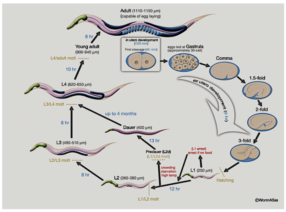
C. elegans pass through 4 larval stages (L1-L4) before the final molt to adulthood
Each larval stage lasts about 9 – 12 hours depending on the growth temperature.
C. elegans growth temperature
Typically, worms are grown at 20C, but development can be slowed by growing them at 15C. If grown above 25C, fitness markedly descreases.
C elegans life cycle diagram
The life cycle of C. elegans. Upon starvation or stress conditions L1 worms can exit the life cycle and enter dauer stage indefinitely. When environmental conditions improve, development resumes.
C. elegans developement
C. elegans is programmed to change its development depending on environmental signals (see Figure 1).
In environments with plentiful food, L1 larvae develop directly through the L2, L3 and L4 stages.
Alternatively, harsh or stressful environments with little food or overcrowded conditions trigger L1s to arrest development at L2 and become dauer larvae, an alternative larval stage. Dauers do not feed and have reduced metabolic activity, but do retain limited mobility and explore the environment in search of food sources.
They are easily identified in a dissecting microscope because they are thin and their thicker exoskeleton (called the “cuticle”) gives them a more refractile appearance. Once a new source of food is found, dauer larvae will molt into the L4 stage and resume reproductive development.
C. elegans as a Model System
transparent, can use light and flourescence microscopy
relatively small, 959 somatic cells, we know complete cell lineage/fate of every cell
entire neural network (connectome) mapped
easy to modify
short generation time
C. elegans Reproduction
The sex of C. elegans is determined by sex chromosomes; hermaphrodites have 2 sex chromosomes (XX) and males have one (XO). The majority of C. elegans are hermaphrodites, which contain both ovaries and spermatheca. Because hermaphrodites can self-fertilize, homozygous hermaphrodites generate genetically identical progeny. Males (XO) arise infrequently (0.1%) as a result of spontaneous chromosomal nondisjunction in the hermaphrodite germ line. In addition, male progeny are produced when males cross-fertilize hermaphrodites. The major phenotypic differences between males and hermaphrodites begin at L2 stage, where the male copulatory apparatus begins to develop, and the shape of their tail changes to blunt and scalloped with sensilla. The specialized tail of males allows them to attach to the hermaphrodite vulva and inject sperm into the vulva.
The adult hermaphrodite produces oocytes for
about 4 days, known during this time as a “gravid” adult, and then lives for a subsequent 10-15 days in a non-gravid state.
A hermaphrodite that self-fertilizes can produce about 300 progeny due to limited sperm number, but this number increases to 1200-1400 if mated to a male, who can father ~3000 progeny.
Adult Anatomy
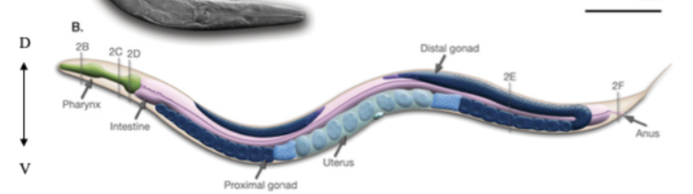
gonad
contains 2 symmetric arms – one in the anterior and one in the posterior. Germ cells are produced (through mitosis) in the distal arm of each arm of the gonad. As more and more germ cells are produced in this distal region, they are pushed to occupy more proximal regions of the gonad and undergo meiosis. Mature oocytes become fertilized as they pass through the spermatheca (the spongy-looking brighter blue rectangle between the dark blue oocyte and light blue fertilized eggs). Fertilized eggs, which exit the gonad through the vulva, can be in various stages of development from 1 cell to about 64 cells.
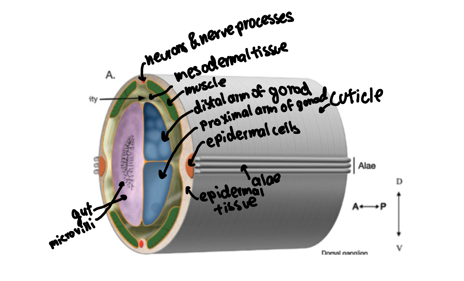
label c. elegans cross section
C. elegans microscropy
Dissecting microscopes are used in the laboratory to view C. elegans for manipulation.
low magnification microscopes with a large working distance and large
field-of-view, which are used to look at the surface of objects in three-dimensions.
Incident light is emitted from an illuminator that is separate from the microscope. Specimen can be illuminated from above, with “epi”-illumination. Alternatively, the light source can be transmitted through the translucent worms from underneath the stage.
C. elegans grown in lab
agar plates or in liquid cultures, with E. coli as the food source. E. coli is spread, or ‘seeded’, onto agar plates and allowed to grow overnight until it forms a thin layer, or “lawn”. C. elegans traveling across this lawn leave sinusoidal tracks in their wake, and an absence of these lawn tracks indicate that the worms have depleted their food source (or are paralyzed mutants!). C. elegans can survive for weeks to more than a month without food, but to ensure you are studying worms exposed to a consistent environment, it is important to transfer them to a fresh plate when E coli is depleted.
Two techniques can be used to transfer worms
‘picking’ and ‘chunking’. By the picking method, a thin platinum (Pt) or platinum-iridium (Pt-Ir) wire is used to gently lift a single worm off a plate and move it to a new plate. The platinum wire cools quickly and can be exposed to a flame in between each worm picked to allow for sterile transfer of worms. To transfer many worms to a new plate, the chunking method is used. In this method, a small square of agar is cut out of a plate crowded with worms and simply placed face-down onto a new plate with a fresh lawn. Worms can crawl out of the chunk and continue to feed and reproduce. Chunking is fast and doesn’t necessarily require that you look in a microscope.
which wire to use for pick
you are making a flattened-tipped pick, use the pure platinum. If you are making the loop-tipped pick, use the Pt-Ir.