Systemic Diseases
1/125
There's no tags or description
Looks like no tags are added yet.
Name | Mastery | Learn | Test | Matching | Spaced | Call with Kai |
|---|
No analytics yet
Send a link to your students to track their progress
126 Terms
Label the clinically relevant anatomy of the bovine head
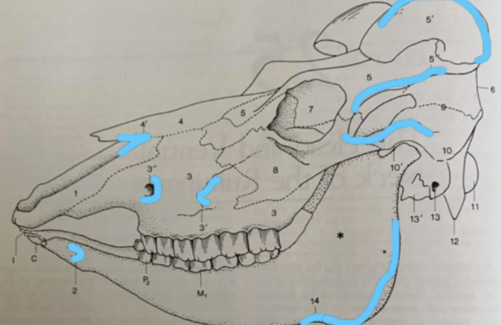
Bones
Mandible (*)
Maxilla (3)
Incisive (1)
Nasal (4)
Frontal (5)
Zygomatic (8)
Temporal (10)
Occipital (11)
Palpable Structures
Temporal line (5”)
Facial tuberosity (3’)
Infraorbital foramen (3”)
Nasoincisive notch (4’)
Mental foramen (2)
Zygomatic arch (8’)
Cornual process/horn (5’)
Ramus of mandible (14+)
3 Sinuses of the skull
Frontal (rostral + caudal → cornual process)
Maxillary
Palatine
Label the clinically relavent soft tissue structures of the bovine head
Veins
Arteries
Nerves
Muscles
Salivary glands
Lymph nodes
Veins
Linguofacial (18)
Facial (4)
Cornual (8)
Arteries
Common carotid (19)
Facial (10)
Cornual (8)
Nerves
Cornual (8)
Facial (5 and 6)
Infraorbital (9)
Muscles
Masseter (1)
Zygomaticus (2)
Salivary glands
Parotid (11)
Submandibular (12)
Lymph nodes
Parotid (13)
Lateral retropharyngeal (14)
Submandibular (20)
Palpable in healthy animal
Lingual fossa
Pocket between the torus and tongue which is a common site for sharp instruments to accidentally enter
Describe the deciduous and permanent dentition of cattle
Deciduous: I 0/3 : P 0/1 : M 3/3
Permanent: I 0/3 : C 0/1 : P 3/3 : M 3/3
Eruption times important for aging and certain diseases
Cattle graze using tongue to grasp pasture (mandibular incisors are not necessary)
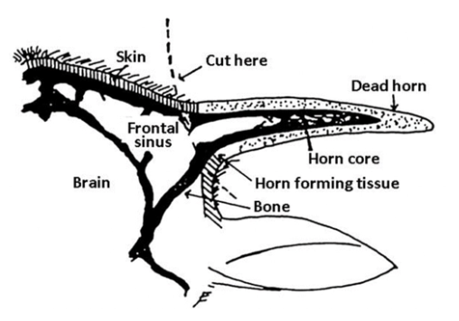
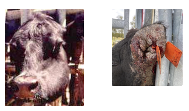
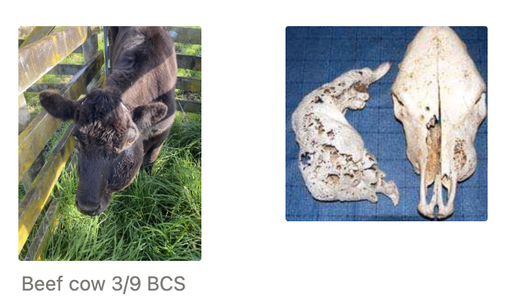
Frontal Sinus Empyema
3 Aetiologies
5 Clinical signs
5 Treatments
Aetiologies: Secondary to
Dehorning → Open sinus formation
Trauma (fighting)
Respiratory disease (uncommon)
Clinical Signs:
Discharge form dehorning site
Nasal discharge (purulent/serosanguinous/epistaxis)
Exophthalmos (extension of infection to retrobulbar space)
Head tilt (extension to vestibular system)
Pyrexia (systemic illness when severe)
Treatment:
Sinus trephination
Sinus lavage
Systemic antibiotics
NSAIDs
± Cosmetic dehorning = Skin flap OR occlude hole
Hope exposed bone and sinus hardens with granulation tissue to fill the defect in the frontal sinus
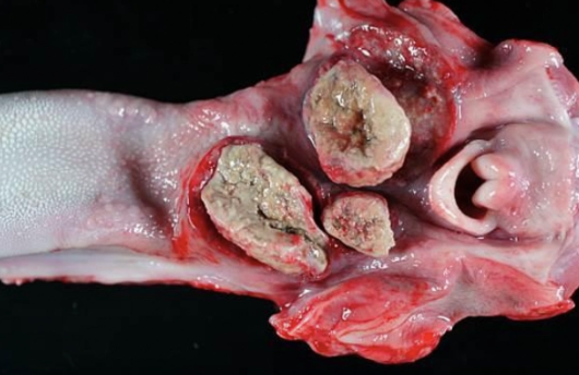
Woody Tongue
Agent
Aetiology and 3 causes
Inflammation
4 Clinical signs
2 Methods of diagnosis
3 Treatments
Agent: Actinobacillus lignierseii
Commensal oral bacteria that causes opportunistic infection due to mucosal trauma
Aetiology: Soft tissue trauma
Erupting teeth
Rough feed
Drenching injuries
Inflammation: Granulomatous swelling of the tongue, cheek, nasal passages
Cutaneous actinobacillosis = Granulomatous lesions on ear, nose, cheek and oesophageal groove → Chronic, friable, non-resolving lesions
Clinical Signs:
Inappetence
Ptyalism
Firm/oedematous submandibular swelling NOT associated with bone
Tongue protrudes from mouth → Firm and painful ± ulcers/erosions
Diagnosis:
Clinical signs and response to treatment #1
± Biopsy for atypical presentations
Treatment:
Streptomycin 3 - 4mL/100kg q24hr IM/SC
Aminoglycoside → Excreted in kidneys and cannot be used in food animals in USA (okay in NZ)
Sodium/potassium iodide (organic farms)
MoA: Breaks down granulomatous tissue to allow immune response to heal animal
Sodium iodide IV or potassium iodide PO in feed
NSAIDs
Lumpy Jaw
Agent
Aetiology
Signalment
5 Clinical signs
3 Methods of diagnosis
4 Methods of treatment
Prognosis
Agent: Actinomyces bovis
Aetiology: Eruption of cheek teeth → Allows bacteria to infiltrate and cause osteomyelitis (bone remodelling)
Signalment: 2 - 3yr
Clinical Signs:
Acute: Asymmetrical, firm swelling of jaw associated with bone (painful when early in disease)
Chronic: Deformed skull
Difficulty eating
Drooling
Weight loss
Diagnosis:
Clinical signs #1
Radiography
U/S
Treatment: As for woody tongue if caught early
Streptomycin
Sodium/potassium iodide
NSAIDs
Euthanasia if signs are severe (avoid pathological fractures and malocclusion)
Prognosis: Depends on severity (guarded to poor)
7 Structures around the jaw
Lymph nodes
Masseter muscle
Mandible
Salivary glands
Base of tongue
Nerves
Vessels
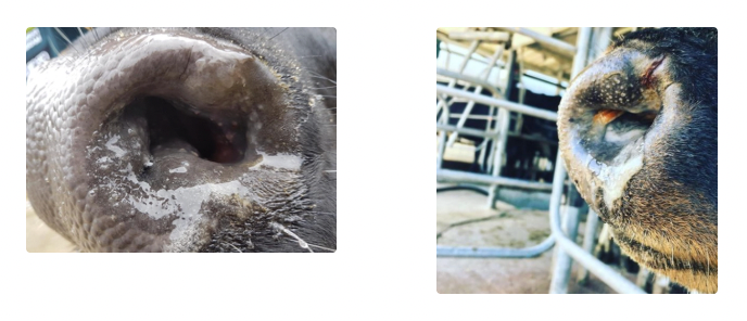
Nasal Catarrh
Prevalence
2 Aetiologies
Pathogenesis
Signalment
4 Clinical signs
Method of diagnosis
4 Treatments
aka. Atopic rhinitis, chronic granular rhinitis, bovine nasal granuloma, allergic rhinitis, stick granuloma
Prevalence: Extremely common in NZ
Aetiologies: Multifactorial
Chronic allergy (eg. pollen, mites, fungal spores) → Seasonal pattern
± Infectious bovine rhinotracheitis (IBR)
Pathogenesis: Allergic rhinitis → Itchy nose → Stick foreign body in nasal cavity → Granulomatous inflammation
Signalment: Jerseys
Clinical Signs:
Swelling of nasal passages → Snuffling and loud breathing
Nasal discharge (serous → mucopurulent/bloody with foreign body)
Nodular lesions on mucosa (chronic)
Severe pruritis → Foreign bodies
Treatment:
Remove foreign body
Steroids? → Abortion of foetus
NSAIDs
± Anti-histamine
Spontaneous resolution if caused by IBR
Oral Necrobacillosis
Agent
2 Locations/forms
Aetiology and 3 causes
5 Clinical signs
4 Methods of diagnosis
3 Treatments
Agent: Fusobacterium necrophorum = G+ anaerobe
Causes MANY diseases ANYWHERE with existing trauma
Locations:
Necrotic stomatitis = Lips, tongue, cheeks
Calf diphtheria/necrotic laryngitis = Larynx and pharynx
Aetiology: Trauma
Aggressive tube feeding (soft ball at tip becomes sharp when grazed against sharp molars)
Poor hygiene
FPTi
Clinical Signs:
Inappetence/dysphagia
Halitosis
Cough
Progressive dyspnoea ± dysphonia
Pyrexia
Diagnosis: Clinical signs and response to treatment #1
Supported with bloodwork
Imaging
Treatment:
Early cases: High dose penicillin 30,000IU/kg IM/SC q12 - 24hr for 5 - 7d
NSAIDs
± Emergency tracheostomy if dyspnoeic
Pharyngeal Trauma
Aetiology and 3 causes
4 Clinical signs
4 Treatments
Aetiology: Iatrogenic → Infection with ANY bacteria (associated with adult cows)
Orogastric tubing
Drench administration
Bolus administration
Clinical Signs:
Ptyalism
Dysphagia
Swelling of throat/neck/face
Change in voice/coughing
Treatment:
Remove foreign bodies
Drain abscess
Systemic antibiotics
NSAIDs
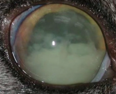
8 Ocular DDx in cattle
Infectious bovine keratoconjunctivitis (pinkeye)
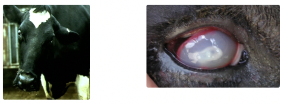
Bovine uveitis and keratoconjunctivitis (silage eye)
Squamous cell carcinoma (cancer eye)
Entropion
Inherited bilateral cataracts (BVD in-utero infection)
Blindness
IBR = Conjunctivitis ± corneal opacity
MCF = Marked scleral congestion and keratitis
Foreign body
Infectious Bovine Keratoconjunctivitis
Agent
Signalment (age)
3 Predisposing factors
Transmission
6 Clinical signs
Method of diagnosis
4 Treatments
4 Methods of prevention
aka. Pinkeye
Agent: Moraxella bovis (G- bacilli)
Pathogenicity depends on presence of pili and haemolytic enzymes
Pili = Projections on the bacterial wall which allow adhesion to the corneal surface and penetration through the intact corneal epithelium
Bacteria without pili and haemolysins carried asymptomatically → Change to pathogenic strain under different conditions (eg. concurrent IBR or increased levels of UV radiation)
May be complicated by viral infection (eg. IBR and Mycoplasma bovis)
Signalment: Younger stock at higher risk
Predisposing Factors:
Dust
Flies (vector of infection)
UV rays (summer #1)
Transmission: Highly contagious (direct OR indirect contact eg. flies)
Clinical Signs: Uni- or bilateral (eg. NH3 toxicity due to overstocking → bilateral)
Blepharospasm
Epiphora → Periocular alopecia due to discharge and rubbing
Corneal oedema (keratitis) and neovascularisation
Corneal ulceration (circular and central) and potential rupture with iris prolapse
Conjunctivitis
Photophobia
Diagnosis: Clinical signs, history ± cytology or culture of conjunctival swab
Corneal ulceration of multiple stock
Treatment:
Topical cloxacillin (Orbenin eye ointment) q48hr to BOTH eyes
Systemic antibiotics (oxytetracycline/penicillin)
Severity determines dose
Avoid subconjunctival as irritating
NSAIDs ± steroids
Protect eye from further damage
3rd eyelid flap or tarsorraphy for severe ulcers
Eye patch
Prevention:
Ear tags impregnated with permethrin to prevent flies
Vaccination with Piliguard to prevent outbreaks (variable response do the different strains) 3 - 6w prior to risk period → Booster q1yr
Vaccination stimulates Ab production against bacterial pili → Prevents bacterial adhesion to the cornea
Reduce dust exposure
Quarantine affected animals (ocular discharge = source of infection)
Function of the 3rd eyelid flap
Prevent rupture of corneal ulcers by protecting globe from UV light and trauma
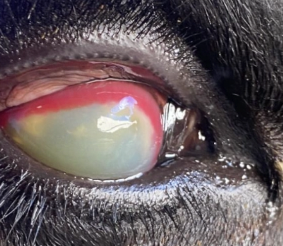
Bovine Uveitis and Keratoconjunctivitis
Agent
4 Clinical signs
Method of diagnosis
3 Treatments
Agent: Listeria monocytogenes
Poorly preserved silage
Affects ENTIRE eye (vs. pinkeye = just anterior portion)
Clinical Signs: Uni- or bilateral
Epiphora
Blepharospasm
Corneal oedema
Photophobia
Diagnosis: Clinical signs and history of silage ingestion
Treatment:
Topical cloxacillin (Orbenin eye ointment)
Topicals have poorer penetration and cannot feasibly be given q2hr to cattle in paddocks
Subconjunctival/systemic antibiotics (oxytetracycline/penicillin) or dexamethasone (overseas)
NSAIDs (± steroids)
Cancer Eye
MPI regulations
3 Treatment options
Regulations: Cannot transport cows with cancerous lesions of eye if
Lesion >2cm
Bleeding or discharging material
Evidence of spread to orbit or lymph nodes
Treatment: Mild cases
Cryotherapy
Surgical removal of 3rd eyelid
Exenteration = Removal of globe and ALL surrounding structures
Ocular surgery in cattle
3 Indications
4 Example surgeries
4 Types of local blocks
Additional drugs
Surgical scrub
Indications:
Treat pinkeye
Remove SCC lesion
Ruptured globe/trauma
Examples:
Temporary tarsorrhaphy (3rd eyelid flap)
3rd eyelid removal
Local mass removal
Enucleation/exenteration
Blocks:
4-point retrobulbar
Auriculopalpebral
Line block/local infusion
Peterson block
Drugs:
NSAIDs
± Antimicrobials
Scrub: Dilute iodine (betadine)
Avoid chlorhexidine/alcohol → Irritation and damage to cornea
3 DDx for blindness in cattle
Hypovitaminosis A
PEM
Lead toxicity
List 7 specific anatomic features of the bovine respiratory tract
NOT athletes
Narrowed trachea (~30% narrower than a horse)
Fewer alveolar capillaries and macrophages
More segmented anatomy
Lack of Pores of Kohn = Collateral ventilation to allow communication between adjacent alveoli
More sensitive to histamine (lung = shock organ)
Healing via fibrin and abscess formation
Larger RIGHT lung volume than left (rumen occupies left)
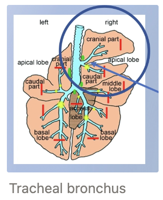
Which lung lobe has highest risk of aspiration pneumonia? Why?
Tracheal bronchus = Supplies the cranial RIGHT lung lobe
Dependent (ventral) in standing animal
Cranial portion of cranial lung lobe (in front of the heart)
Caudal portion of the cranial lung lobe (behind heart)
5 Features to assess on URT examination
Tracheal and laryngeal palpation (abnormal: cough on palpation)
Auscultate over trachea
Auscultate over nasal passages
Percuss sinuses (abnormal: dull/pain → sinusitis)
Examine nares for discharge, bilateral airflow, ozena, foreign bodies/material
5 Abnormal LRT sounds (+ Cause)
Crackles = Opening of collapsed alveoli
Wheezes = Narrowing bronchioles
Friction rubs = Fibrinous inflammation of pleural surface rubbing
Absent/muffled sound = Congestion, pulmonary consolidation, abscess, pleural effusion
Expiratory grunt
List 8 further diagnostics tests which can be performed for respiratory examination
Rebreathing examination = Allows for deeper inspiration and evaluation of collapsed airways
U/S = Visualise surface of lungs for pleural effusion, congestion, consolidation, abscesses if present near the surface
Radiography = Calves for distribution of lung pathology (NOT practical for adult cattle on farm)
Nasal swab = Diagnosis of acute viraemia (NOT helpful for bacteria)
Transtracheal wash = Culture/cytology
Bronchoalveolar lavage = Cytological evaluation of inflammatory airway disease
MDB = Presence of inflammation, poor gas exchange or electrolyte abnormalities
Serology = Confirmation of infectious systemic diseases (eg. BVD)
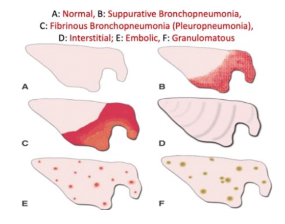
3 Types of pneumonia
Definition
Distribution
Subtypes
Bronchopneumonia = Bacterial with predisposing viral aetiologies
Distribution: Cranioventral
Types: Consolidation, abscessation, fibrinous adhesions
Interstitial pneumonia = Non-infectious, toxic, hypersensitivity, parasitic
Distribution: General/diffuse → Rib impressions
Types: Interstitial oedema, emphysema, fibrosis
Embolic/metastatic = Septic embolic, abscess, granulomatous
Distribution: Multifocal associated with vessels
3 Risk factors for respiratory disease (+ examples)
Season
Change in weather
Large swings in temperature (eg. warm day → cold night)
Allergens and fungal spores
Types of feed available (eg. dusty)
History
Trucking/transport (shipping fever)
Yarding = Dry and dusty environment
Oral drenching
Mixing cattle from different groups
Movement onto new pasture
Immunosuppression/stressful events
Age/Signalment
4 Reasons why pneumonia is LESS common in NZ
Extensive pastoral management
Temperate climate
Lack of housing and reduced intensity of management
Fewer feedlot
Massive issue overseas due to association with significant use of metaphylactic antibiotics
Aspiration Pneumonia
Aetiology
3 DDx in calves
4 DDx in adult cattle
7 Clinical signs
2 Treatments
Prognosis
Aetiology: Individual cow disease due to aspiration of saliva, fluids, rumen contents, oral medication
Calves:
Tube feeding
Faulty bottles/nipples
Oral Drenching
Adults:
Lateral recumbency (eg. milk fever and GA)
Bloat/choke
Oral drenching
PKE
Clinical Signs: Severity depends on contents aspirated (volume, type of material and bacteria involved)
Acute death (large volumes)
Severe pneumonia and toxaemia (caustic feeds with high bacterial load)
Dyspnoea and tachypnoea
Feed/material observed coming from nose
Fever within hours
Auscultation = Cranioventral crackles, pleuritic friction rubs and dullness
± Chronic suppurative bronchopneumonia with survival of acute stage
Treatment:
NSAIDs
Broad-spectrum antibiotics 5 - 7d
Prognosis: Poor
3 DDx for interstitial pneumonia
Fog fever (acute bovine pulmonary oedema and emphysema)
Farmer’s lung (hypersensitivity pneumonitis/diffuse fibrosing alveolitis)
Verminous pneumonia
Fog Fever
5 Sources of toxin
Seasonality
MoA
7 Clinical signs
2 Methods of diagnosis
Treatments
Sources: Change from poor quality → Lush and rapidly growing forage within 5 - 10d
Lush pasture
Lucerne
Kale
Turnips
4-ipomeanol in mouldy sweet potatoes causes the same syndrome
Seasonality: Autumn (foggy) and aftergrowth from recently cut silage
MoA: Pneumotoxic metabolite → Acute Bovine Pulmonary Oedema and Emphysema
L-tryptophan → Indole-acetic acid → 3-methylindole by rumen microbes (Lactobacillus)
Peak 4 - 5d and drops off after 6 - 7d
3-methylindole in blood → Lungs
Activation of pneumotoxic metabolite (3-methyleneindolenine) by type II pneumocytes (Clara cells) → Cellular damage and necrosis of type I pneumocytes (required for gas exchange)
Proliferation of type II pneumocytes
Decreased gas exchange surfaces and thickened septa with interstitial oedema
Clinical Signs: 4 - 5d after ingestion
Sudden tachypnoea and expiratory dyspnoea
Reluctance to move and weakness
Auscultation = Respiratory grunt and occasional soft crackles
Normothermic
Orthopnoea = Head extended, nostrils dilated, open-mouth breathing and elbows abducted
Subcutaneous emphysema may occur with rupture of bulla
Acute deaths or within 1-2 days
30 - 50% morbidity
30% mortality
Diagnosis: History of exposure to lush feed or diet change with sudden respiratory signs →
Measure L-tryptophan in feed
PM examination = Heavy wet lungs with foam in trachea and interlobular oedema/emphysema
Histology of lungs = Interstitial pneumonia
Treatment:
Remove from feed if <5d (care to avoid exertion → sudden death)
Frusemide to enhance water elimination
NSAIDs
Prevention:
Provide supplemental feed to prevent large intake of pasture
Graze pastures with cattle <15 months (less susceptible)
Monensin/ionophores 200mg/cow/d begun a few days prior to movement onto lush pasture → Reduced production of 3-methyl-indole in rumen

Farmer’s Lung (hypersensitivity pneumonitis/diffuse fibrosing alveolitis)
Pathogenesis
5 Clinical signs
Diagnosis
2 Treatments
Pathogenesis: Type III hypersensitivity reaction to dust and mould in grain/hay
Clinical Signs: Acute and chronic forms
Frequent coughing
Increased RR and effort
Auscultation = Diffuse crackles and squeaks of mucus
CHF due to pulmonary fibrosis (pulmonary hypertension)
BAR and normothermic
Diagnosis: History and clinical signs → Eosinophilia
Treatments:
Steroids
Cull
Bovine Tuberculosis
Aetiology
5 Modes of transmission
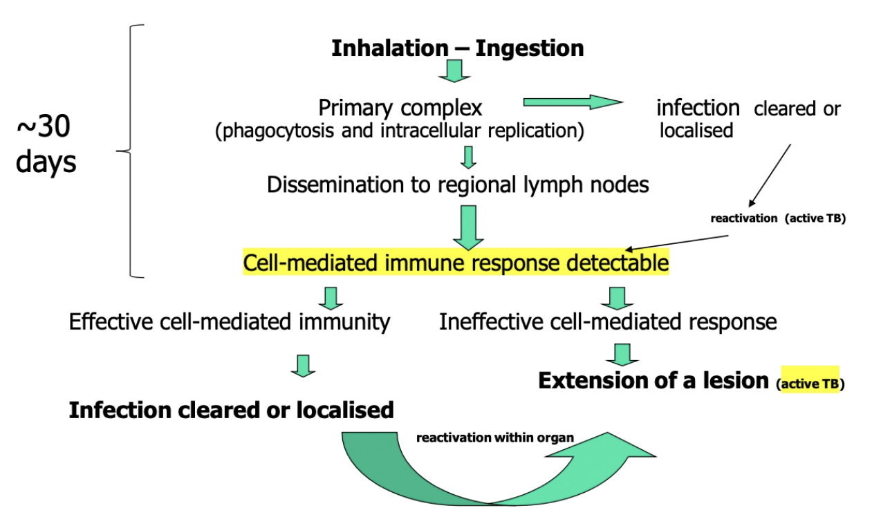
Pathogenesis
Aetiology: Mycobacterium bovis
Aerobic, non-spore forming bacilli with acid-fast cell wall → Enables growth at low pH
Facultative intracellular pathogen which survives in macrophages by inhibiting phagosome-lysosome fusion
Survives well in environment, faeces and pasture
Natural host = Cattle
Other hosts = Humans (5% of human TB), swine, carnivores, deer, possum, ferret ± sheep
Transmission: Inhalation (± oral)
Major NZ reservoirs: Infected cattle and possum
Cow ⇔ cow
Cow ⇔ possum = most important route in NZ (responsible for many TB breakdowns)
Cow → calf
Possum → possum
Cow → pig (through ingestion of cow milk/mild serum)
Predators/scavengers, spillover hosts and sporadic transmitters: Ferrets, pigs, wild deer, cats
Pathogenesis:
Ineffective CMI and weak humoral response due to intermittent, low-intensity antigenaemia
Methods of bovine TB diagnosis
Method
Advantages
Disadvantages
Direct microscopy of smears from lesions with Ziehl-Neelsen stain
+ve: Low cost
-ve:
Low DSn and DSp
Must have externally affected lymph node
Culture from lesion
+ve: Gold standard
-ve:
Slow growth (≤12w)
Some TB lesions may be sterile → Culture multiple lesions
Histology at PM = Caseous granulomatous lesions (poor DSp)
PCR
+ve: Fast
-ve: Lower DSn than culture
Tuberculin skin test = #1 AM diagnostic test for eradication and surveillance
Method: Intradermal injection of M. bovis PPD (purified protein derivative) that elicits delayed (type IV) hypersensitivity reaction in MOST infected animals
Caudal fold injection for cattle in NZ (cervical in some countries)
Cervical fold injection for deer
Positive Test: Lump at injection site 72hr post-injection
-ve:
DSn: Imperfect due to false negatives due to…
Recent TB infection (CMI not detectable for ~30 days post-exposure)
Individual-animal anergy (no immune response) in advanced TB usually due to old age
Occur in % of cows up to 6 weeks post-calving
DSp: False positives due to…
Cross-reactivity (non-specific sensitisation with other mycobacteria such as Johne’s infection or vaccination) OR other factors
Up to 40 - 50 days after previous intradermal tuberculin skin test?
Desensitisation = Animal’s skin reactivity to tuberculin is reduced for some time after the skin test
Comparative intradermal test = TWO intradermal injections with M. avium and M. bovis PPD at least 15cm apart on lateral neck
Read after 72 hours (skin fold thickness difference measured using tables)
M. bovis PPD > avian PPD = Reactor animal
Avian PPD ≥ M*. bovis* PPD = Non-reactor (due to false-positive)
+ve: Distinguish infected from false-positive due to previous M. bovis PPD test
-ve: Must wait between 42 - 60 days from first intradermal test to for desensitisation to wane (if another injection is administered too soon after the first without sufficient time for the animal’s immune system to recover, it can result in failure to react)
Interferon gamma assay = Rapid, blood-based assay of cell-mediated immunity for the diagnosis of bovine TB infection
Test rationale: When exposed to M. bovis, infected animals produce more IFN-ɣ from lymphocytes
Test principle: Measures amount of IFN-ɣ released by lymphocytes from whole blood sample after overnight incubation with added M. bovis PPD COMPARED with control
NOT a serological test (tests CMI)
+ve: Do NOT need to wait 60 days from last tuberculin skin test
-ve:
No international consensus about test performance (vs. intradermal test)
Increasing use in UK to release comparative intradermal cervical tests
High cost
4 Methods of bovine TB control (+ vaccination)
Test and slaughter
Animal movement restrictions
Action on wild reservoirs (depopulation)
THREE negative herd tests over ≥2 yrs → TB-free status
TB VACCINE
NOT used because:
Effectively delays clinical disease onset but does NOT reduce infection prevalence
Vaccine interferes with intradermal testing
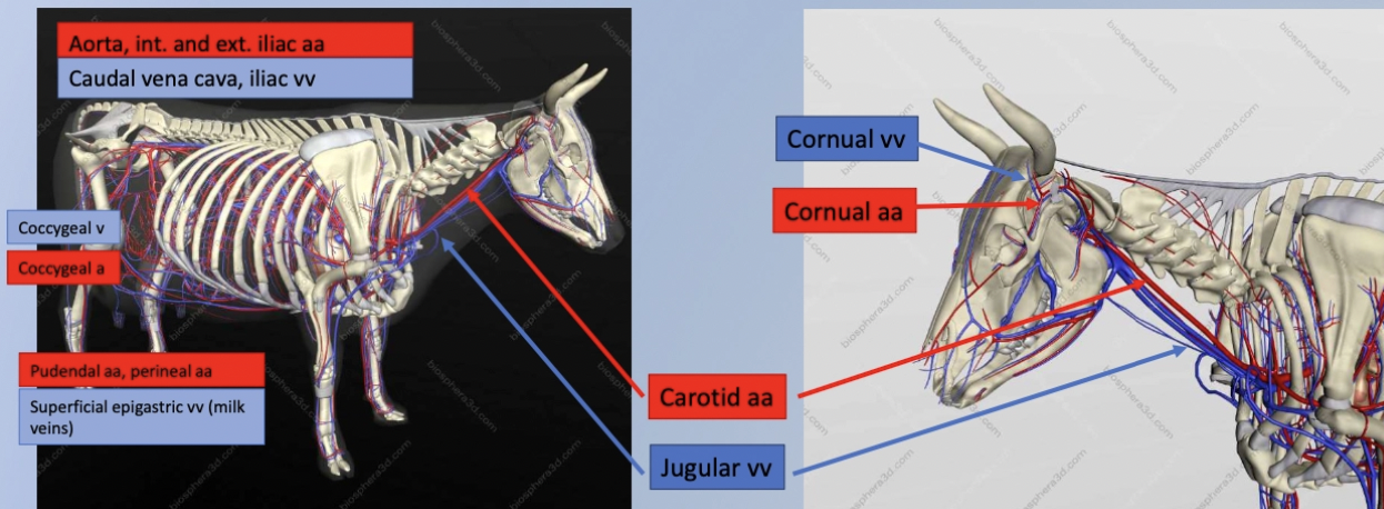
List 7 important blood vessels in a cow
Aorta and caudal vena cava
Internal and external iliac arteries + iliac veins
Coccygeal vein + artery = Tail blood sample
Pudendal artery and perineal artery
Superficial epigastric vein (aka. milk veins) = Supplying and draining udder
Cornual vein and artery
Carotid arteries and jugular vein = Blood sample or IV injections
List 7 clinical signs of cardiac dysfunction
Dull, weak, lethargic and painful
Low BCS, weight loss and decreased appetite/production
Brisket oedema due to vascular congestion → Increased hydrostatic pressure + force of gravity
Elevated jugular pulse >1/3 - 1/2 up neck
Physiological = Occlude top of jugular vein and wipe blood into heart → Cording SHOULD disappear
Jugular cording = Distension/congestion of vein
Jugular fill = Volaemic status
Increased RR
Endocarditis
Prevalence
Aetiology
3 Agents
Pathogenesis
History
5 Clinical signs
3 Methods of diagnosis
Prognosis
Treatment
Prevalence: Sporadic and under-reported in adult cattle
Aetiology: 2˚ bacterial infection of heart valves due to haematogenous spread of septic emboli from primary bacteraemia caused by
Rumenitis
Metritis
Pneumonia
Liver abscess
Agents:
Trueperella pyogenes
Streptococcus spp.
E. coli
Pathogenesis: Insufficiency/stenosis of heart valves
Right AV » Left AV > Semilunar valves
History: ± Previous history of bacterial infection
Clinical Signs:
Weight loss and depression
Intermittent pyrexia
Heart murmur ± palpable thrill
Intermittent positive Wither’s pinch test
Signs of RCHF (30 - 50%)
Diagnosis:
CBC and biochemistry
± Inflammatory leukogram (inconsistent)
± Elevated globulin and fibrinogen (inconsistent) + liver enzyme elevation in terminal RCHF
U/S = Vegetative lesions on AV valve (R > L)
PM findings
Prognosis: Poor to grave depending on timing of diagnosis due to pulmonary or coronary blockages (eg. pulmonary infarcts)
Treatment: Sustained treatment with antibiotics for 4 - 6 weeks
Common to relapse within 7 days after treatment stops
Sudden death or acute collapse during treatment common due to myocardial/pulmonary infarcts
Do NOT attempt with signs of CHF present
4 Aetiologies of myocardial disease (+ 3 - 4 example DDx)
Myocarditis
Bacterial = Staphylococcus aureus, Clostridium chauvoei, Histophilus somi
Viral = FMD
Parasitic = Toxoplasmosis, cysticercosis, sarcocystis
Cardiomyopathy
Se/vitE deficiency → White muscle disease
Chronic copper deficiency
Inherited DCM in Holstein-Friesians and Australian polled Herefores
Neoplasia = Lymphoma (EBL)
Toxicity
Yew (Taxus spp.) = Taxine alkaloids → Neurological signs and acute cardiac failure
Oleander (Nerium oleander) = Oleandrin = Cardiac glycoside
Rhododendron (Rhododendrons spp.) = Grayanotoxin → ONLY causes of vomiting in ruminants
Ionophores (lasolocid and monensin) → Free radical cytotoxicity of cardiac and skeletal muscle
3 Congenital cardiac defects (+ clinical signs)
Ventricular septal defect = #1 congenital cardiac defect in large animals
Clinical signs: Depends on size of defect = Clinically normal → Sudden death
Sudden death
Failure to thrive
Harsh heart murmur (bilateral)
Grade ≠ severity
Palpable thrill through chest wall
Patent ductus arteriosus (PDA) = Failure of closure of ductus arteriosis at birth
Often with other defects
Clinical signs:
Exercise intolerance
Lethargy
Continuous machinery murmur L > R
Atrial septal defect or patent foramen ovale
Signalment: Common in calves often with PDA
Clinical signs:
Asymptomatic
Left heart base murmur
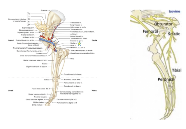
List the 4 important forelimb nerves and 5 important hindlimbs nerves (+ supply)
Brachial plexus = C6 - T2
Median
Radial
Ulnar
Common digital
Lumbosacral plexus = L4 - S2
Obturator
Femoral
Sciatic
Peroneal/tibial
Common digital (axial and abaxial)
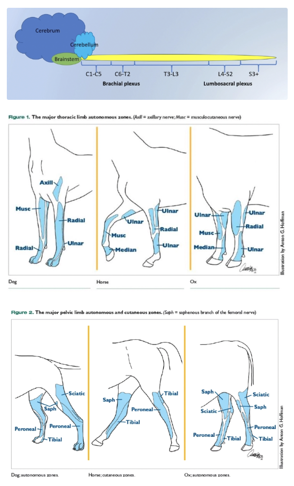
Most common neurological DDx for:
Neonates
Juveniles
Adults
Neonates = Hypocalcaemia, congenital defects, bacterial meningitis
Juveniles = Toxin exposure
Adults = Abscess, trauma, metabolic disorders
List 10 DDx for cerebral disorders
Polioencephalomalacia (PEM)
Lead toxicity
Salt/water toxicity (see pigs)
Calves = Improper mixing of calf milk replacer
Bacterial meningitis
Nervous ketosis and hepatic encephalopathy
Hypomagnesaemia
Sporadic bovine encephalomyelitis (SBE)
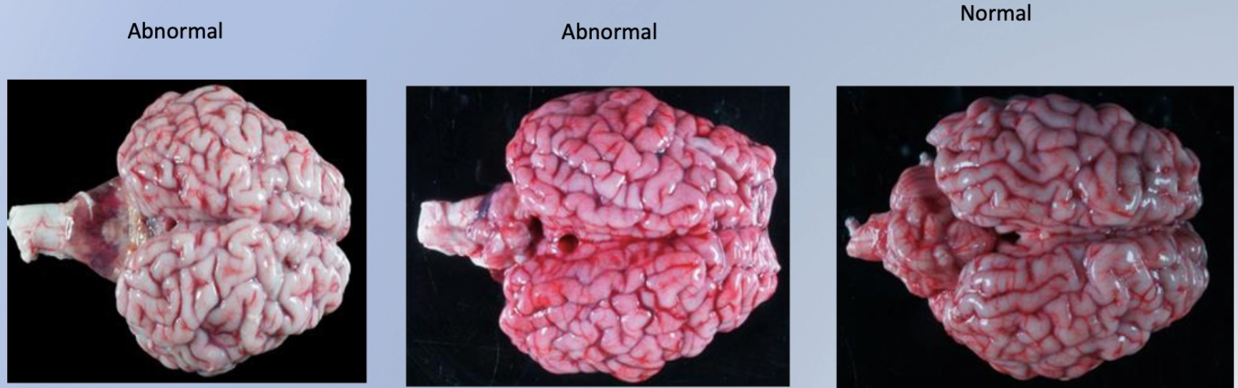
Malignant catarrhal fever (MCF)
Trauma/abscessation
Nervous coccidiosis
Polioencephalomalacia (cattle AND sheep)
Aetiology and pathogenesis
Signalment
4 Causes
Aetiology/Pathogenesis:
Thiamine deficiency
Thiamine is a rate-limiting cofactor for transketolase (involved in production of ATP required for the NA/K ATPase pump in neurons)
No ATP available to pump Na+ out of cell via Na/K ATPase pumps
Na+ accumulates in neurons → Osmosis
Acute cerebral oedema and laminar necrosis
Visual cortex at the caudal aspect of the cerebrum is squashed
Signalment: Recently weaned calves/lambs ± adults
Causes:
Altered rumen microbes (produce thiaminases eg. Bacillus spp.)
Recently weaned calves (1 - 2 animals affected)
Sudden change in quality/quantity of feed within 7 - 10d
Adult ruminant with anorexia (concurrent illness)
Bracken fern toxicity (contains thiaminases)
Thiamine analogues (eg. amprolium = coccidiostat)
Sulphur toxicity (eg. high S in soils and water)
8 Clinical signs of polioencephalomalacia
Central blindness
Palpebral and PLR present
Menace response absent
Nystagmus
Staggering and incoordination
Dehydration and poor rumen fill
Head-pressing and depressed mentation
Lateral recumbency and opisthotonus (star-gazing)
Dorsomedial strabismus when head is elevated (normal = ventrolateral strabismus)
Seizure
Death
Polioencephalomalacia
2 Methods of diagnosis
4 Treatments
2 Methods of prevention
Diagnosis: History and clinical signs (rule out other DDx eg. exposure to lead)
Response to treatment #1
PM = Laminar necrosis of the grey matter
Treatment:
Thiamine (vitB1) IV = Aggressive/early high dose @ 10mg/kg → 5mg/kg IM q6 - 12hr for 3 - 5d
NSAIDs
Rumen transfaunation (0.5L for calf and 5L for ow)
Fluid therapy if dehydrated
Prevention:
Reduce sudden dietary changes
Avoid brassicas on high sulphur soils
Lead toxicity
Sources
MoA/pathogenesis (+ 3 outcomes)
Sources: Paint, lead shots, sinkers, batteries and soil
Small amount absorbed through GI → Excreted in bile, faeces, urine, milk, sweat and saliva
MoA:
Curious calves experience the world orally
eg. lead batteries or paint
Lead rapidly absorbed through GI
Outcomes:
Disruption of heme synthesis → Basophilic stippling and nRBC (dogs)
Capillary damage, neuronal necrosis, demyelination and altered neurotransmission → Neurological signs
GIT signs of irritation
3 Clinical signs of lead toxicity in cattle (vs. dogs)
Cattle (3 - 4d duration)
Sudden death in calves
CNS (as for PEM):
Staggering, bellowing, chomping
Fine fasciculations around muzzle and nose
Blindness, opisthotonus, head-pressing and seizures
GI: Hypersalivation, ruminal atony, bruxism, abdominal pain, diarrhoea, anorexia, dehydration
Dogs
CNS signs
GI: vomiting, diarrhoea/constipation, anorexia
5 Methods of lead toxicity diagnosis
History of lead exposure and clinical signs →
LONGER response to thiamine supplementation (vs. PEM)
Blood smear = nRBCs and basophilic stippling with no regenerative signs (DOGS)
Measure lead in whole blood (heparinised tube), liver, kidney or bone
Pb > 0.35 - 0.5mg/L
Radiography to detect lead foreign body
PM findings are non-specific → Collect brain for histology (cerebral lesions indistinguishable from PEM)
MAY find paint flakes in rumen
Treatments for lead toxicity
Remove lead from GI where possible
Antidote (chelation) = Ca-EDTA 110mg/kg slow IV EOD x3 or D-penicillamine
Treat cerebral oedema and seizures with hypertonic IVFT and diazepam
Thiamine supplementation
± Gastroprotectants
Prognosis of lead toxicity
Depends on age and amount ingested
Clinical signs improve in 2 - 3d of treatment
Blindness may persist up to 3w
Slower response than PEM as lead → necrosis (vs. PEM → Cerebral oedema)
Salt/water toxicity in cattle (vs. pigs)
3 Causes
2 Methods of diagnosis
3 Treatments
3 Methods of prevention
Causes:
Improperly mixed milk replacer
Incorrect electrolyte use (eg. adult formulation given to calves)
Bottle feeding water at weaning
Diagnosis: History and clinical signs (iatrogenic) →
Serum [Na] > 170mmol/L
PM findings as for PEM ± gross congestion of omasal and abomasal mucosa
Treatments:
Prevent free water consumption (minimise water available)
Isotonic/hypertonic electrolytes PO to gradually transition back to water
Isotonic 0.9% NaCl
Mannitol 20% 1 - 2mg/kg IV for cerebral oedema
Thiamine supplementation
Prevention:
Educate client on milk replacer formulation
Frequent monitoring of water sources
Bacterial meningitis
Aetiology (+ 4 agents)
2 Predisposing factors
4 Clinical signs
2 Methods of diagnosis
5 Treatments
Prognosis
Aetiology: Common sequela of ascending naval infection and bacteraemia
Agents:
E. coli
Klebsiella
Salmonella
H. somni (adults)
± SBE (Chlamydia pecorum)
Predisposing Factors:
FPTi
Poor hygiene → Navel infection/neonatal diarrhoea
Clinical Signs:
Depression, collapse, coma, death
Pyrexia
Seizures
± Concurrent systemic infection
Navel/joint ill and scours
Endotoxaemia/SIRS → Injected scleral vessels and petechiae
Diagnosis:
Evidence of systemic infection
CSF tap → Bacteria and inflammatory cells
Treatments:
Aggressive penicillin, ampicillin or TMPS → Penetrate into CSF and effective against most pathogens
Most AB penetrate due to inflammation of the BBB
G- → 3rd/4th gen cephalosporins
NSAIDs
IVFT
Parenteral nutrition
Anti-convulsive therapy
Prognosis: Fair (suckling) otherwise guarded to poor
Sporadic Bovine Encephalomyelitis
Aetiology
Signalment
5 Clinical signs
Treatment
Aetiology: Chlamydophila pecorum
Usually abortion, pinkeye and venereal diseases in sheep
Signalment: Calves <6m (multiple calves)
Clinical Signs:
Depression
Weakness and hindlimb ataxia
Urinary incontinence and poor tail tone
Opisthotonus
Blindness
Treatment: Highly effective oxytetracycline q48hr x2
List 2 DDx for cerebellar disorders
Cerebellar hypoplasia (congenital BVD)
Ryegrass staggers (see “Alternative Forages”)
→ Incoordination, hypermetria and intention tremours
Assess coordination, gait and tremours
Cerebellar Hypoplasia
Aetiology
Signalment
6 Clinical signs
Aetiology: In utero infection with BVDV 60 - 180d of gestation
Unknown herd BVD status
Signalment: Single neonate <7d
Clinical Signs: Present at birth
Recumbent
Strabismus and horizontal nystagmus
Reduced PLR
Tremours and convulsions
Ataxia
Torticollis and opisthotonus
List 4 DDx for brainstem disorders
Listeriosis
Otitis media/interna (Mycoplasma bovis)
Trauma
Facial paralysis syndrome (FPS)
→ Head tilt, circling/leaning, vision/hearing/balance deficits, facial asymmetry/paresis/paralysis, ataxia/vestibular signs (nystagmus)
Listeriosis (cattle AND sheep)
Agent
Pathogenesis
Agent: Listeria monocytogenes = G+ facultative anaerobe
ZOONOTIC!
Pathogenesis:
Soil contamination or poor quality silage (pH > 5)
Proliferation of Listeria monocytogenes
Tooth eruption or oral wounds allows ascending infection along the trigeminal nerve (CN V) to the brainstem
UNILATERAL microabscessation of mid-brain = CN V, VII, VIII and IX
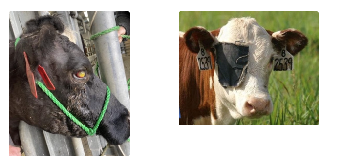
Listeriosis
Signalment
6 Clinical signs of the encephalitic form
4 Other manifestations
5 DDx
Signalment: Single animal ± outbreak
Clinical Signs:
Changes in mentation
± Pyrexia
Unilateral CNS deficits = Head tilt, dropping ear, eyelid, unable to close eye, facial asymmetry, dysphagia
Assess angle of eyelashes
Circling
Recumbency
Death
Other Manifestations:
Abortion
Silage eye (anterior uveitis = vascular engorgement, blue corneal opacity, no ulceration and less severe conjunctivitis than pink eye)
Septicaemia (neonates)
Enteric form (rare)
DDx:
Brain abscess/meningitis
Neoplasia
Trauma to facial nerve
Otitis media/interna
Listeriosis
2 Methods of diagnosis
Prognosis
4 Treatments
Diagnosis: History and clinical signs →
CSF tap = Monocytosis
PM = Multifocal abscessation of the brainstem around the cranial nerve nuclei
CBC and biochemistry unrewarding
Prognosis: Good (standing) but poor if recumbent
Treatment:
Procaine penicillin high dose BID x4
Oxytetracycline IV once → IM/SC EOD x4
NSAIDs
Fluids and good quality feed
Otitis media/interna
Aetiology
5 Clinical syndromes
3 neurological signs (+ signalment)
Diagnosis
3 Treatment options
Aetiology: Mycoplasma bovis (no cell wall) → Otitis media/interna
Syndromes:
Mastitis
Arthritis
Polyserositis (neonatal calves)
Pneumonia (older calves)
± Late-term abortion or premature birth
Clinical Signs: Calves < 6m
Unilateral head tilt and ear droop
± Ocular/nasal discharge = Keratoconjunctivitis
Depression and decreased appetite
Diagnosis: Call MPI (under surveillance) →
Fresh and fixed samples
Culture ± serology
Treatment: Labelled against Mycoplasma
Tulathromycin
Gamithromycin
Enrofloxacin
Facial Paralysis Syndrome (FPS)
Aetiology
Signalment
5 Clinical signs
Treatment
Prognosis
Aetiology: Unique to Franklin in NZ
Linked to usage of slag by-product for fill in sheds/tracks → High levels of vanadium = toxicity
Signalment: Calves 1 - 3m
Clinical Signs:
Unilateral/bilateral facial paralysis
Mild transient fever and depression
Respiratory discharge and mucopurulent occulonasal discharge
Infection of periodontal tissue and tooth loss + jaw deformity
Treatment: ONLY for 2˚ infection
Prognosis: Spontaneous recovery in 70% (takes ≤8w)
Remaining 30% have continued signs:
Continued ptyalism
Reduced GR
Persistent facial nerve dysfunction
2˚ bacterial infections
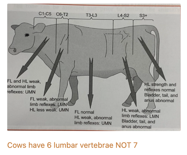
List 6 DDx for spinal cord disorders
Vertebral body osteomyelitis after navel ill
Associated with mineral deficiency (vitD, Ca, Cu) affecting vertebral bodies
Ankylosing spondylitis
Spinal tumours
Verminous meningoencephalomyelitis
Iatrogenic
Epidural anaesthesia
Segmental spinal anaesthesia (eg. paravertebral anaesthesia)
Regional nerve blocks
Tetanus
Aetiology
4 Predisposing factors
7 Clinical signs
5 Treatments
Aetiology: Clostridium tetani = Saprophytic G+ anaerobe
IP = 10 - 14d (longer if further away from spinal cord)
External wound contaminated with spores → Germination → tetanospasmin and tetanolysin
Predisposing Factors:
No vaccination
Castration, external wounds/punctures, disbudding/dehorning
After yarding/transport (bruising)
± Dystocia or rumenitis
Clinical Signs:
Muscle stiffness = Opisthotonus and extensor spasticity
Mild bloat
Anxious facial expression (sardonic grin) with erect ears, lock jaw and prolapse of 3rd eyelid
Sawhorse stance
Hyperaesthesia
Tail held in pump handle position
→ Collapse, respiratory failure, convulsions and death
Treatment:
Tetanus antitoxin (TAT) 10,000 - 20,000 units half IV and half IM
Often equine origin
Binds free toxin ONLY (does not affect bound toxin)
Antibiotics against G+ anaerobes = Procaine penicillin 30mg/kg IM BID 5 - 10 days (off-label)
Anticonvulsant/spasmolytic
Emergency rumenotomy to correct bloat
Fluids and feeding
List 3 neurological DDx which are exotic to NZ
Bovine spongiform encephalopathy
Rabies
± Botulism (not reported in NZ)
Bovine Spongiform Encephalopathy (BSE)
Aetiology
Signalment
5 Clinical signs
Aetiology: Prion disease acquired through ingestion of ruminant-derived protein
Signalment: 2 - 8yr (highest incidence 4 - 6yr)
IP = Long
Clinical Signs:
Change in behaviour (aggression/depression)
Ataxia
Tremours/twitching/hyperexcitability
Recumbency
Wasting/low production
List 3 other DDx for PEM (+ how to distinguish)
Lead toxicity
± PLR (may be absent)
PEM = PLR and palpebral present
GI irritation
Slow response to thiamine supplementation
Salt/water toxicity = PEM (assess history)
Meningitis
Fever
Systemic infection (eg. hypopyon)
EXAMPLE QUESTIONS: NO ANSWERS PROVIDED
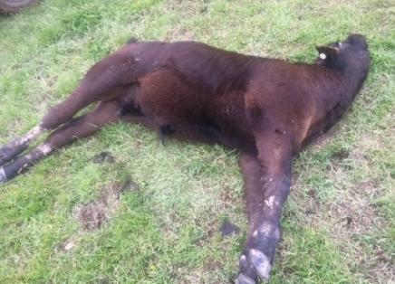
You see a calf showing signs of blindness, lateral recumbency and opisthotonus
Which part of the nervous system is likely involved?
List 3 differential diagnoses for the signs listed above. Chose one of these DDx and explain the pathophysiology
Discuss how you would differentiate a calf with PEM versus Lead Toxicity on clinical examination or through diagnostic testing
What are clinical signs associated with cerebellar disease?
What viral infection may cause in utero brain pathology?
A farmer has reported he has had to euthanise 3 R2 Friesian heifers due to broken legs in the past week. What mineral deficiency is associated with spontaneous humeral fractures in cattle?
What other body system should always be considered with neurological DDx
Musculoskeletal (difficult to distinguish)
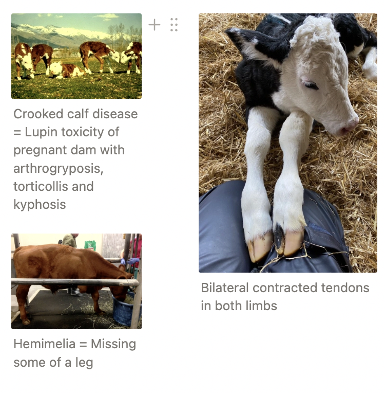
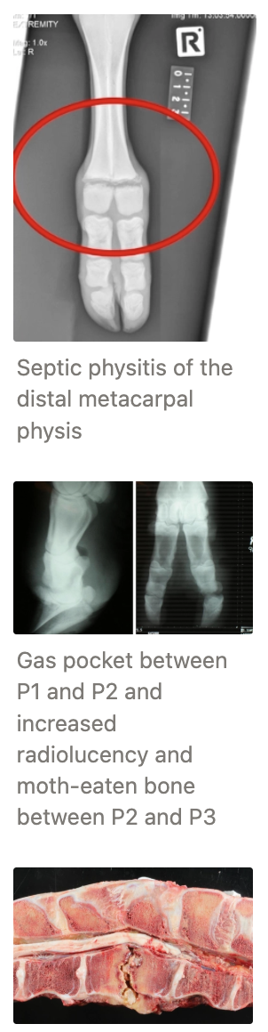
List 7 congenital MSK diseases
Contracted flexor tendons
Treatment: Splint and physiotherapy ± surgical tenotomy
Angular limb deformities
Multiple calves = Toxicity or deficiency in utero (eg. Crooked Calf Disease = Lupin toxicity of pregnant dam → Arthrogryposis, torticollis and kyphosis)
Arthrogryposis = Joints seized and curled)
Polydactyl/syndactyl
Amelia/hemimelia
Myostatin mutation = Belgian Blue double muscle
Myotonia congenita = Fainting goats
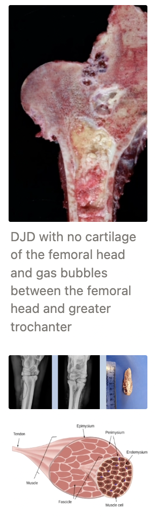
List 4 infectious MSK diseases (+ causes)
Septic arthritis (DDx = Fractures)
Neonates = Carpus/hock/stifle/vertebrae (neonates) 2˚ to navel ill
Treatment: High dose antibiotics, NSAIDs ± joint lavage → Euthanasia
Adults = Deep digital sepsis 2˚ to WLD/ulcer
Treatment: Claw amputation or euthanasia
Osteomyelitis/physitis
Causes:
Haematogenous
Fracture complication
Actinomycosis (Lumpy Jaw)
Clostridial myositis (aka. Blackleg or Malignant Oedema)
Case: 2˚ to vaccination or bruising
Suppurative discospondylitis
List 3 inflammatory MSK diseases (+ aetiology)
Degenerative joint disease
2˚ to congenital dysplasia (OCD, deformation, trauma)
Osseous sequestration
Bone fragment loses blood supply after trauma/infection → Necrotic fragment acts as a foreign body → Abscessation and periosteal reaction
Ischaemic myopathy (aka. compartment syndrome)
Complication of prolonged recumbency (Downer Cow Syndrome) → Increased CK and AST
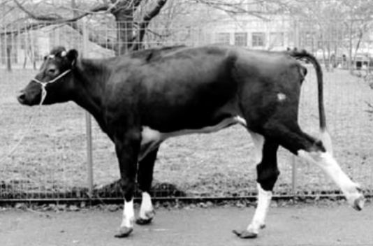
What is this?
Aetiology
Signalment
Clinical sign
Treatment
Bovine Spastic Paresis = Unilateral/bilateral increased muscle tone/permanent spasm of the gastrocnemius ± quadriceps
Aetiology: Heritable and progressive neuromuscular disease of the gastrocnemius, quadriceps and nerves
Signalment: ALL breeds
Treatment: Surgical neurectomy or tenectomy
Fractures
4 Traumatic Causes
3 Predisposing factors
Traumatic Causes:
Running into fences and fighting → Cervical spine
Misadventure and calves stepped on by cow → Limbs
Tail break
Knocked down hip → Fracture to tuber coxae
Predisposing Factors:
Pathological fractures 2˚ to osteomyelitis
Copper deficiency (± Mo excess) → Enzootic swayback in deer and humeral fractures in heifers
VitD deficiency (esp. fodder beet) → Rickets/osteodystrophy
Treatment of fractures
Closed fracture distal to carpus/tarsus
Closed fracture of tibia/radius/ulna
Closed fracture proximal to stifle/elbow
Open fracture
Closed fracture distal to carpus/tarsus = Immobilisation and restricted movement (good response)
External fixation (eg. cast or splint above and below affected joint)
Phalangeal fractures → Use block to elevate healthy claw
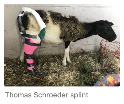
Closed fracture of tibia/radius/ulna = Thomas Schroeder splint
More difficult to immobilise with higher risk of complications
Closed fracture proximal to stifle/elbow = Surgical fixation, amputation or euthanasia
-ve: Ruminant cortical bone is thinner than other species with higher risk of sepsis, osteomyelitis and neurovascular damage
Open fracture = ≥10 - 14d antibiotics
Fracture heals in 4 - 6w
Neonates → Change bandage q2 - 3w to prevent pressure necrosis from growth
Soft Tissue Trauma
Aetiology
Causes
4 Example diseases
Prognosis
Aetiology: Any ligament, tendon or muscle strain/rupture/trauma → Dysfunction
Causes: Fighting, restraint in foot crush, falling/struggling
Examples:
Cruciate disease
Patellar luxation/upward fixation
Gastrocnemius rupture → Cannot extend hock (collapsed)
Peroneus tertius rupture
Prognosis: Difficult to repair due to massive size and lack of hardware
5 Types of superficial trauma (+ definitions)
Seroma = Fluid-filled swelling, non-painful, normal temperature and sterile (diffuse hypoechoic on U/S)
Haematoma = Blood-filled swelling, may be painful and sterile (flocculant on U/S)
Abscess = Consequence of poor aseptic technique when performing FNA of swelling
Bursitis = Inflammation of the synovial structures around bony points ± pain
Predator attacks = Calves, small ruminants and pet pigs
Crushing injuries often get worse before they get better
List 3 nutritional deficiencies causing MSK disease
VitE/Se deficiency = White muscle disease
Copper deficiency
VitD deficiency (fodder beet low in P) = Rickets
List 2 parasites causing MSK disease
Sarcocystis spp. = Protozoal parasite that encysts in smooth muscle and striated muscle
Usually asymptomatic but occasional clinical cases of myositis and myocarditis
Taenia (Cysticercus) ovis = Sheep measles in sheep and goats
Tissues cysts at slaughter → Incidental finding but carcass condemnation
6 History questions to cow with diarrhoea
Onset: Acute, subacute, or chronic?
How many animals affected? One age group or multiple?
Any clustering, different herds?
Additional signs besides diarrhoea?
Recent management or feed changes?
Any sick workers?
ALWAYS consider Salmonella first until proven otherwise (#1 cause of adult diarrhoea)
List 8 DDx for adult cow diarrhoea
Salmonella
Johne’s Disease
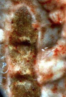
Mucosal Disease (MD)
Malignant Catarrh Fever (MCF)
Parasites = Liver Fluke/Rumen Fluke
Winter Dysentery
Dietary/Toxic/Minor causes
Theileria
Agents of salmonellosis
Common and important
Typhimurium (enteric)
Bovis-morbificans (enteric)
Transmitted from sheep
Hindmarsh (enteric)
Brandenburg (abortions in South Island)
Rare and unusual
Montevideo
Newport
Exotic to NZ (present in AUS and AUK): Dublin
Emerging strain: Give (since 2019)
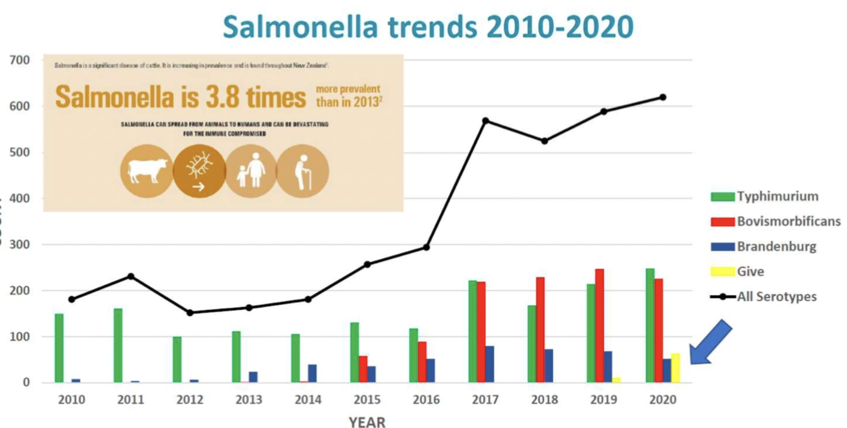
Increased variability of Salmonella strains over time BUT dropped back dramatically in last 3 years
Increased prevalence of salmonellosis overtime
3 Presentations of salmonellosis and clinical signs
Peracute = Septicaemia and death
Acute = Enteritis/dysentery, bacteraemia and pyrexia
S. Brandenburg and S. Dublin (exotic) → Abortion
Chronic = Diarrhoea ± dysentery, weight loss and inappetence → Carrier state
S. Bovismorbificans
Calves: 58.2% morbidity and 27.7% mortality
Adults: 8.5% morbidity and 1.6% mortality
Salmonellosis
Transmission
5 Sources of infection
3 Risk factors
Transmission: Faecal-oral
Sources: Persists in environment
Carrier/chronically infected animals (≤15% cows)
Contaminated PKE, water and pasture
Fomites
Vectors (eg. wild birds, rodents and vets)
Aborted material (sheep)
Risks:
Shedding precipitated by stress (eg. transport and mixing)
Diet change → Change in gut pH and microbiome
Pelletised magnesium oxide → Increased GI pH → Reduced dose of ingested Salmonella required to cause disease
Salmonellosis
3 Treatments
4 Methods of control
Treatments:
Antibiotics = Oxytetracycline and potentiated sulphonamides (consider carrier status, resistance and drug selection)
Whole herd antibiotic treatment may be required (eg. milk powder)
Avoid fluoroquinolones and ceftiofur
Fluids (oral equipment must stay on farm to prevent spread)
NSAIDs for sick animals
Control:
Reduce stocking rate → Reduce spread of infection
Isolate infected or aborting cows
Prevent contamination of feed/water supplies
Vaccination of calves with Salvexin B
Calf infection is inevitable unless switch to milk powder → Begin prophylactic antibiotic oxytet powder in milk and begin vaccination from 2 weeks of age
Timing:
1st year = Sensitiser ≥ 6 weeks before risk period → Booster 4 weeks after (≥2 weeks before risk period)
Annual = Booster ≥2 weeks before risk period
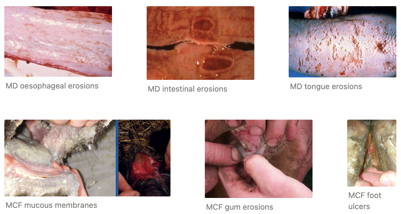
Mucosal disease (MD) vs. Malignant catarrhal fever (MCF)
Feature | MD | MCF |
|---|---|---|
Age | ||
Other cases | ||
Pyrexia | ||
Sheep contact | ||
Nasal/eye signs | ||
Lymph nodes | ||
Skin lesions | ||
CNS signs |
Feature | MD | MCF |
|---|---|---|
Age | 15–18 months | Any |
Other cases | Likely | Sporadic |
Pyrexia | No | Yes |
Sheep contact | No | Yes |
Nasal/eye signs | Rare | Common |
Lymph nodes | Normal | Enlarged |
Skin lesions | No | Yes |
CNS signs | No | Possible |
Winter Dysentery
Definition
Agent
Signalment
Treatment
Prevention
Definition: Acute, apparently contagious, foul-smelling enteritis occurring during colder months of the year
High morbidity and low mortality
Agent: Likely coronavirus
Signalment: Housed cattle
Treatment: Supportive (fluids ONLY)
Prevention: NONE
Yersiniosis
Agent
Risk factors
Transmission
Clinical signs
Treatment
PM findings
Histological findings
Agent: Yersinia pseudotuberculosis
Common isolate of intestinal tract healthy ruminants
Risk Factors:
Wet and cold weather → Survival of Yersinia on pasture
Poor feeding
Parasites
Mineral deficiencies
Transmission: Faecal oral
Clinical Signs: High morbidity and low mortality (chronic > acute)
Diarrhoea ± blood/mucus
Poor growth, stunting or wasting
NOT pyrexia
Treatment: Oxytetracycline
PM: Fluid intestinal contents and external evidence of diarrhoea
Characteristic multifocal, ulcerative enterocolitis
Microabscessation of mucosa with lots of G- coccobacilli
4 Types of wasting (empirical approach)
Ingestion = Cannot/will not eat
Digestion/absorption
Assimilation = Efficient use of absorbed nutrients
Excretion = Excessive protein loss from the body
Ingestion → Wasting
2 Methods of diagnosis
4 DDx for “cannot eat”
9 DDx for “will not eat”
Diagnosis:
Observe for quidding = Dropping food from mouth while chewing cud
Push grass into mouth and see if cow will chew and swallow
Cannot Eat:
Teeth = Heifers changing teeth OR old cows losing teeth
Tongue = Woody tongue/damage/ulceration
Swallowing/chewing = CN V/VII, trauma to jaw/lumpy jaw
Oesophagus = Stricture/blockage/foreign body
Will Not Eat:
Unpalatable diet
Uraemia
Ketosis (LDA, RDA, fat cow syndrome)
Toxic (mastitis, metritis, peritonitis)
Pyrexia
Pain (lameness, abomasal ulcers)
CHF
Chronic infection (tuberculosis, MCF)
Hydrallantois (DDx for abdominal distension and wasting)
9 DDx for poor digestion/absorption → wasting
Chronic traumatic reticuloperitonitis
LDA (or RDA)
Vagal indigestion
Actinobacillosis of reticulum/rumen
Chronic Salmonellosis
Johne’s disease
Intestinal leukosis/tumours
Villous atrophy post-enteritis (calves)
Post-acidosis rumen damage
List 4 causes of poor assimilation of nutrients → wasting (+ example DDx)
Nutritional imbalance
Chronic hypomagnesaemia (Taranaki anaemia)
Cobalt deficiency (Bush sickness)
Trace element deficiency
Cu deficiency
Mo excess
Liver damage
Cirrhosis post-FE or ragwort
Abscesses post-navel ill/acidosis
Chronic poisoning
Lead
Ragwort
Causes of protein loss → wasting
3 Kidney DDx
2 Bladder DDx
6 Intestine DDx
Kidneys
Amyloidosis
Pyelonephritis
Haematuria/haemoglobinuria
Bladder
Tumour
Cystitis
Intestine
Johne’s disease
Chronic salmonellosis
Worms and liver fluke
Melaena
Abomasal ulcers
Mycotoxins
List 7 important skin diseases of cattle
Photosensitisation (facial eczema and spring eczema)
Parasites (see “Parasitology”)
Lice
Mites
Ticks
Ringworm
Warts
Dermatophilus
Pseudolumpy skin disease
MCF
3 Types of photosensitisation (+ examples)
Primary = Direct ingestion of photodynamic compounds
eg. Spring eczema = St John’s Wort, Ngaio, Alligator weed, Musky Storksbill

Secondary = Facial eczema, ragwort toxicity, leptospirosis
± Rape scald, turnip toxicity, HR swedes (glucosinolates)
Congenital eg. Bovine congenital porphyria
5 Reasons why facial eczema is so important
Major animal welfare concern (esp. lifestyle blocks or naive farms → clinical cases)
Sporadic disease = Worse in some years and highly seasonal → Vets and farmers can become complacent and be caught out in a bad FE season
~$500M/yr lost in production
Underestimated and ignored (sparse research in last 20yr)
Climate change will significantly extend risk of FE throughout NZ
Occasional cases of Zn toxicity and potential for residues in soil/animal products
Aetiology of Facial Eczema
Agent
Region
Seasonality
4 Risk factors
Agent: Sporidesmin mycotoxin produced by spores of Pseudopithomyces chartarum
Saprophytic fungi which grows on dead litter at the base of pasture
Region: North Island ± top of South Island
Seasonality: Jan - May (esp. Feb - April)
Risk Factors:
Weather = Fungus requires warm, moist, humid conditions for growth
100% humidity
4 consecutive warm nights >12˚C
Small amount of rain (~4mm) 48hr apart → Triggers sporulation
Forage Type = Densely-growing grass with lots of dead litter at the base
eg. Ryegrass, cocksfoot, browntop
Clover, chicory, plantain and brassicas safe
Grazing Conditions = Hard grazing → Spores more concentrated at base of sward
Warm sheltered paddocks or areas within paddocks (eg. sheltered gullies) = Higher risk vs. colder (south-facing) or very windy paddocks
Animal Factors = Genetic susceptibility
Describe the pathogenesis of facial eczema
Fungus makes toxin in mycelium
Toxin concentrates in spores at sporulation
Animal ingests sporidesmin spores while grazing
Toxin released from spores into rumen (water-soluble) and absorbed into bloodstream
Toxin removed by liver and kidneys → Concentrates in bile duct (10x) and urine
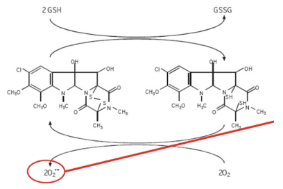
→ Release of free radicals which damage lining of bile ducts and bladder
ONE molecule of sporidesmin can create many free superoxide free radicals
Glutathione reduces disulphide bridge of sporidesmin toxin
O2 reoxidises bridge → Formation of superoxide free radical
Cyclical
→ Cholestasis and cystitis
Occluded bile ducts cannot eliminate waste products from chlorophyll metabolism (phylloerythrin)
Phylloerythrins build up in bloodstream and react with sunlight in non-pigmented skin and mucocutaneous junctions
→ Photosensitisation
Diagnosis of facial eczema (vs. spring eczema)
Biochemistry = Marked increase in GLDH, GGT and hyperbilirubinaemia
vs. Spring eczema = Rare liver damage
Clinical signs of facial eczema
Onset
5 Early clinical signs
3 Late clinical signs
2 Chronic clinical signs
Onset: 2 weeks spore exposure
Early:
SC oedema and erythema
Sheep = Face and ears with no wool cover → Floppy ears
Cattle = White patches, vulva and teats (most noticeable in afternoon milking)
Acute liver damage = Depression, inappetence and drop in milk production (~0.14kgMS/d)
Hunched due to liver damage and visceral pain
± Transient diarrhoea
Shade-seeking (photophobia) and irritation
Kicking at udder (DDx: Colic)
± Haemolysis with haemoglobinuria and anaemia
Late:
Decreased eating and BCS/weight (not grazing in sun)
Solar dermatitis
Eyelids glued shut due to damage
Damaged skin peels
±2˚ bacterial infection
Severe = Anorexia and death
Chronic: Cirrhosis → Cannot reverse clinical signs as liver already damaged
± Reduce production during physiological stress periods
± Neoplastic keratoma transformation of skin lesions
How is photosensitivity the “tip of the iceberg” in sheep?
5% of sheep with visible clinical signs → ≥50% of sheep have liver damage
Many beef cattle (eg. black Angus) do not show clinical signs due to dark pigment BUT liver damage still occurs