M3.1 - Assessment of the Ankle/Foot
1/30
There's no tags or description
Looks like no tags are added yet.
Name | Mastery | Learn | Test | Matching | Spaced | Call with Kai |
|---|
No analytics yet
Send a link to your students to track their progress
31 Terms
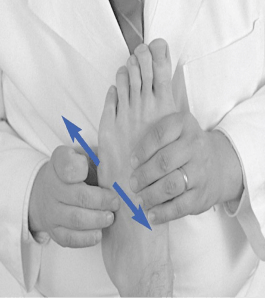
Long axis distraction of tibiotalar joint
ID assessment
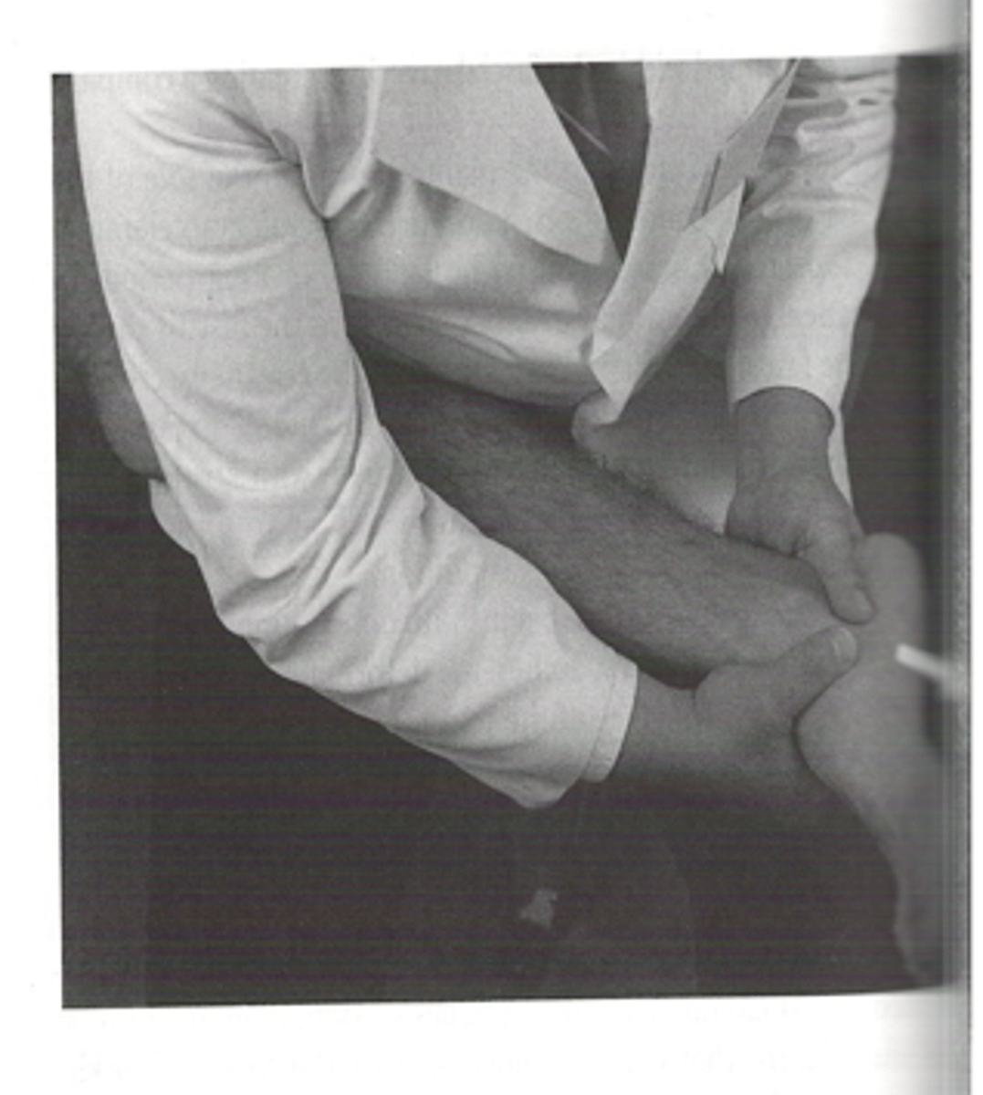
- Supine
- Knee flexed to ~90˚ with hip flexed/abducted
Long axis distraction of tibiotalar joint
- Patient position

Sits on table between legs facing footward
Long axis distraction of tibiotalar joint
- Doctor position

Web contact
Long axis distraction of tibiotalar joint
- Doctor contact

- Dome of the talus
- Superior aspect of the calcaneus
Long axis distraction of tibiotalar joint
- Patient contact

Apply distraction force through both hands
Long axis distraction of tibiotalar joint
- LOD

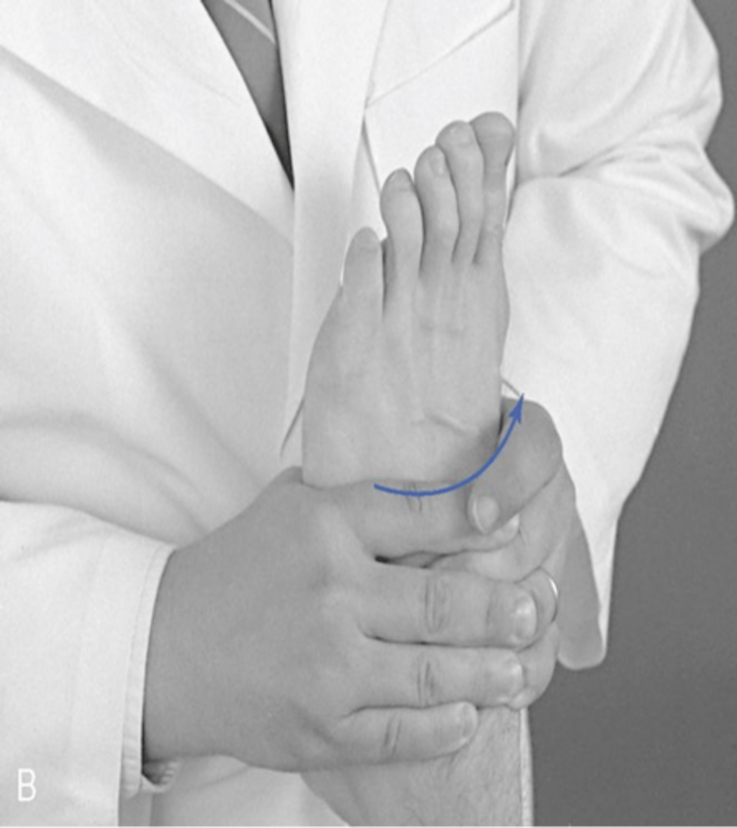
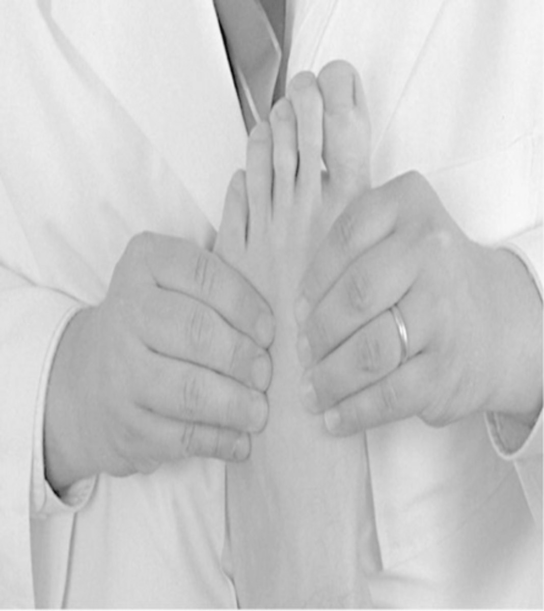
A-P/P-A glide of tibiotalar joint
ID assessment

- Supine
- Knee/hip flexed
- Calcaneus on table
A-P/P-A glide of tibiotalar joint
- Patient position

Web contact (cephalad/caudal hands)
A-P/P-A glide of tibiotalar joint
- Doctor contact

- Cephalad hand: anterior distal tibia
- Caudal hand: dome of talus
A-P/P-A glide of tibiotalar joint
- Patient contact

Apply force in opposite directions using both hands
A-P/P-A glide of tibiotalar joint
- LOD
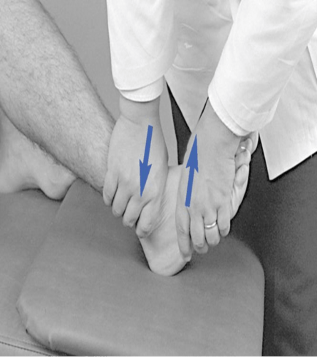
Medial to lateral glide (inversion) of tibiotalar joint
ID assessment

Supine
Medial to lateral glide (inversion) of tibiotalar joint
- Patient position
At foot of table
Medial to lateral glide (inversion) of tibiotalar joint
- Doctor position

- Contact dome of talus with fingers of both hands
- Both thumbs contact plantar surface of foot
Medial to lateral glide (inversion) of tibiotalar joint
- Doctor/patient contact

Stress talus in M-L direction
Medial to lateral glide (inversion) of tibiotalar joint
- LOD

Lateral to medial glide (eversion) of tibiotalar joint
ID assessment

Supine
Lateral to medial glide (eversion) of tibiotalar joint
- Patient position

At foot of table
Lateral to medial glide (eversion) of tibiotalar joint
- Doctor position

- Contact dome of talus with fingers of both hands
- Both thumbs contact plantar surface of foot
Lateral to medial glide (eversion) of tibiotalar joint
- Doctor/patient contact

Stress talus in L-M direction
Lateral to medial glide (eversion) of tibiotalar joint
- LOD
A-P/P-A/M-L/L-M glide of subtalar joint
ID assessment

- Prone
- Knee flexed to ~60˚
- Toes resting against examiner's abdomen
A-P/P-A/M-L/L-M glide of subtalar joint
- Patient position

- Interlace fingers
- Contact calcaneus
A-P/P-A/M-L/L-M glide of subtalar joint
- Doctor/patient contact

Use both hands to create A-P, P-A, M-L, L-M movements
A-P/P-A/M-L/L-M glide of subtalar joint
- LOD

A-P/P-A glide of the tarsals (cuboid, navicular, cuneiforms)
ID assessment
Contact tarsal with thumb and index while stabilizing the proximal tarsal
A-P/P-A glide of the tarsals (cuboid, navicular, cuneiforms)
- Doctor/patient contact

A-P/P-A glide of intermetatarsal joints
ID assessment

- Contact with thumb and finger
- Stabilize adjacent metatarsal joint
A-P/P-A glide of intermetatarsal joints
- Doctor/patient contact
A-P, P-A, M-L, L-M, Internal/External Rotation, Long Axis Distraction of metatarsophalangeal interphalangeal joints
ID assessment

- Contact with thumb and index
- Stabilize proximal with thumb and index
A-P, P-A, M-L, L-M, Internal/External Rotation, Long Axis Distraction of metatarsophalangeal interphalangeal joints
- Doctor/patient contact
