Nervous System pt 3 (LAB)
1/11
There's no tags or description
Looks like no tags are added yet.
Name | Mastery | Learn | Test | Matching | Spaced | Call with Kai |
|---|
No analytics yet
Send a link to your students to track their progress
12 Terms
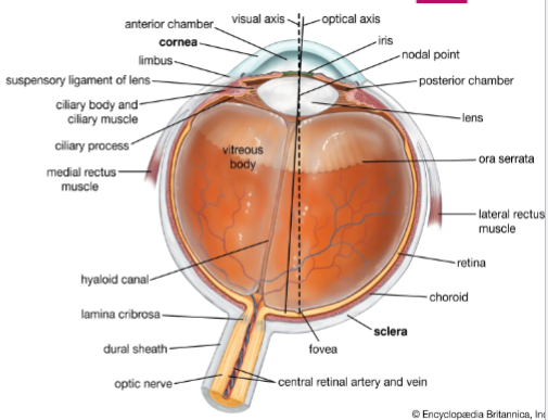
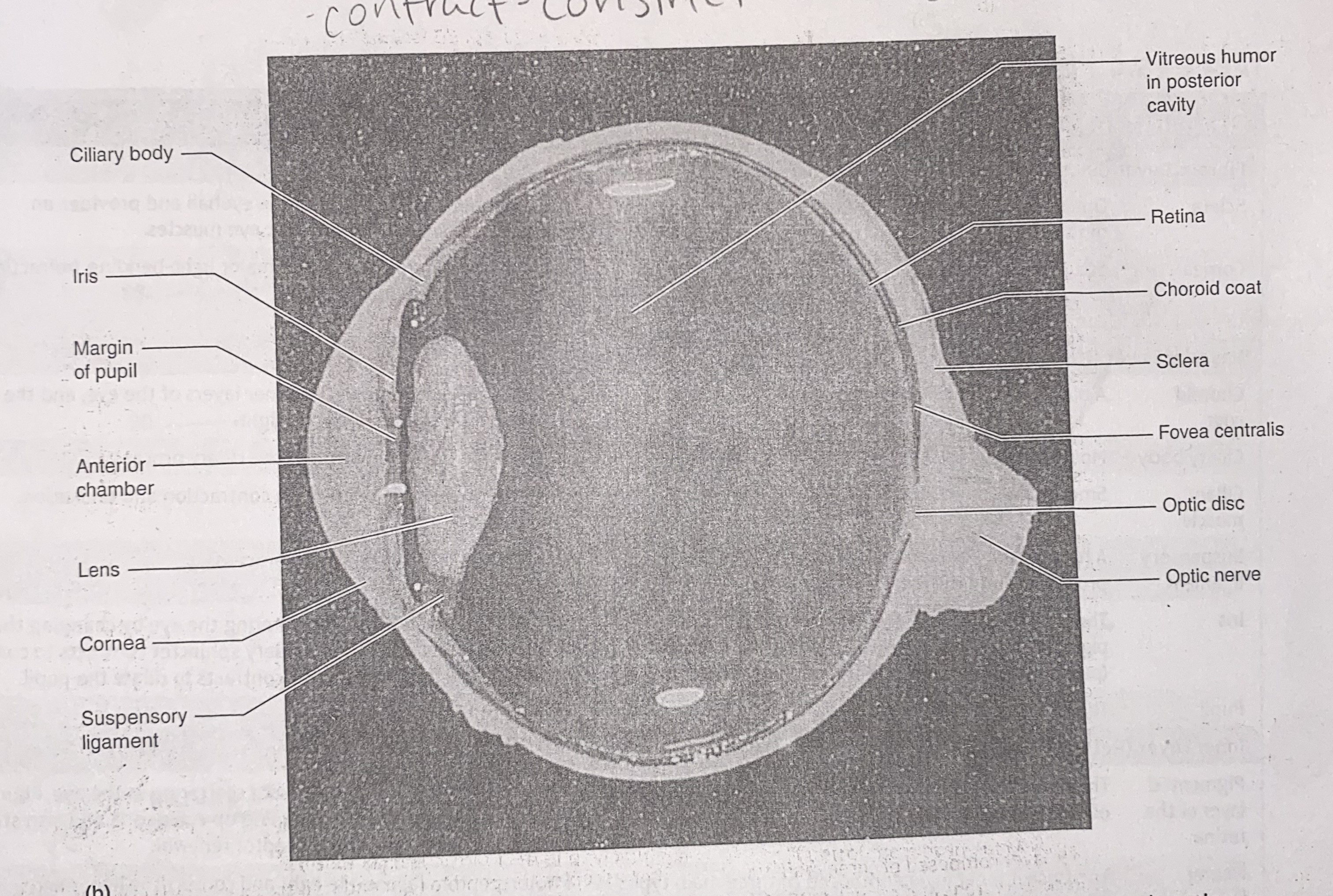
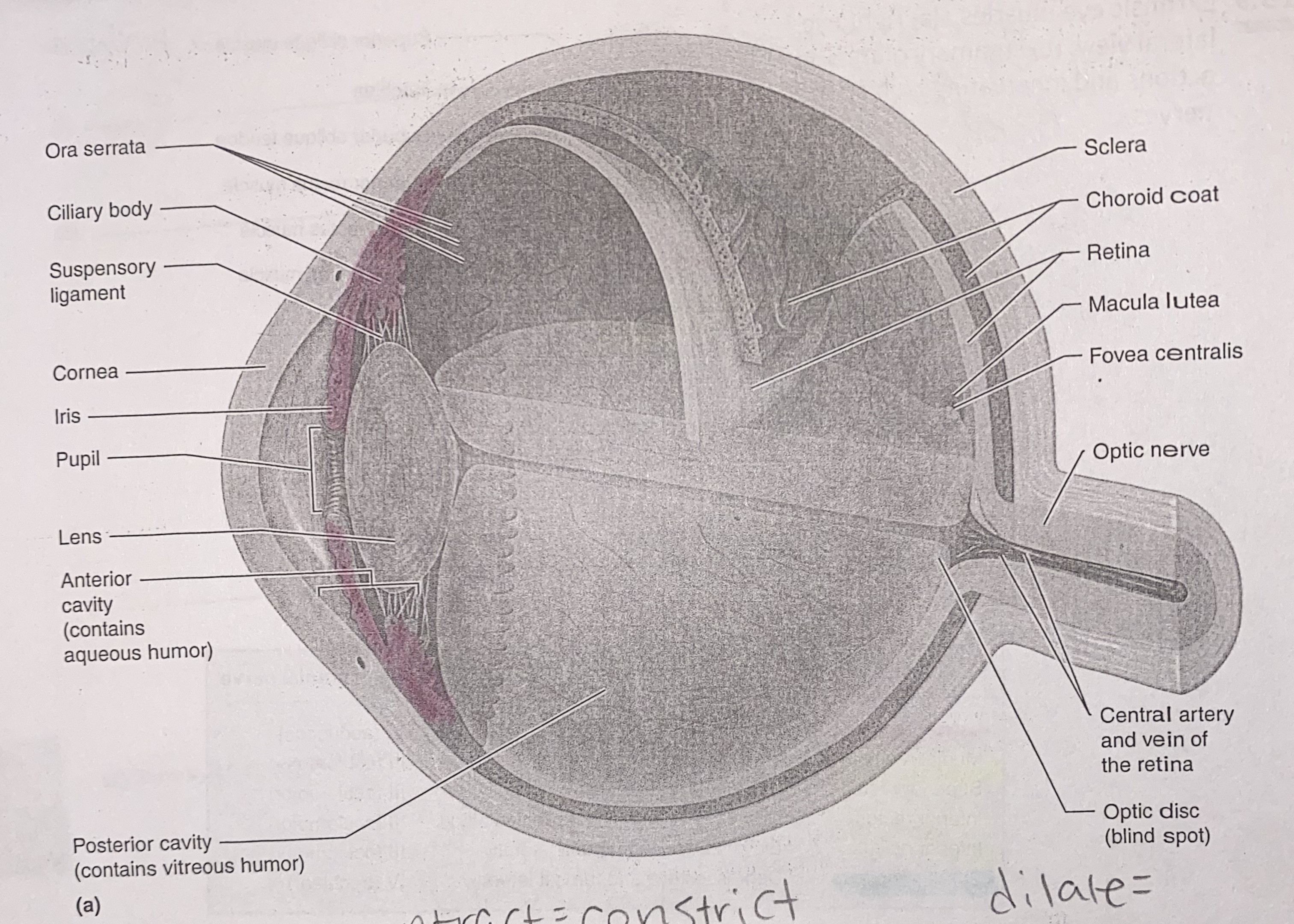
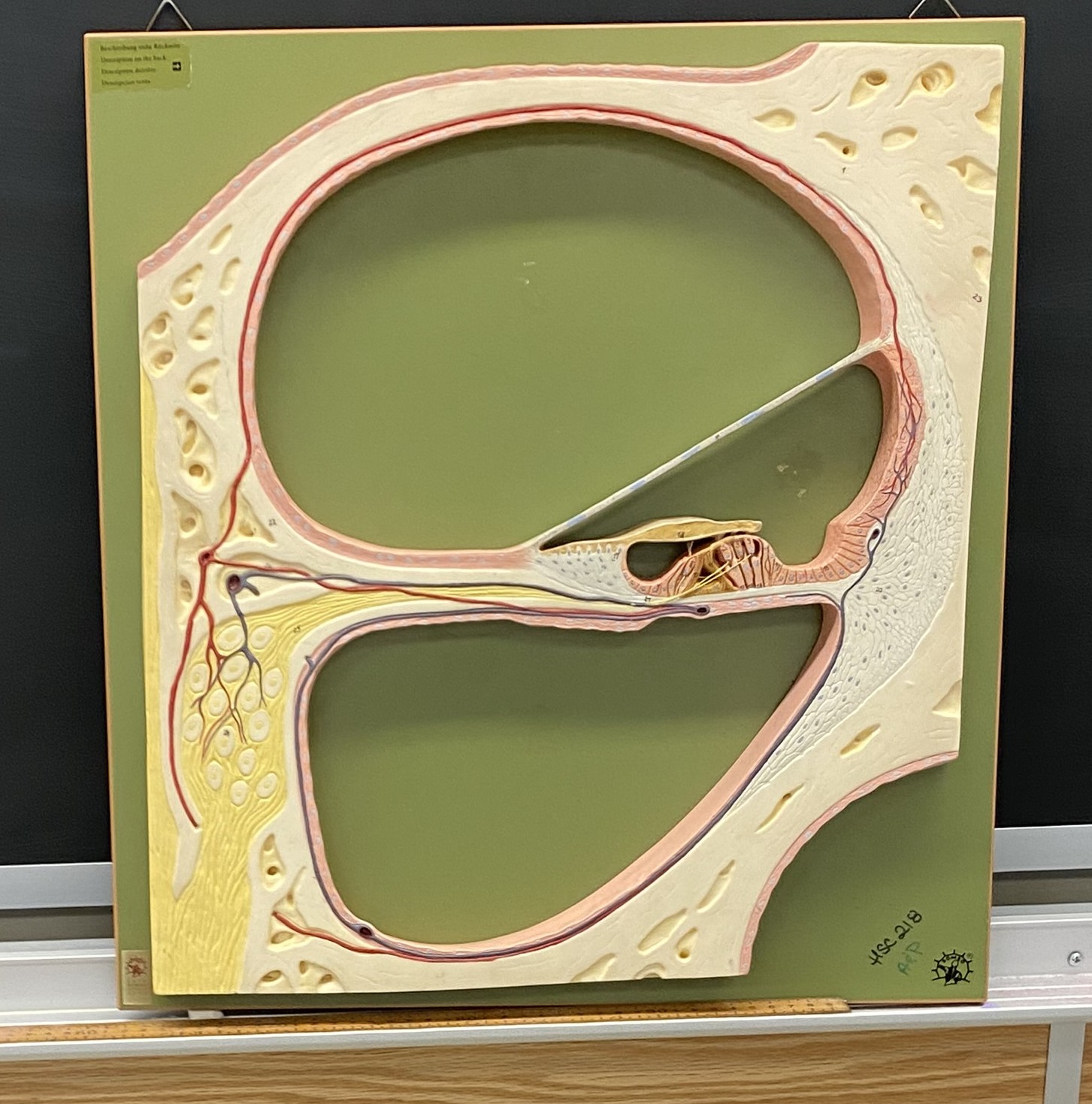
Eye
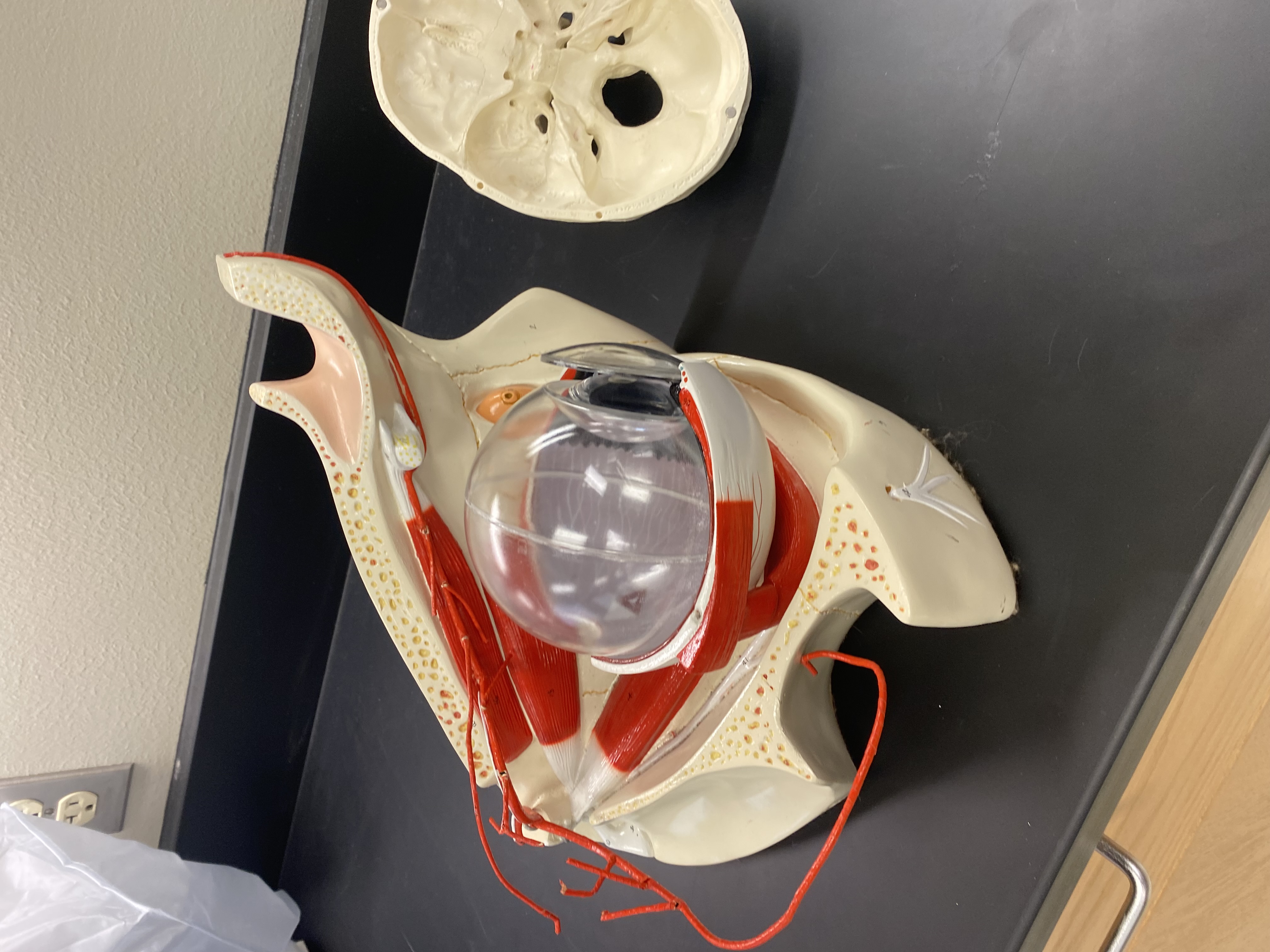
anterior cavity
posterior cavity
visual axis and fovea
blind spot
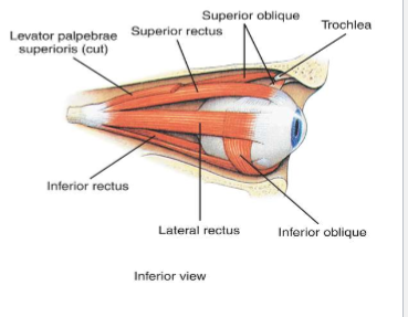
Eye Muscles
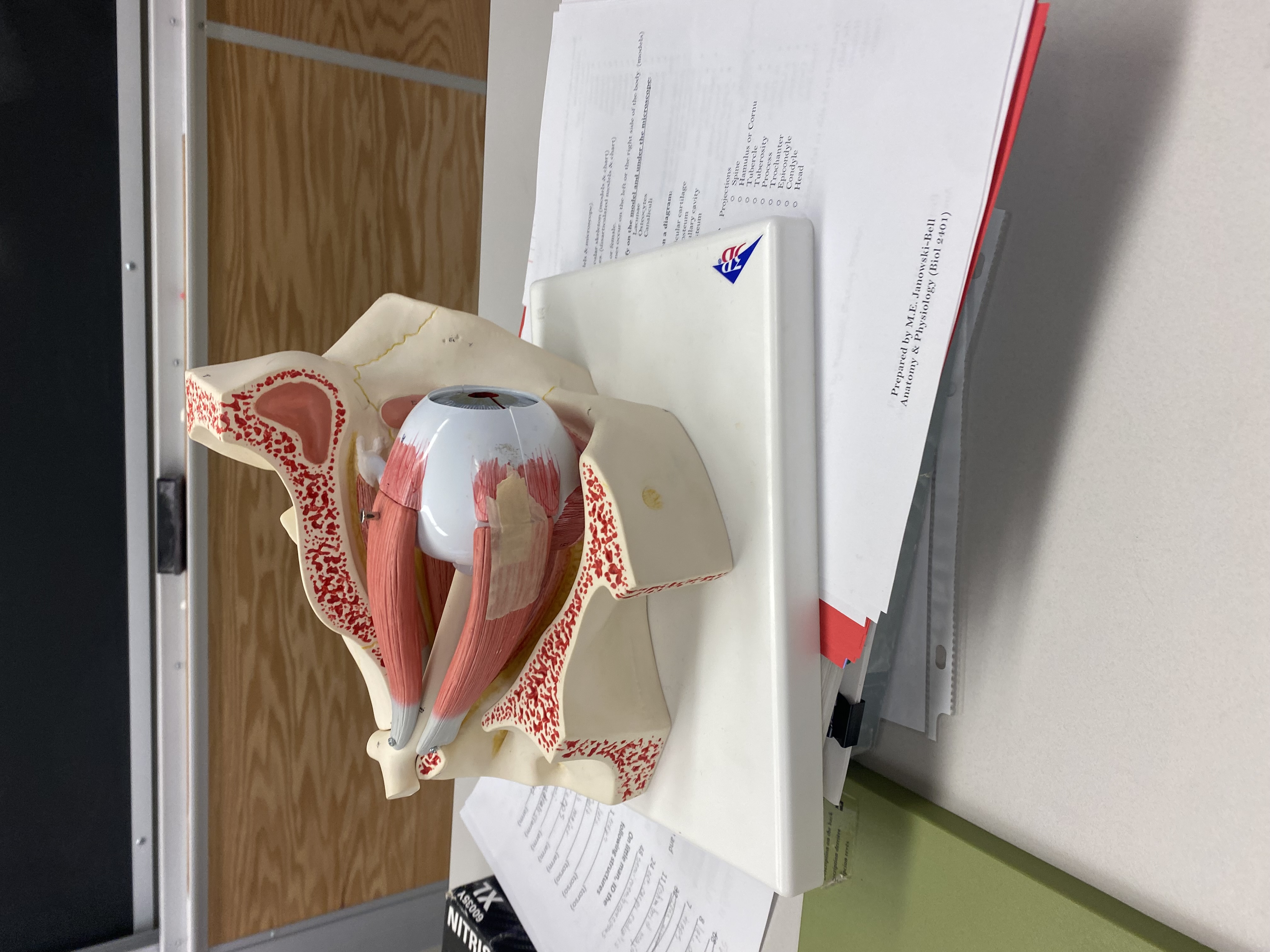
6 muscles
LR6
S04
R3 (parasympathetic but skeletal muscle)
Layers and structures of eyeball
fibrous
outer layer
collagen
sclera and cornea
vascular
middle layer
everything
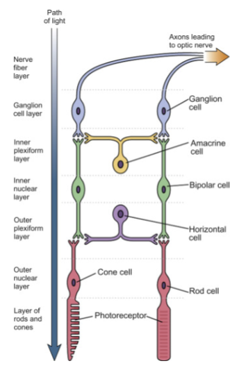
nervous/retina
retina and photoreceptors
visual axis to forea (highest concentration of photoreceptors)
Chambers
anterior chamber
aqueous fluid
in front of lens
thin
Posterior chamber
vitreous fluid
behind lens
thick
Important stuff
choroid coat- pigmentation; causes eyes to glow in animals
pupillary constrictor- cranial nerve 3
pupil- hole in vascular layer
light hits lens and bends
fovea centralis -A tiny depression in the center of the retina, located within the macula lutea. Contains the highest concentration of cone photoreceptors — no rods. Responsible for sharpest visual acuity and color perception.
Rods vs. Cones
🌟 Photoreceptors of the Retina: Rods vs. Cones📌 Overview
Photoreceptors are specialized neurons in the retina that detect light and convert it into neural signals.
The two primary types are rods and cones, each designed for distinct visual functions.
🔦 Rods
Function: Specialized for low-light (scotopic) vision
Color Sensitivity: None — detect grayscale only
Visual Detail (Acuity): Low; blurry but sensitive
Location: Mostly in the peripheral retina
Number: About 120 million in the human eye
Adaptation: Slow to adjust between light levels (think stepping into a dark room)
Extra Insight: Crucial for night vision and motion detection, but easily saturated in bright light
🎨 Cones
Function: Specialized for bright-light (photopic) and color vision
Color Sensitivity: Yes — trichromatic system (red, green, blue)
Visual Detail (Acuity): High; sharp and focused
Location: Densely packed in the fovea centralis (center of retina)
Number: About 6 million
Adaptation: Quick to adjust to brightness
Extra Insight: Vital for detail-oriented tasks like reading and recognizing faces
Intrinsic Eye Muscles and their response to light
Bright light=circular muscles contract= small
dim light= eyes dilate= big
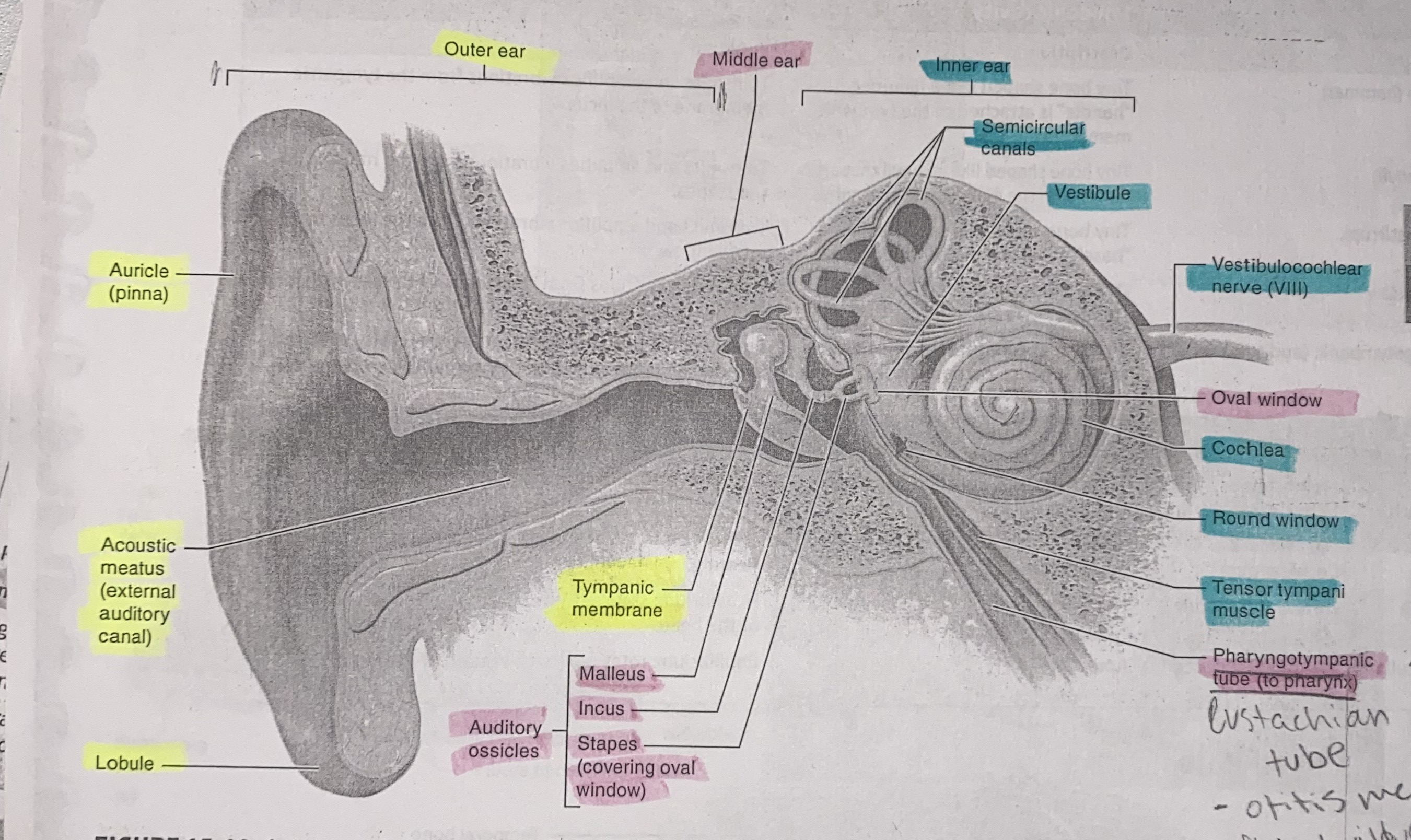
Outer Ear
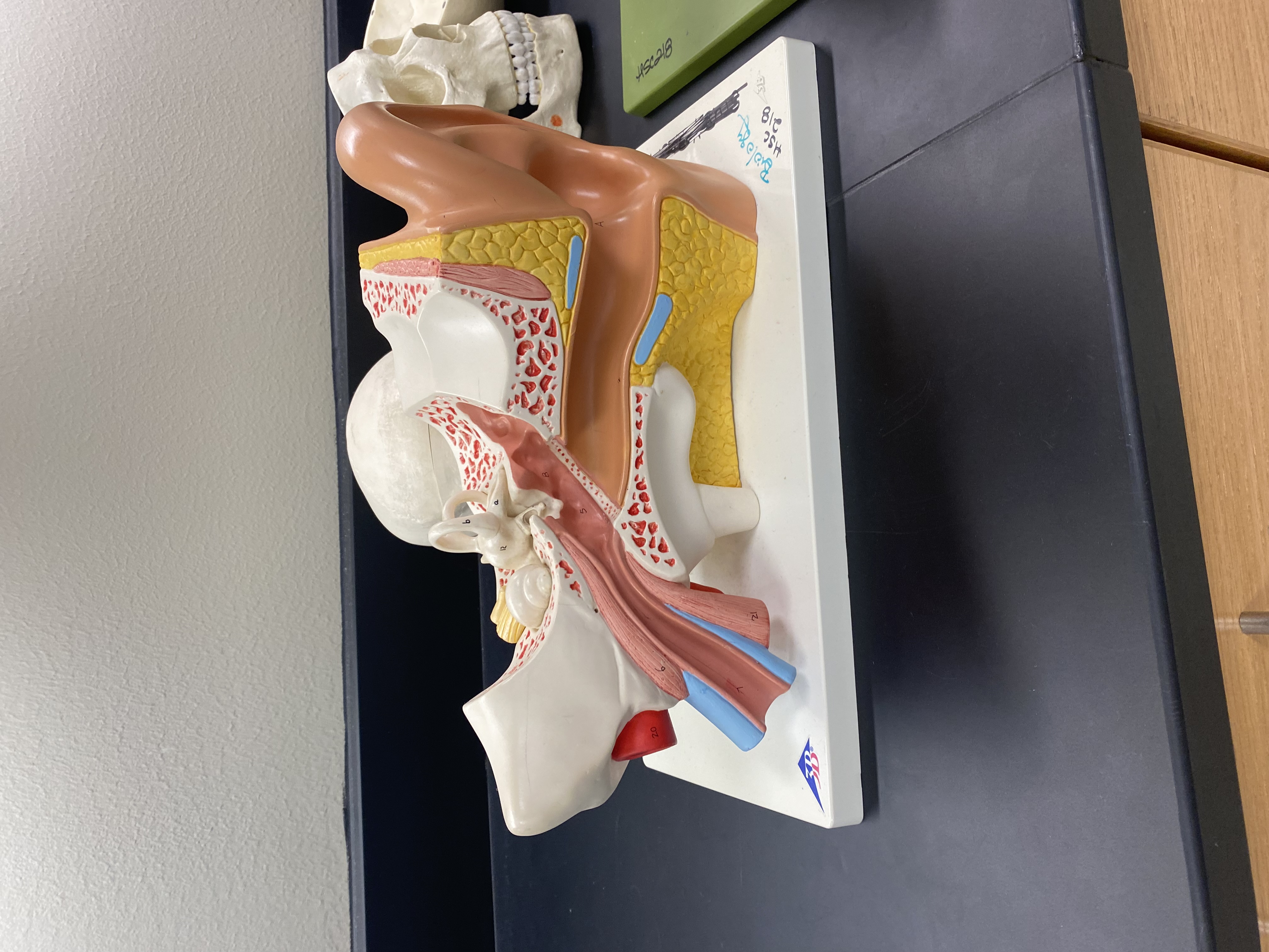
Outer Ear
pinna/auricle
external auditory meatus
tympanic membrane
Middle Ear
auditory ossicles
malleus, incus, stapes
oval window
pharyngotympanic tube
Tensor tympani
Inner Ear
semicircular canals
vestibule
Nerve 8
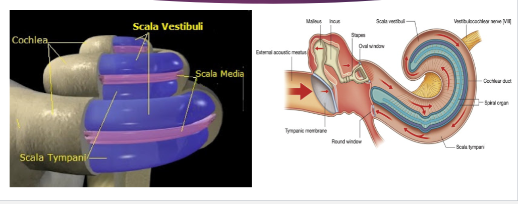
Cochlea
Round Window
located in petrous portion of temporal bone
vestibulocochlear nerve hole
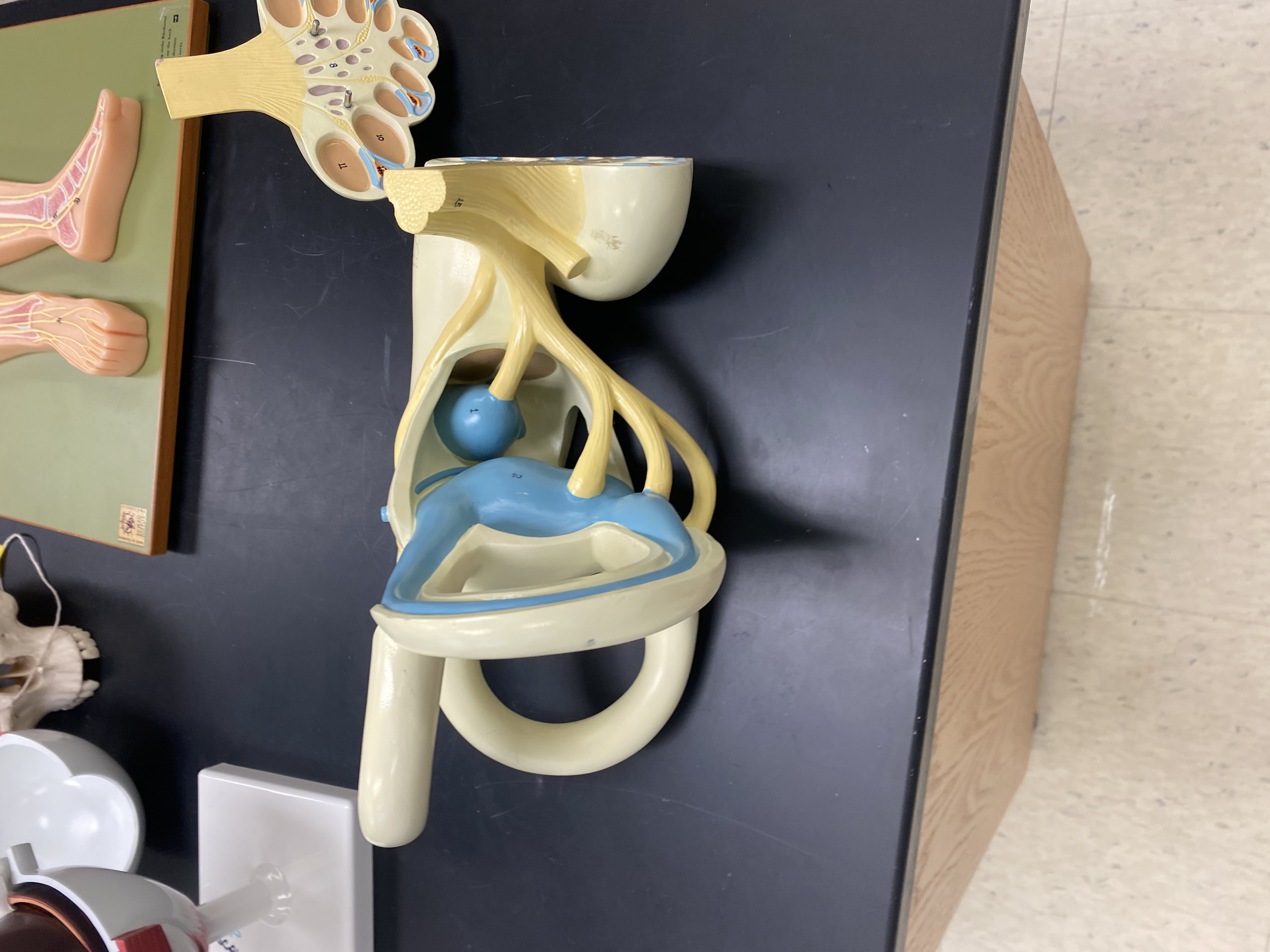

Static Equilibrium
vestibule
utricle and saccule
each have hair cells that are filled with otolithic membrane
When your head tilts or you experience linear acceleration (like riding an elevator), gravity causes the otolithic membrane to shift.
This movement bends the stereocilia on the hair cells, triggering electrical signals
static equilibrium and linear acceleration of the head
POSITION


Dynamic Equilibrium
semicircular ducts
ampullae
When your head rotates, the fluid (endolymph) inside the canals lags behind due to inertia. This causes the cupula to bend, which in turn bends the stereocilia on the hair cells. That bending generates nerve impulses
rotational acceleration of the head
MOVEMENT
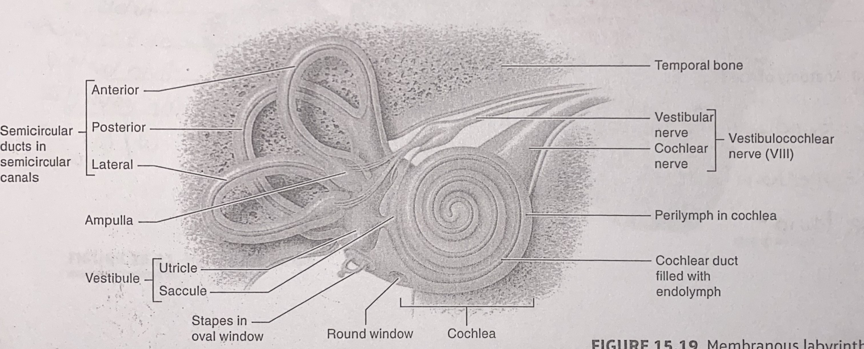
Hearing
Cochlea
organ of corti
action potentials
Contains hair cells (inner and outer) with stereocilia that detect sound vibrations.
When sound waves cause fluid movement in the cochlea, the basilar membrane vibrates, bending the stereocilia.
This bending opens ion channels, triggering electrical signals.
These signals are sent via the auditory nerve to the brain, where they're interpreted as sound.
Order of how we interpret sound
Pinna (Auricle): The outer ear collects sound waves and funnels them into the ear canal.
External Auditory Canal: Channels the sound waves toward the tympanic membrane (eardrum).
Tympanic Membrane (Eardrum): Vibrates in response to sound waves, converting them into mechanical energy.
Auditory Ossicles (Middle Ear Bones):
Malleus receives vibrations from the eardrum
Incus passes them along
Stapes pushes against the oval window, amplifying the signal
Oval Window: Transfers vibrations into the cochlea of the inner ear.
Cochlear Fluid Movement: Vibrations cause fluid waves inside the vestibular duct, which ripple through the cochlear duct and tympanic duct.
Basilar Membrane & Hair Cells: Fluid movement causes the basilar membrane to vibrate, bending stereocilia on hair cells in the organ of Corti.
Neurotransmitter Release: Bending of stereocilia opens ion channels, triggering neurotransmitter release and generating electrical signals.
Auditory Nerve (Cochlear Branch of CN VIII): Carries the electrical signals to the brainstem, then to the temporal lobe, where sound is interpreted.