Gastrointestinal 3 - Viscera
1/44
There's no tags or description
Looks like no tags are added yet.
Name | Mastery | Learn | Test | Matching | Spaced | Call with Kai |
|---|
No analytics yet
Send a link to your students to track their progress
45 Terms
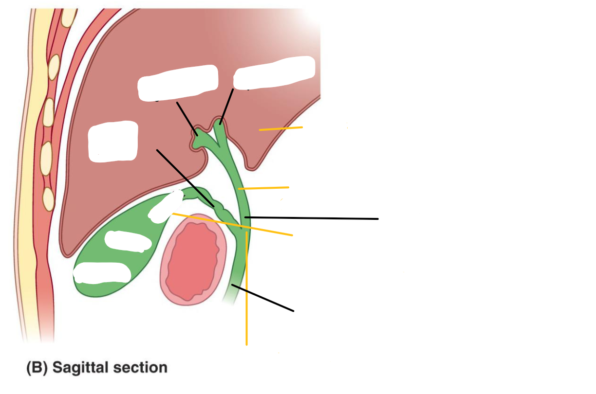
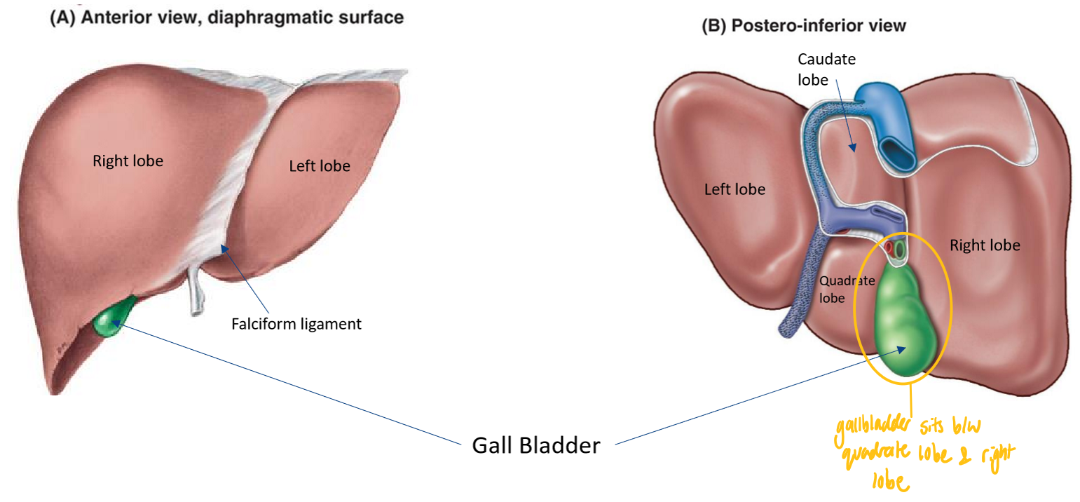
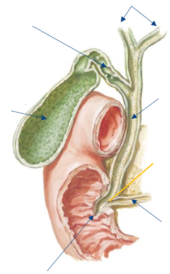
Label the following diagram of the gallbladder
(Ignore the yellow lines)

What is the gallbladder?
Pear-shaped sac on visceral surface of liver, wedged in a fossa b/w the right & quadrate lobes
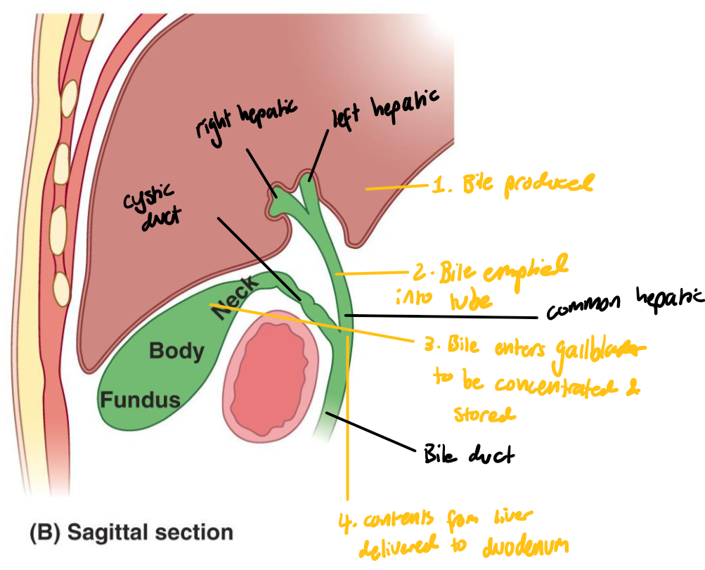
What is the role of the gallbladder?
Receives, concentrates & stores bile from the liver
What are the 3 major components of the gallbladder?
Fundus: concentrates bile
Body
Neck: site of retrograde bile flow into gallbladder if bile is not needed
What does the gallbladder empty into?
The cystic duct, a component of the bile duct system
Describe the movement of bile from the liver to the gallbladder
Bile is produced in the liver
Bile is flows into the right hepatic & left hepatic ducts
Bile flows into the common hepatic duct & then into the cystic duct → gallbladder for storage
Bile flows into the bile duct to be delivered to the duodenum

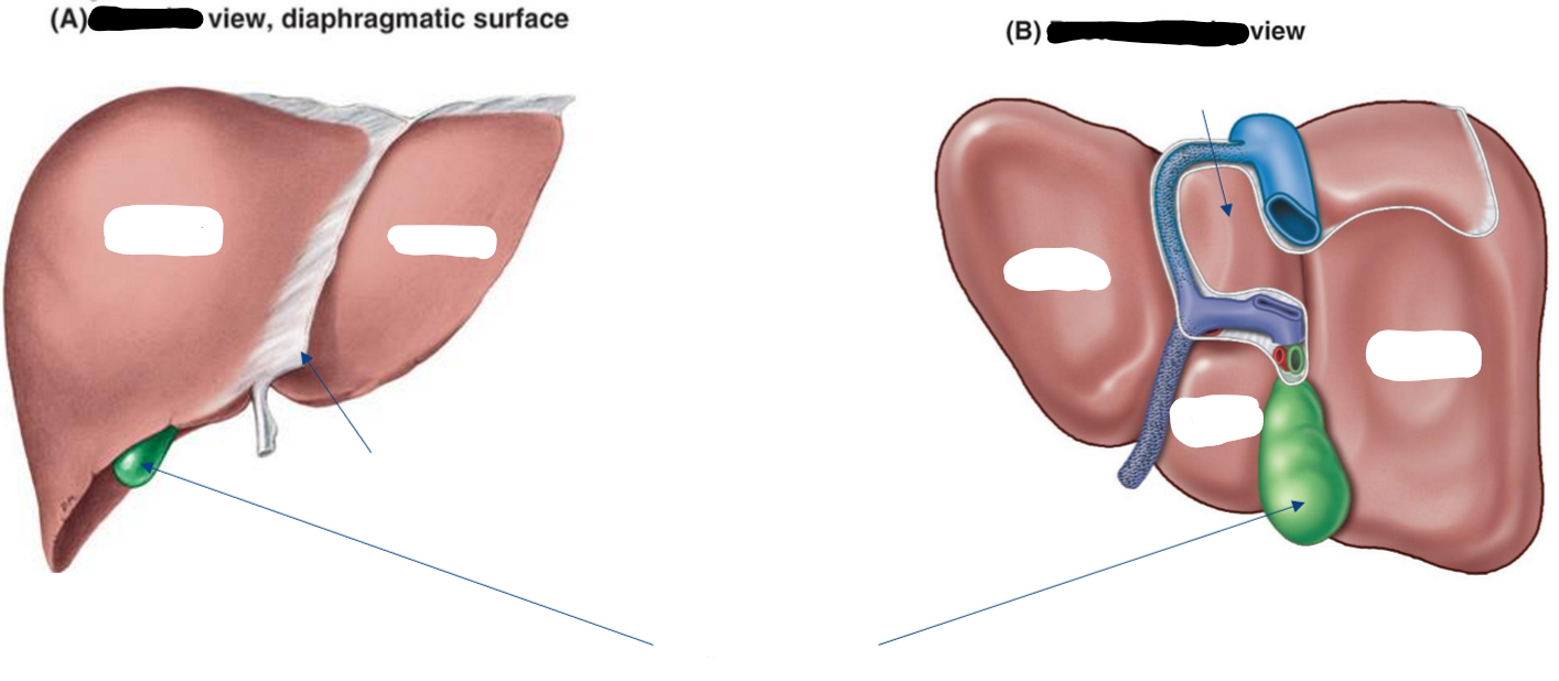
Label the following diagram to show the positioning of the gallbladder
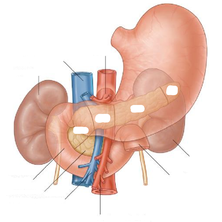
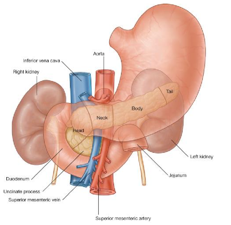
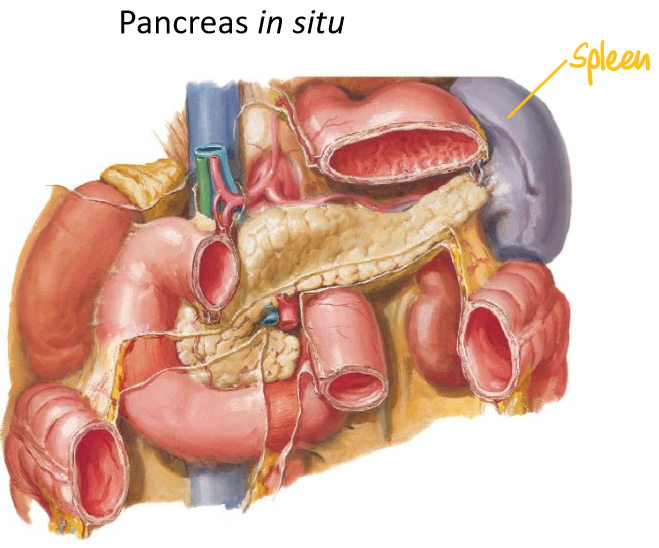
Label the following diagram of the pancreas & associated structures

For the pancreas, state:
What type of organ it is
Whether it is more superficial or deep, & retroperitoneal or intraperitoneal
Major components
Associated structures
Appearance
Exocrine & endocrine glandular tissue
Mostly exocrine in GIT; → signalling molecules + enzymes into ducts
Very deep & retroperitoneal
Major components:
Head (w/ uncinate process): surrounded by duodenum
Neck: near superior mesenteries (large blood vessels)
Body
Tail (near spleen)
Associated structures:
Duodenum
Stomach
Spleen
Superior mesenteric vessels
Bile duct
Distinct lobulated appearance w/ no capsule; looks very fatty
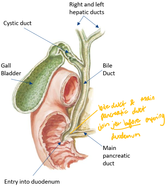
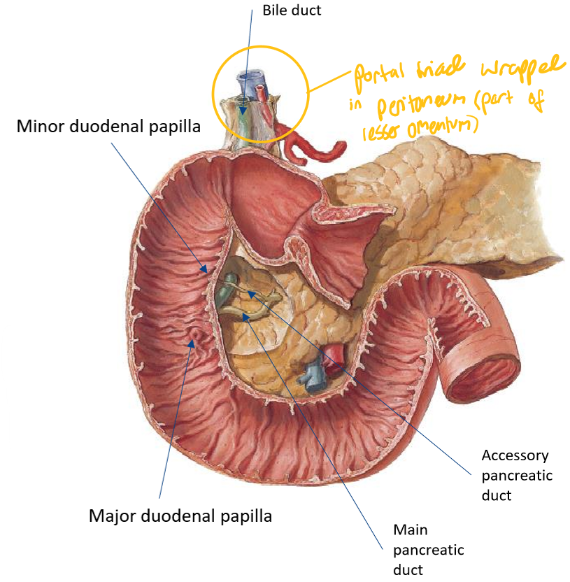
Label the following diagram of the pancreatic ducts

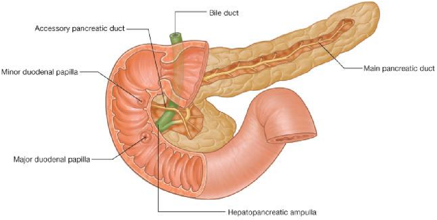
What does this diagram show about the relationship between the bile & pancreatic duct?
They join just before entering the duodenum

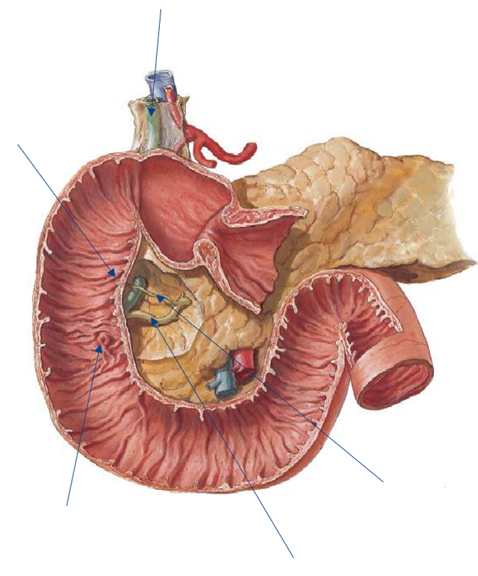
Where do the minor duodenal papilla & major duodenal papilla of the pancreas enter?
Enter the second part of the duodenum
Where are the minor duodenal papilla & major duodenal papilla positioned with respect to the abdominal wall?
Fixed on the abdominal wall → more stable
First part of the duodenum bends & twists (comes off abdominal wall)
Label the following diagram of the bile & pancreatic ducts
What is the portal triad wrapped in?
Peritoneum; part of the lesser omentum

Label the following diagram of the pancreatic & bile ducts
What is the spleen & what is its role?
Vascular organ & the largest lymphatic viscera but is NOT part of the digestive pathway
Role: produces white blood cells (but its role changes throughout development, puberty & adulthood)
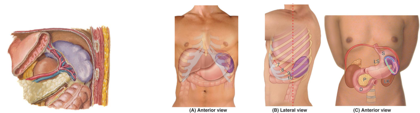
For the spleen, state:
Where it is located & implications of this location
Shape
Whether it is intra- or retroperitoneal
Close anatomical relationship to another structure
Deep in upper left quadrant to left ribs 9-10/11
Risk of injury from rib fractures → life threatening due to possibility of major blood loss
Variable in shape
Intraperitoneal
Splenic hilum close to pancreatic tail

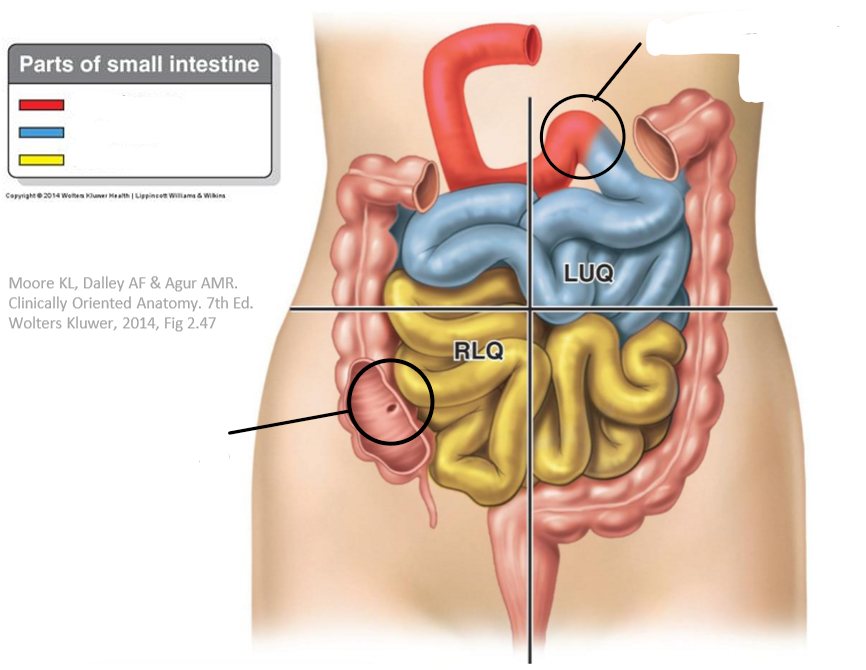
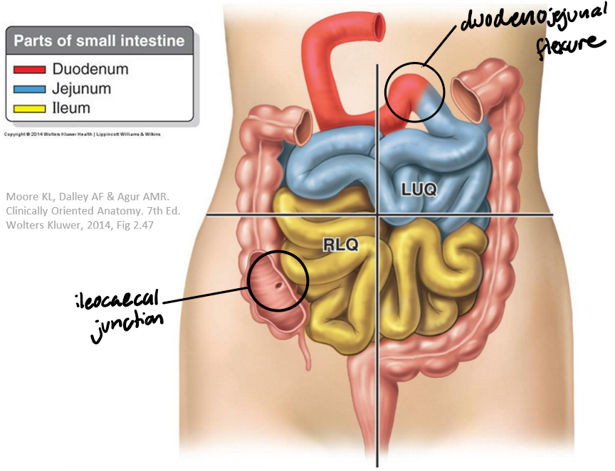
Label the following diagram
For the jejunum, state:
Where it begins
Where it is positioned
What is comprises
Diameter & wall thickness
Key feature
Begins @ duodeno-jejunal flexure
Largely in left upper quadrant
Comprises proximal 40% of small intestine
Large diameter + thick wall
Prominent plicae circulares (luminal folds) → ^ SA for absorption
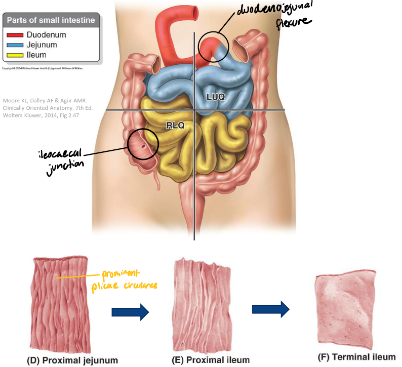
For the ileum, state:
Where it begins
Where it is positioned
What is comprises
Diameter & wall thickness
Key feature
Where it ends
Poorly defined transition from jejunum to ileum
Largely in right lower quadrant
Comprises distal 60% of small intestine
Narrow diameter + thin wall
Less prominent & prevalent plicae circulares (b/c there is less stuff to absorb as food travels through the SI)
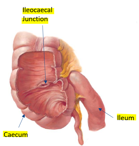
Ends @ ileocaecal junction → empty contents into initial large intestine

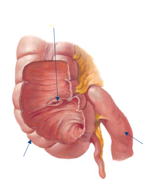
Label the following diagram
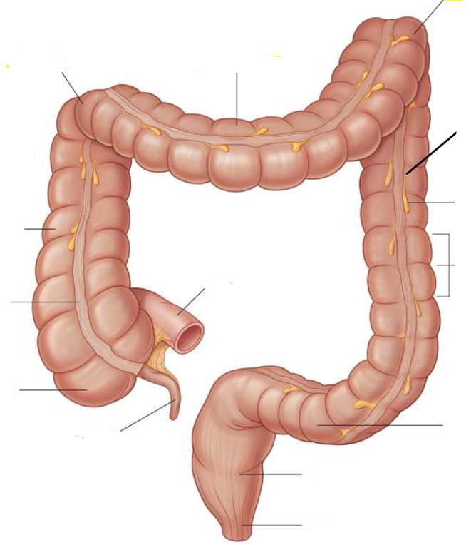
Label the following diagram

How does the diameter & length of the large intestine compare to the small intestine?
Larger diameter of LI
Shorter length of LI
What does the large intestine begin as?
The caecum at the ileocaecal junction
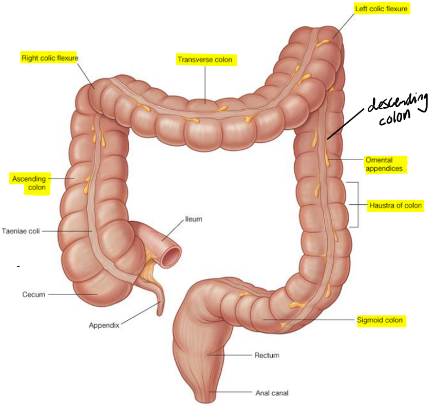
What are the 5 key components of the large intestine?
Caecum (+ appendix)
Ascending colon
Transverse colon
Descending colon
Sigmoid colon

What are the 2 major flexures of the large intestine
Hepatic (right colic) flexure
Splenic (left colic) flexure

Name 3 distinctive features of the large intestine
Taenia Coli: longitudinal bands of smooth muscle
Haustra: sacculations (small pouches) of the colon
Omental appendices: small pouches of peritoneum filled w/ fat

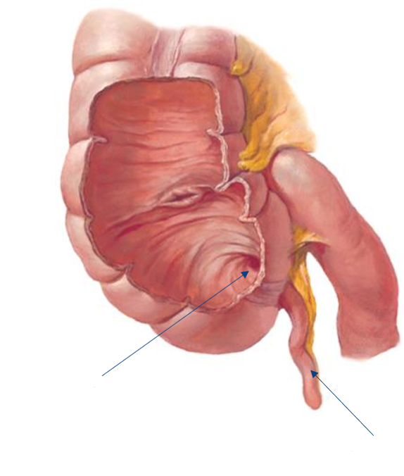
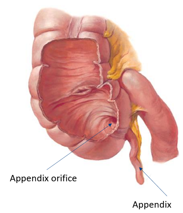
Label the following diagram

For the appendix, state:
Shape
Orientation
Clinical implications
Key feature
Vermiform (worm-like)
Variable orientation w/ its own mesentery (mesoappendix)
Prone to sudden implications → appendectomy
High proportion of lymphoid tissue
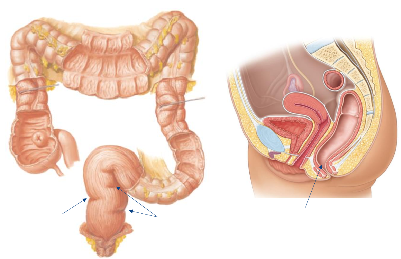
Label the following diagram
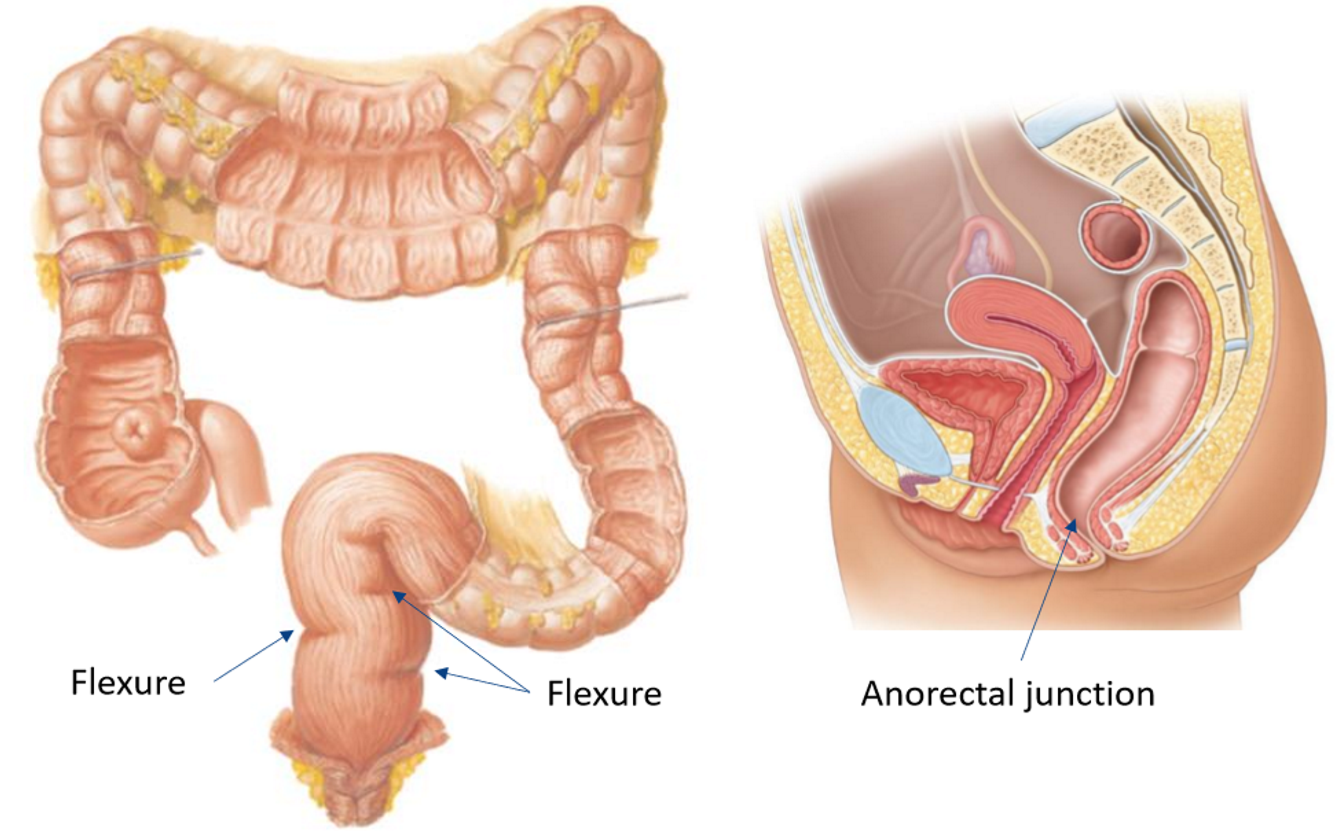
For the r3ctum, state:
Where it begins
What it follows & where it finishes
Change in musculature
What can typically be seen from an anterior view
Begins at rectosigmoid junction
Follows convexity of sacrum & finishes at tip of coccyx (→ @nal canal)
Change in external musculature for sigmoid colon to r3ctum
Taeniae coli blending into broad, continuous, longitudinal bands
Anterior view → 3 distinct flexures

Label the following diagram

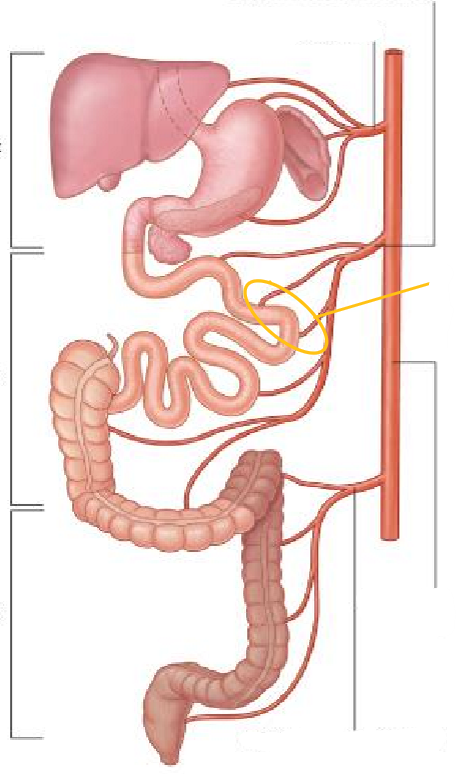
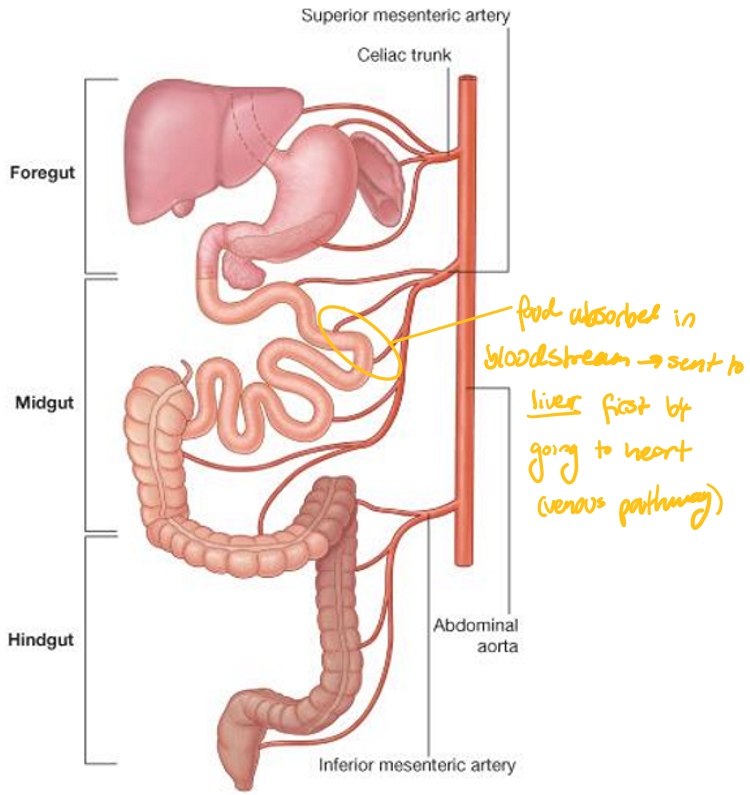
Name the major arterial supply to the abdominal viscera & the names of the 3 branches
3 major unpaired branches from abdominal aorta:
Celiac trunk
Superior mesenteric artery
Inferior mesenteric artery

What does the celiac trunk supply?
Foregut:
Liver
Gallbladder
Spleen
Stomach
Pancreas
Duodenum

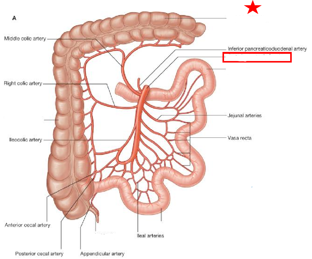
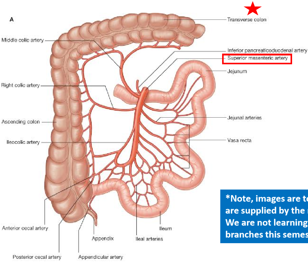
What does the superior mesenteric artery supply?
Midgut:
Pancreas
Duodenum
Jejunum
Ileum
Caecum
Appendix
Ascending colon
Transverse colon

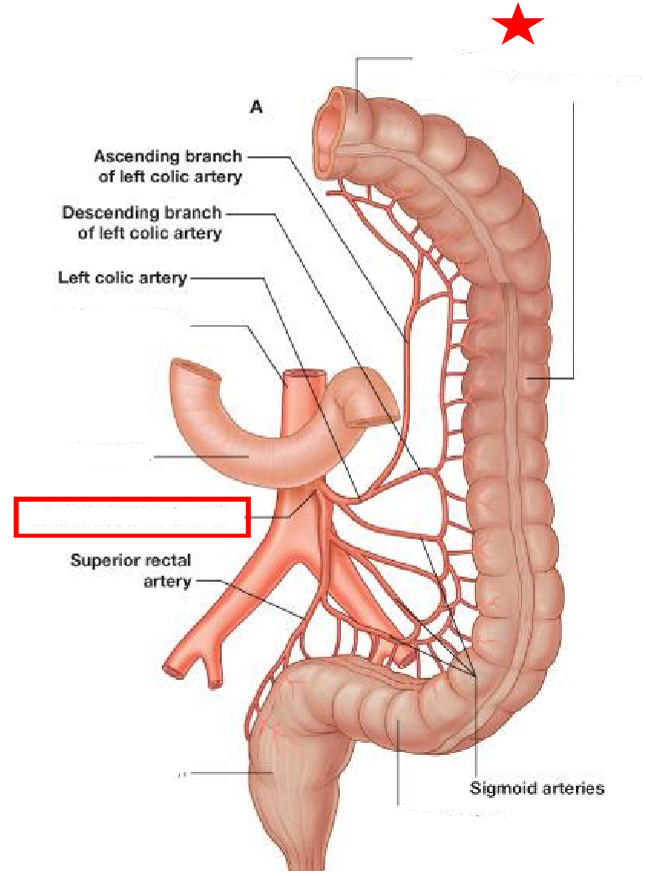
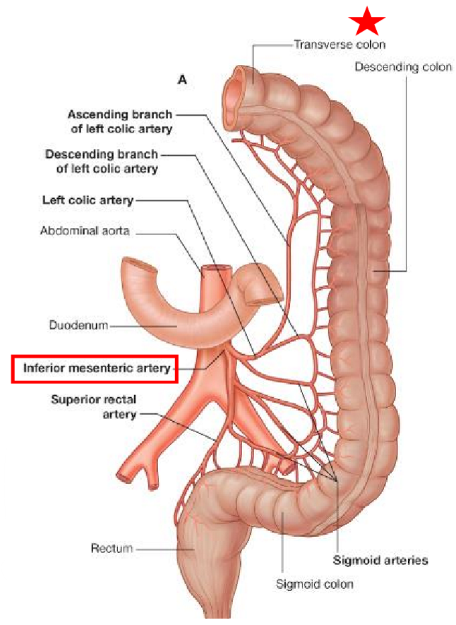
What does the inferior mesenteric artery supply?
Hindgut:
Transverse colon
Descending colon
sigmoid colon
R3ctum
Which abdominal viscera receive blood from 2 branches of the abdominal aorta, & which branches are they?
Pancreas + duodenum: receive blood from celiac trunk + superior mesenteric artery
Transverse colon: receives blood from superior mesenteric artery & inferior mesenteric artery

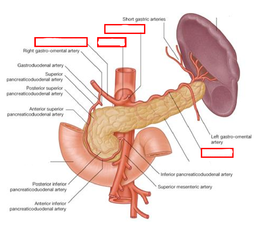
Label the following diagram of the celiac trunk, its branches, & surrounding structures
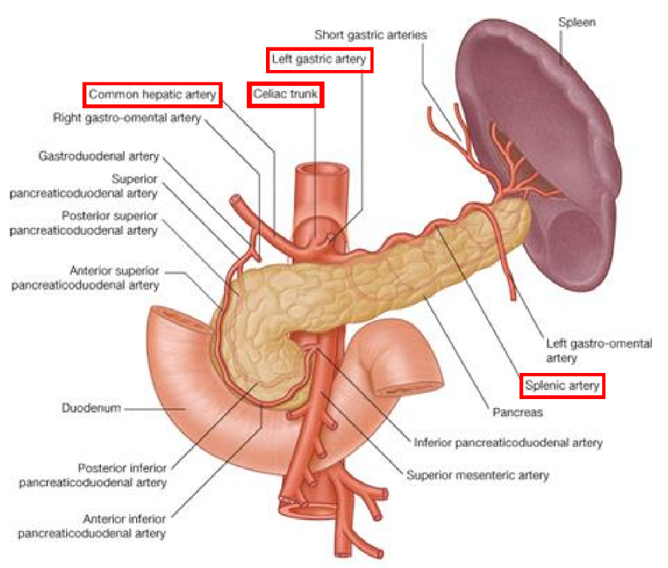
Name the 3 major branches of the celiac trunk & what they each supply, & where they are found
Splenic artery: pancreas, stomach, spleen
Tortous appearance
Moving towards upper left quadrant
Common hepatic artery: stomach, duodenum, liver & gallbladder
Moving towards upper right quadrant
Left gastric artery: stomach & oesophagys
Shorter branch

Label the following diagram
Label the following diagram
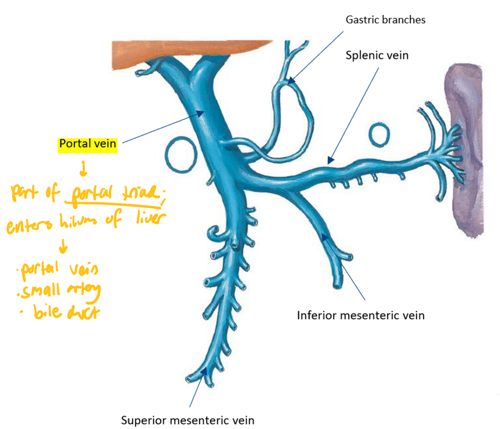
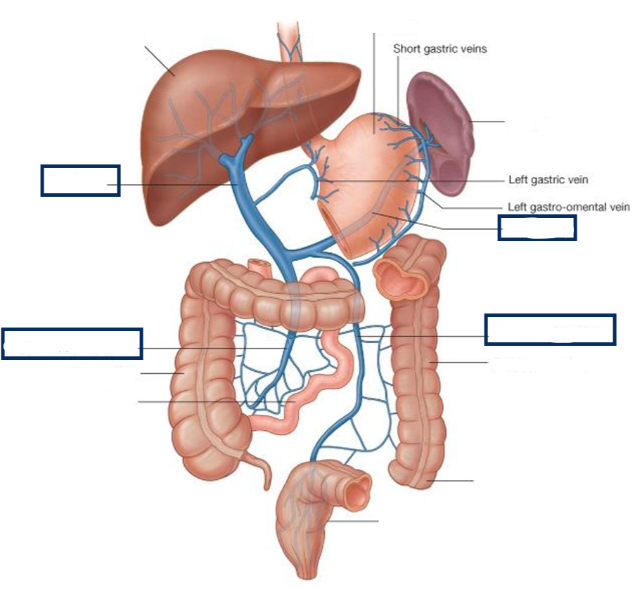
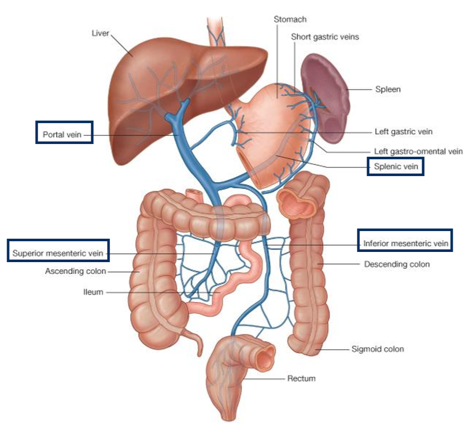
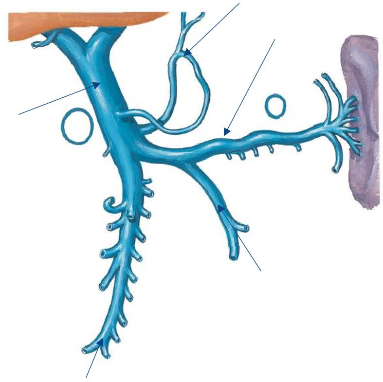
Compare the venous & arterial system of the abdominal viscera
Both have 3 major unpaired branches:
Celiac trunk (arterial) & splenic vein (venous)
Superior mesenteric (both)
Inferior mesenteric (both)
Arteries originate from abdominal aorta
Veins drain into portal vein (NOT directly into IVC)
Label the following diagram
Label the following diagram