8 myoglobin and hemoglobin
1/50
There's no tags or description
Looks like no tags are added yet.
Name | Mastery | Learn | Test | Matching | Spaced | Call with Kai |
|---|
No analytics yet
Send a link to your students to track their progress
51 Terms
what is the function of a protein determined by
the structure
what is the structure of myoglobin
•Monomer
• 1 subunit/1 polypeptide chain - 153 aa and 1 domain (bc less than 200)
• has only tertiary structure - doesnt have quaternary
has 8 alpha helices and irregular structures (loops)
has heme prosthetic group
has hydrophobic pocket between helix E and F for heme since its hydrophobic but amphipathic
what is the structure of hemoglobin
•Oligomer - atleast more that 1 subunit
• 4 subunits - Each subunit has 1 domain
• also has quaternary structure - heterotetramer (atleast two are different, 2 alpha and 2 beta)
what is the main function of red blood cells
has hemoglobin (doesnt have mitochondria) that binds to oxygens in the lungs and releases it in the tissues that need it
O2 structure is not polar and not super soluble in water which is why you need help transporting it
what is the function of myoglobin
• Act as local reserve of O2 during intense exercise
• Store O2 in aquatic animals
cytoplasmic protein found in muscle
• Facilitate O2 diffusion through muscle tissue - by allowing it to move quicker from plasma membrane to inner mitochondria membrane in aqueous solution
• Inactivating Nitric oxide and removes reactive O2 species because their unstable
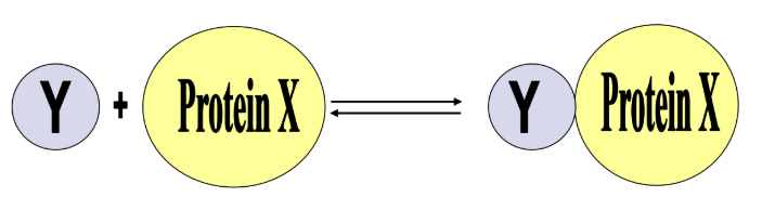
what does the function of proteins also depends on
depends on their ability to bind other small molecules (ligands) reversibly
The greater the affinity of Protein X for ligand Y the more XY we will have at any concentration of Y or X - the interactions that link them is h bond, ionic interaction, hydrophobic, dipole-diople - the more you interact the better you can bind protein to ligand
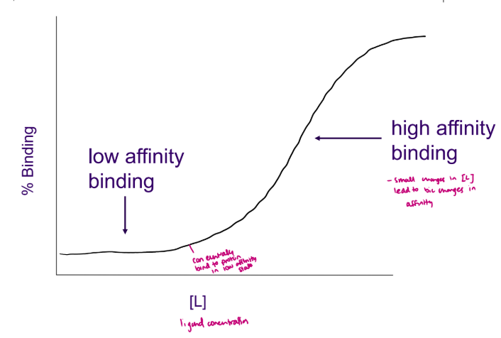
what do ligand binding curves look like
hyperbolic curve, as the concentration of the ligand increases a lot the binding plateaus because all binding sites are full
as affinity increases the Kd lowers
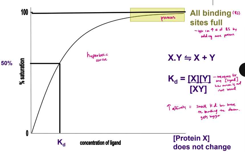
what is Kd
measures for any [ligand] how much is and isnt bonded
![<p>measures for any [ligand] how much is and isnt bonded </p>](https://assets.knowt.com/user-attachments/4933c487-40c9-4966-8bf3-1809a7278fcd.png)
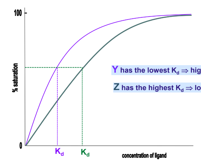
how does the ligand binding curve change depending on the affinity
Y has the lowest Kd → highest affinity
Z has the highest Kd → lowest affinity
lower affinity and higher Kd shifted to the right
Why does myoglobin need a prosthetic heme group
to carry oxygen bc side chains cant and important for structure
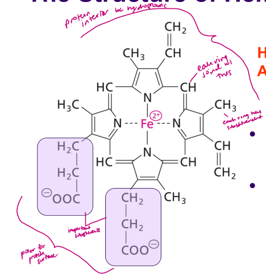
what is the structure of heme
circular and planar - doesnt have a protein
In heme, the porphyrin ring contains an Fe2+ ion coordinated between the four N atoms - can form 6 coordination bonds which are covalent and 1 atom donates both electrons - these keep iron tightly bound
has to be Fe 2+ to bind to oxygen
Note that the two substituents at the bottom of the ring are polar propionyl groups whereas the rest are non-polar aliphatic groups.
it is amphipathic
see slide 11
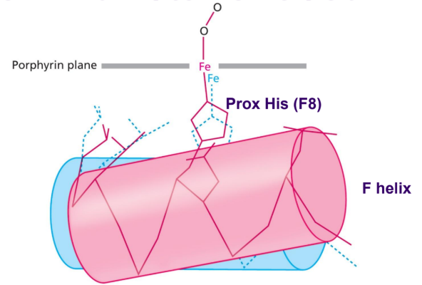
which histine does heme form a coordination bond
His F8 aka His 93 - proximal histidine which permanently attaches heme to globin
on fifth coordinate position of Fe2+ ion
how is the porphyrin ring help in place
by hydrophobic interactions and by coordination bond between Fe2+ and a histidine (aa 93) His F8 which is proximal
where does oxygen bind to the heme group
on 6th coordinate position on the Fe2+ ion - always binds at an angle
the distal histidine or His E7 assists in oxygen binding by forming H bond with it - only interacts with oxygen not iron, doing this helps carbon monoxide not being able to bond
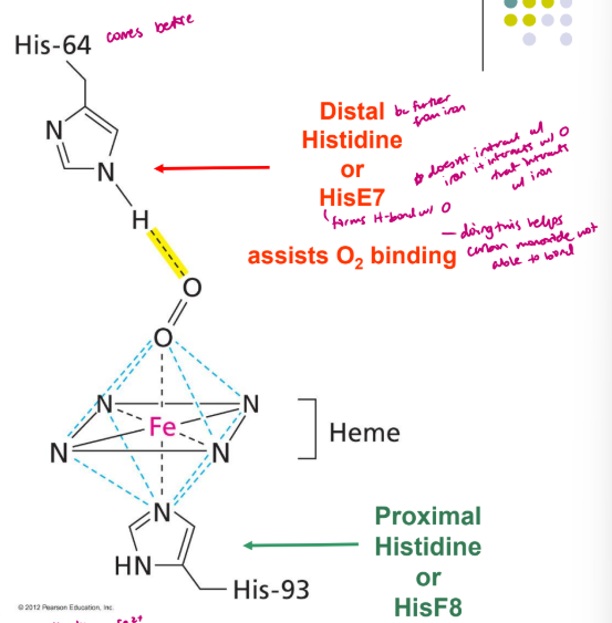
what is prevented when heme binds to polypeptide
Heme binding to the polypeptide helps prevent oxidation of Fe2+
what are two qualities that the oxygen binding sit in myoglobin exhibits and examples
Binding sites are designed precisely to optimize binding affinity and specificity
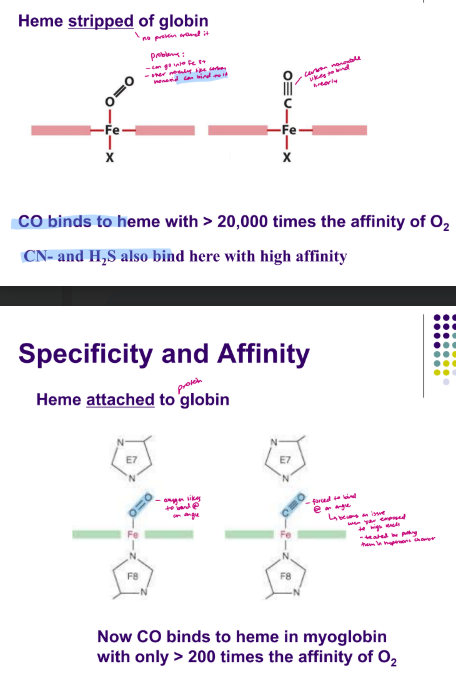
when heme is not with globin - CO binds to heme with > 20,000 times the affinity of O2, CN- and H2S also bind here with high affinity
when heme is attached to globin - Now CO binds to heme in myoglobin
with only > 200 times the affinity of O2 - because oxygen likes to bond at an angle
see slide 19
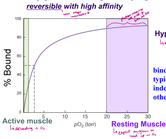
what curve does oxygen binding to myoglobin have
hyperbolic curve that tells you binding is independent of other molecules
lowest affinity when active muscle to offload oxygen, and resting muscle has plateau because you want myoglobin to load up with oxygen
how does pH effect myoglobin binding
small changes dont effect ability of myoglobin binding to oxygen but it does for hemoglobin
what is an apoprotein
known as this with out prosthetic group
when it has prosthetic group it is known as holoprotein
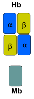
what subunits does the quaternary structure of hemoglobin have
Tetramer with two types of globin: its a heterotetramer
2 identical aplha subunits
2 identical beta subunits
each subunit has a prosthetic group
has 4 binding sites for oxygen (4 hemes interact with oxygen) but myoglobin only has 1 site
how many helices, pp chains, and location of hemoglobin and myoglobin
Hemoglobin (Hb) - found in RBC so its a cytosolic protein (doesnt have disulphide bridges)
4 polypeptide (pp) chains, 2 alpha chains, 2 beta chains
Each pp chain has 8 alpha-helices, loops and 1 heme and one hydrophobic pocket, Hb binds 4O2
In erythrocytes
Myoglobin (Mb)
1 pp chain with 8 alpha-helices, loops and 1 heme
Mb binds 1O2
In myocytes - cardiac and skeletal muscle cells
what are conservative substitutions
-these have minor effects on structure - not being a big change, changing one AA for another
• Leu to Ile, Thr to Ser (both have hydroxyl group and similar size
what are critical substitutions
can change structure and function- depending on location (His - Lys)
• Ser - Val
when choosing you look for the biggest change
what do the tertiary structures of beta-globin, alpha-globin, and myoglobin have in common
All 3 polypeptides comprise 8 alpha- helices with a heme binding pocket between helices E and F (hydrophobic)
alpha and β subunits are ~40% identical in 1o sequence
alpha subunit and MB are ~18% identical in 1o sequence
mainly conservative substitutions
Homologous proteins - have similar secondary and tertiary structures but not similar primary structures
Predict the structure of neuroglobin, a monomeric protein
- 8 helices, no sheets, has E&F, hydrophobic pocket, heme prosthetic group
how does the alpha subunit and beta subunit of Hb bind to oxygen
bind O2 in exactly the same manner as myoglobin
⚫ Each binds oxygen at the 6th coordination position of an Fe2+ ion in a heme ring.
Several critical residues in the oxygen binding sites are invariant (never change) among the three polypeptides - critical for function of proetin
his F8 - proximal
his E7 - distal
what is a hyperbolic curve indicative of
indicative of constant affinity generally high
Ligand affinity (Kd) does not change - E.g. Myoglobin.
typically, monomers
▪ One binding site for ligand that rarely changes
what is a sigmoidal curve a diagnostic of
diagnostic
of cooperative binding affinity.
Ligand affinity changes as more ligand binds - E.g. Hemoglobin (4 binding sites)
typically, oligomers - more than 1 subunit
▪ More than one binding site for the ligand
what type of binding does a sigmoidal curve represent
oxygen binding to hemoglobin
high affinity state in lungs to pick up oxygen
in tissues ther hemoglobin goes into low affinity and offloads oxygen for resting muscles
in active muscle the hemoglobin offloaded a lot of oxygen so myoglobin acts as a reserve for oxygen
Cooperative process
Necessary for efficient O2 delivery
Reflects a change in binding affinity
**theres no myoglobin in lungs
At 20-40 Torr Hb and MB must have different affinities for O2
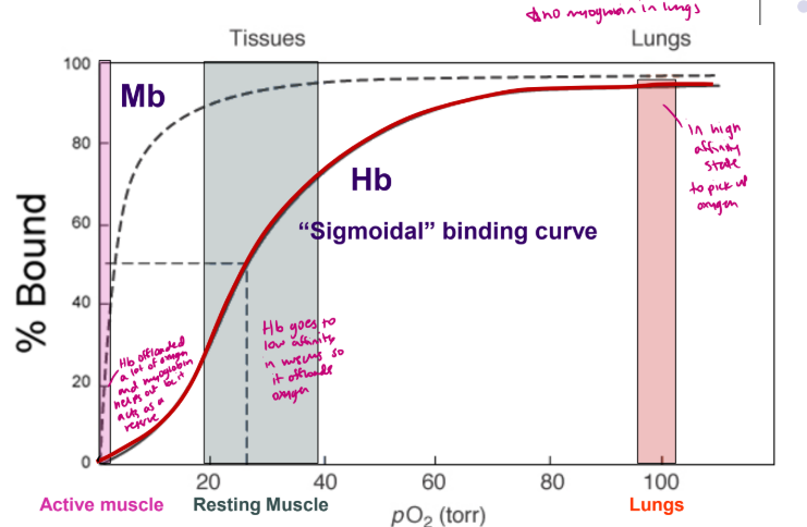
what are the similarities and differences of the functions in Mb and Hb
Similar functions
Reversibly bind/release O2 - myoglobin holds it tighter bc its a reserve
Different functions
Mb: O2 transport/storage within tissue
Hb: O2 transport from lungs to tissues
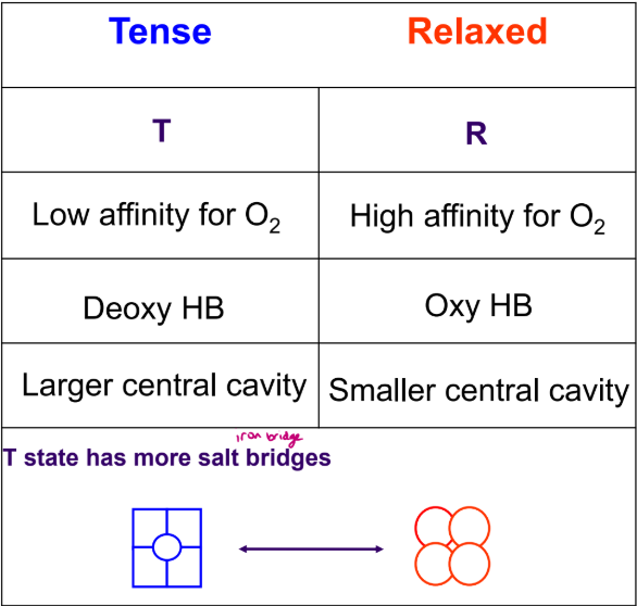
what are the two distinct structure for hemoglobin
tense T state - low affinity, has large central cavity
relaxed R state - high affinity, small central cavity
In deoxyhemoglobin, a His residue on the beta subunit fits between a Thr and a Pro residue in the alpha subunit - T state
Upon oxygenation, the hemoglobin changes shape and the His residue is now located between two Thr residues on the alpha subunit - R state
conformational change in Hb structure
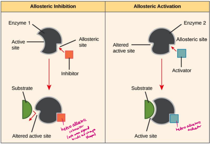
what is allostery
The binding of a ligand at one site on a protein affects the binding of ligands at other sites
can be effectors that are homoallosteric or heteroallosteric
what are effectors
compounds which, upon binding, alter affinity at other binding sites
can increase or decrease but you dont know
what is homoallosteric and heteroallosteric
Homoallosteric – binding of the effector affects further binding of the same compound - Typically referring to ligands increasing its own affinity
Sigmoidal curve in presence of ligand
Heteroallosteric – binding of the effector affects further binding of a different compound
Activators – increase binding affinity of ligand
Inhibitors – decrease binding affinity of ligand
**see slide 40-42
what are the events in oxygen binding to hemoglobin
T-state (no O2 bound)
O2 binds to a subunit
Fe2+ moves into plane of heme
Histidine F8 moves with iron
Helix F moves - gets pulled
Subunit interface changes
Subunit interface change affects other subunits
Helix F/His F8/Fe2+ movement into plane of ring
Oxygen binding site becomes high-affinity (R)- Fe2+ moves into plane of heme rings
Oxygen binds more readily to these sites.
what bonds break when you shift from T to R
The shift from T to R breaks the salt bridges that hold BPG in place
happens in lungs
space in central cavity decreases
what are the 4 allosteric effectors for hemoglobin
O2 - favours R state, positive effector/activator
**BPG (2,3-bisphosphoglycerate) - favours T state, negative effector/inhibitor
H+ - favours T state, low pH = negative effector/inhibitor
CO2 - produced in large amounts during high cellular activity, acts indirectly** and directly to stabilize the T state, favour T state
what does BPG stabilize
BPG is essential in stabilization of the T state of Hb
negative heteroallosteric effector (inhibitor) of oxygen binding
has 5 negative charges
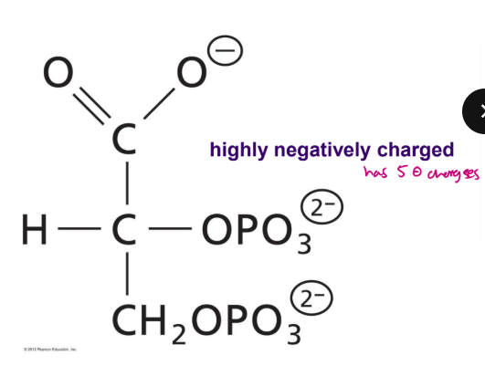
what does BPG bind to
1 BPG binds in the central cavity of deoxyhemoglobin (T state) (1 binding site for BPG and 4 for oxygen)
The negative charges on BPG interact with positively charged groups on the protein that are directed into the central cavity.
The central cavity in oxyhemoglobin (R state) is too small to accommodate BPG
when you increase BPG the S curve shifts away from the y axis to the right
what is the indirect effect of CO2
H+ ions facilitate formation of the T state- enhances BPG binding
The effect of [H+] (pH) on Hb’s O2 binding behaviour is called the Bohr effect - so more CO2 produced = more H+ produced = histidine protinated
CO2 is not very soluble in aqueous blood so most gets taken back to lungs by converting it to HCO3-
when pH decreases the histidine become protinated and forms a salt bridge with BPG
![<p>H+ ions facilitate formation of the T state- enhances BPG binding</p><ul><li><p>The effect of [H+] (pH) on Hb’s O2 binding behaviour is called the Bohr effect - so more CO2 produced = more H+ produced = histidine protinated</p></li><li><p>CO2 is not very soluble in aqueous blood so most gets taken back to lungs by converting it to HCO3-</p></li><li><p>when pH decreases the histidine become protinated and forms a salt bridge with BPG</p></li></ul><p></p>](https://assets.knowt.com/user-attachments/8393d396-0997-4d19-b421-ee9f1f816d4d.png)
what is the bohr effect
Metabolism generates protons (lowers pH; more [H+]):
ATP + H2O → ADP + Pi + H+
CO2 + H2O → HCO3- + H+
Lowering pH leads to protonation of side chains:
His + H+ → His+
His’s associated with BPG binding become protonated
Enhance BPG binding, Reduce O2 binding bc your stabilizing T state
Subunit interface is affected
New electrostatic interactions form
how does capillaries and lungs use CO2
lungs convert biocarbonate ion and H+ ions into water and carbon dioxide
histidine is deprotinated bc pH is more that pKa and wont interact with BPG so well
capillaries in muscle take carbon dioxide and water into biocarbonate ions and H+ ions
histidine become protinated
how does the saturation curve change in the lungs and skeletal muscle
pH of blood in lungs is 7.6 (above physiological pH of 7.4) so theres high pH and low hydrogen ion concentration - high affinity so it shifts to the left (closer to y-axis)
pH of blood in skeletal muscle is 7.2 (below physiological pH) so gives low affinity for oxygen, high hydrogen concentration, low pH, histidine is protinated and helps stabilize T state
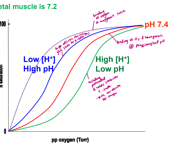
what are the 5 combined effects of oxygen, BPG, and pH on Hb function
In the lungs and in the tissues, any given molecule of Hb can exist in either its T form or its R form.
The proportion of molecules that are in either form (the position of the equilibrium) depends on the presence of CO2, 2,3-BPG, on the [H+] ions, and on the ppO2 (main driving force if youre R or T)
The proportion of molecules that are in either form (high or low affinity) determines how much oxygen is bound or released.
The lungs have a high pp O2 and a relatively high pH (low [H+]), The R state is thus favored, and when oxygen binds it triggers the switch to the R form.
Actively respiring tissues have a relatively low pH (high [H+]), high levels of CO2 and a low ppO2, The T state is favored, and oxygen is released.
![<ul><li><p> In the lungs and in the tissues, any given molecule of Hb can exist in either its T form or its R form.</p></li><li><p> The proportion of molecules that are in either form (the position of the equilibrium) depends on the presence of CO2, 2,3-BPG, on the [H+] ions, and on the ppO2 (main driving force if youre R or T)</p></li><li><p> The proportion of molecules that are in either form (high or low affinity) determines how much oxygen is bound or released.</p></li><li><p> The lungs have a high pp O2 and a relatively high pH (low [H+]), The R state is thus favored, and when oxygen binds it triggers the switch to the R form.</p></li><li><p> Actively respiring tissues have a relatively low pH (high [H+]), high levels of CO2 and a low ppO2, The T state is favored, and oxygen is released.</p></li></ul><p></p>](https://assets.knowt.com/user-attachments/01919838-16c9-4c3e-9367-213aa7c21827.png)
describe the pH, BPG binding, and O2 binding in lungs and skeletal muscle
In Lungs:
High pH → low BPG binding to HB → high O2 binding
In Skeletal Muscle:
Low pH → high BPG binding to HB → low O2 binding
what are two physiological disease/adaptation with AA substitution
Amino acid substitutions may be disastrous or physiologically significant
Sickle cell anemia – a genetic disease
Fetal hemoglobin – a physiological adaption
what are sickle red blood cells
beta chain Glu6 is replaced with Val (critical bc youre going from charged to hydrophobic)
RBC have to be mobile and squeeze through smaller capillaries so sickled ones cant
what is the effect of the sickle cell mutation
In Hb there is a small hydrophobic surface patch which is exposed between the E and F helices during the transition from R to T form.
The hydrophobic Val binds here, causing the Hb molecules to aggregate into long polymers/fibres - which becomes rigid and sickle shape and not mobile enough, recessive disease, found in areas with high malaria
what is fetal hemoglobin
2 alpha and 2 gamma subunits - can be a treatment of sickle cell
gamma-subunit homologous to the adult beta-subunit.
substitution of His143 for a Ser.
This is one of the His residues that is involved in binding BPG.
In its absence, BPG binds with a lower affinity, and so the T state is less stable at any given ppO2 and [H+] ions.
In pregnancy, there is a 30% increase in 2,3-BPG
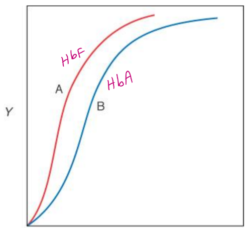
HbF (fetal) vs HbA (adults) affinity for oxygen
HbF has a higher
affinity for O2
than HbA
what are 3 specific roles for His residues in Hb function
His F8 aka proximal
Attachment of heme as prosthetic group to globin
His E7 aka distal
Assist O2 binding by forming h bond with it
Decreases affinity of CO
Geometry of substrate binding site
4 His in central cavity
BPG binding