9.4 - Structure & function of the nervous system
1/23
There's no tags or description
Looks like no tags are added yet.
Name | Mastery | Learn | Test | Matching | Spaced | Call with Kai |
|---|
No analytics yet
Send a link to your students to track their progress
24 Terms
What are the two divisions of the human nervous system?
Central nervous system
Peripheral nervous system (all neurones that are not part of the CNS)
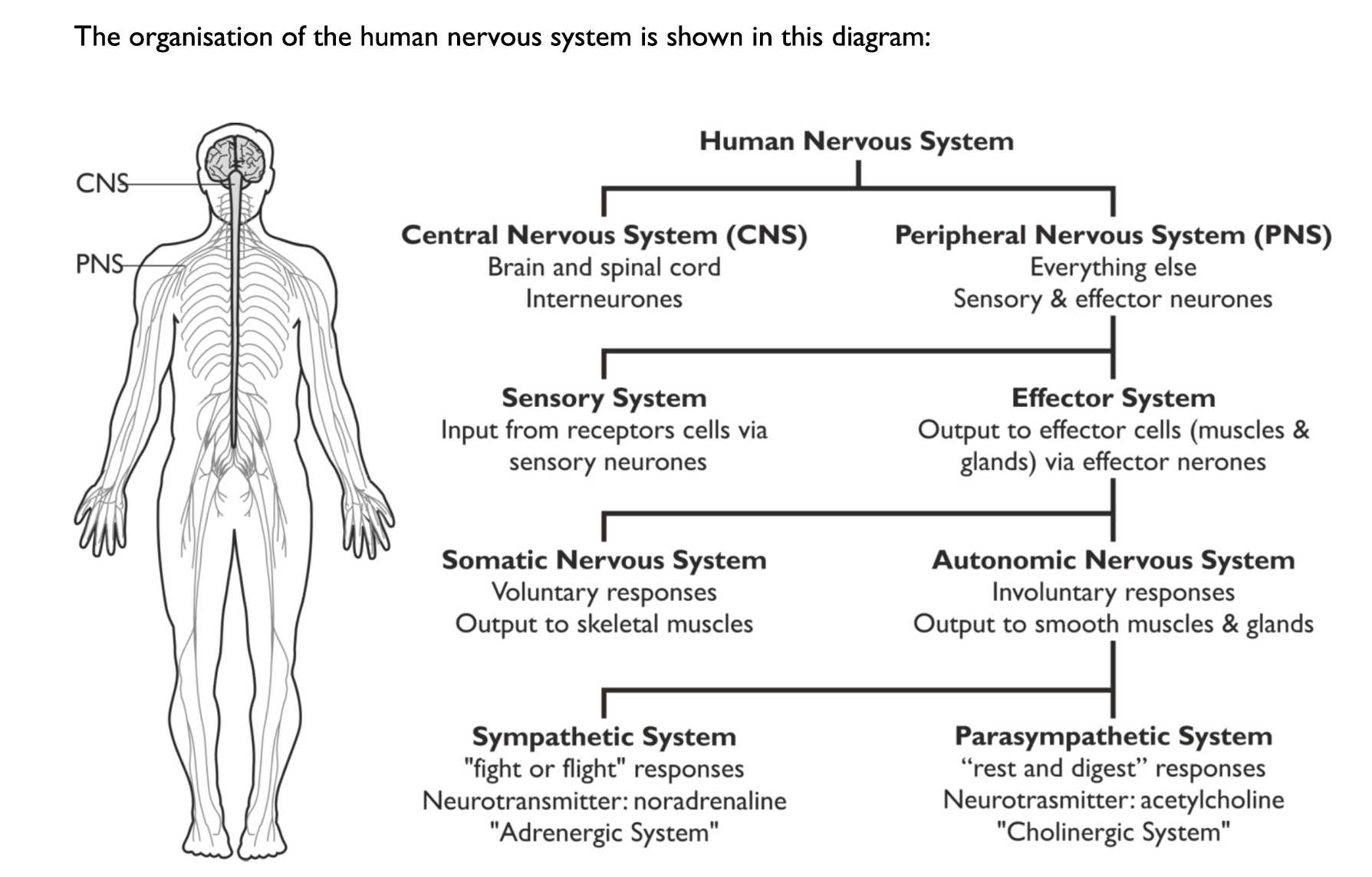
Describe the central nervous system
Comprised of the brain & spinal cord:
specialised system of nerve cells that processes stimuli & propagates impulses
What are the two divisions of the peripheral nervous system?
Somatic nervous system
Autonomic nervous system
What is the difference between the somatic & autonomic nervous system?
Somatic nervous system: controls voluntary responses (e.g. walking) & the output is to the skeletal muscles
Autonomic nervous system: controls involuntary responses (e.g. heart rate) & the output is to smooth muscles & glands
What are the two divisions of the autonomic nervous system?
Sympathetic nervous system
Parasympathetic nervous system
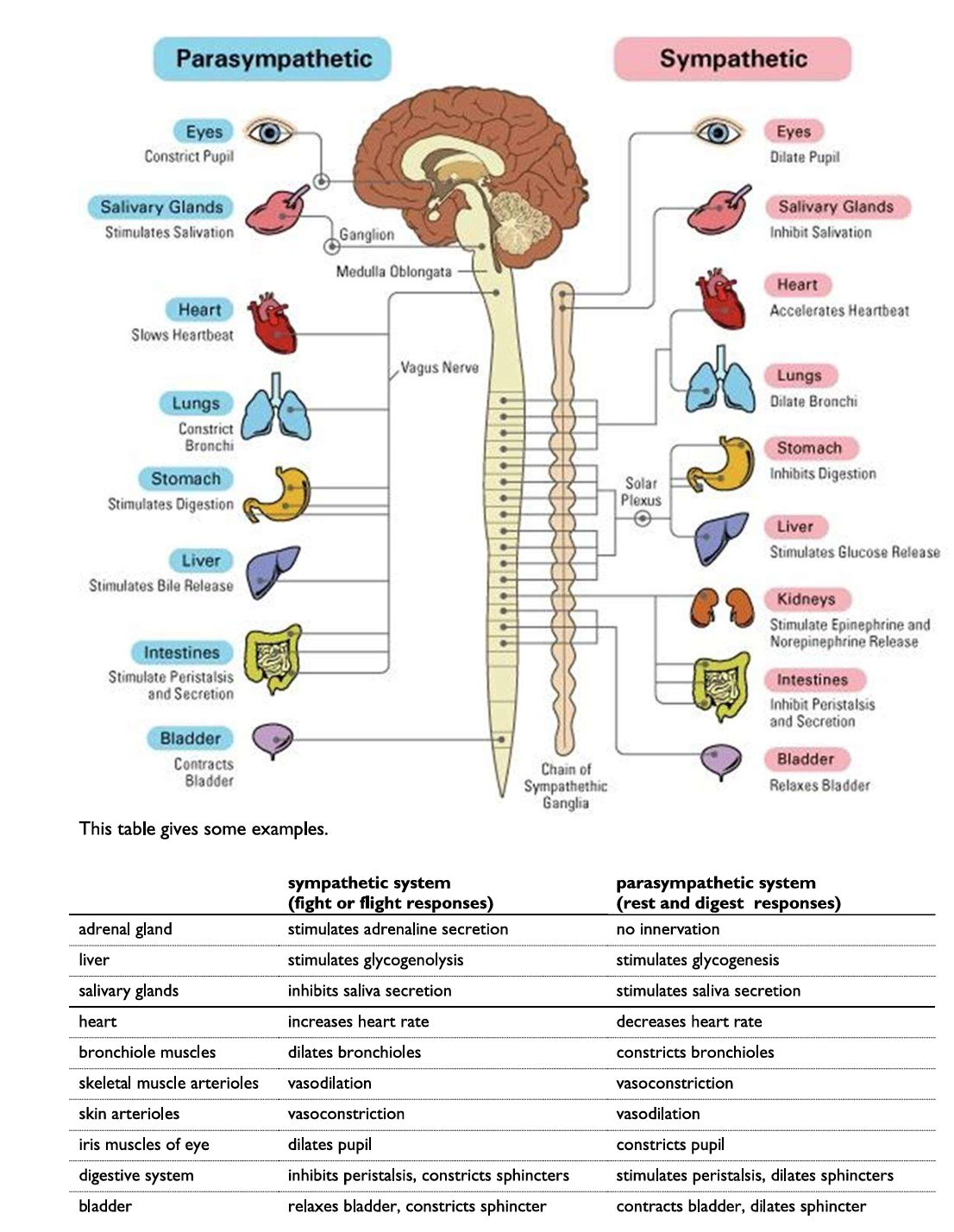
What are the overall roles of the sympathetic & parasympathetic divisions?
Sympathetic nervous system: responsible for the ‘flight or fight’ response & getting the body physiologically aroused; noradrenaline (hormone) is secreted
Parasympathetic nervous system: responsible for the ‘rest & digest’ response & returning the body back to its usual resting state; acetylcholine (neurotransmitter) is secreted)
Explain, using specific examples, why the sympathetic & parasympathetic systems largely have antagonistic effects
The sympathetic & parasympathetic nervous systems have contrasting responsibilities:
the sympathetic nervous system increases heart rate, dilates pupils & inhibits salivation during the ‘fight or flight response’
the parasympathetic nervous system decreases heart rate, constricts pupils & stimulates salivation
What gives grey matter its name?
Grey matter (central zone consisting of unmyelinated interneurones) appears grey under a light microscope
What is white matter made of?
Sensory & effector neurones
What is the central canal?
A narrow, fluid-filled channel that runs through the centre of the spinal cord

How many vertebrae make up the human spine & why must there be gaps between the vertebrae?
33 bony vertebrae:
the gaps between the vertebrae allow the spine to flex & the branches of the spinal cord to extend out to the periphery of the body
Through which ‘root’ do the sensory neurones always enter the spinal cord & the motor neurones always exit the spinal cord?
Sensory neurones always enter the spinal cord through the dorsal root, which contains the dorsal root ganglion
Effector/motor neurones always leave the spinal cord through the ventral root (their cell bodies are at the end of the cell in the grey matter of the spinal cord)

What is a dorsal root ganglion?
A swelling to hold all the sensory neurone cell bodies
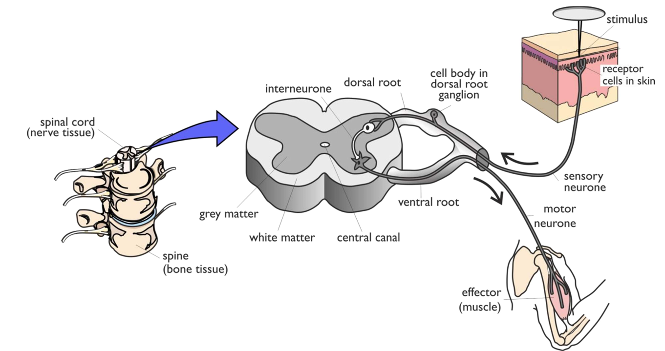
What is the definition of reflex?
A specific response to a specific stimulus, which protects the body from damage
Why are reflexes rapid & involuntary?
They involve few neurones, so have few synapses & the brain is not involved
What is a reflex arc?
The path taken by nerve impulses & involves only three neurones
What technique can be used to study the function of the different brain regions?
Magnetic Resonance Imaging (MRI)

What is the location & function of the medulla oblongata?
Location: base of the brain
Function: controls breathing & heart rate
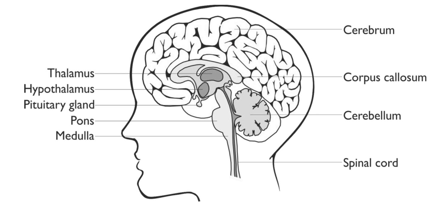
What is the location & function of the cerebellum?
Location: found at the back of the brain, below the cerebrum & behind the brainstem
Function: controls balance & coordination of movement

What is the location & function of the cerebrum?
Location: covers the upper half of the brain & is divided into two cerebral hemispheres
Function: initiates movement & is responsible for all voluntary functions

What is the location & function of the hypothalamus?
Location: base of the brain, below the thalamus & above the pituitary gland
Function: controls thermoregulation, osmoregulation & the release of hormones by the pituitary gland (e.g. sex hormones & the kidney hormone, ADH)
What is the somatosensory area?
The largest sensory area in the brain, responsible for the skin:
the sensory areas receive & organise sensory input from receptor cells → there are different sensory areas for each organ (e.g. visual, auditory, smell, touch, etc)
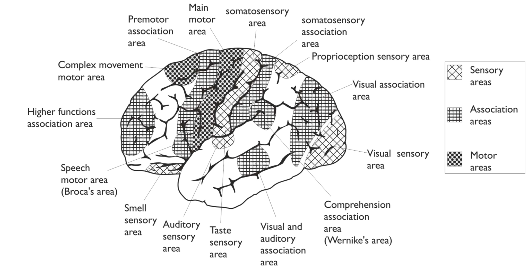
What is an association area?
Compares sensory input with previous experiences & so makes decisions:
they are involved in advanced skills (e.g. visual recognition, language comprehension, speech, writing & memory retrieval)

What is the role of the motor areas?
Organise & send motor output to skeletal muscles:
there is just one motor area, alongside the somatosensory area, which sends impulses via motor neurones to skeletal muscles
