Case 7: Neil Wartson Pt 1 - Hearing Loss
1/45
There's no tags or description
Looks like no tags are added yet.
Name | Mastery | Learn | Test | Matching | Spaced | Call with Kai | Chat |
|---|
No analytics yet
Send a link to your students to track their progress
46 Terms
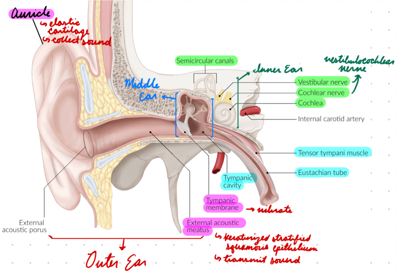
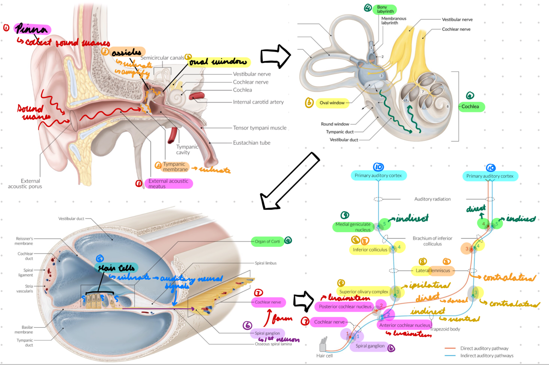
Ear: Parts
Outer ear
Middle ear
Inner ear
Outer Ear
External portion
Contains:
Auricle/Pinna
External auditory meatus
Tympanic membrane
Outer Ear: Auricle/Pinna
Visible portion
Elastic cartilage
Function: Collect sound waves into auditory canal

Outer Ear: External Auditory Meatus
Auditory canal
Keratinized stratified squamous epithelium
Contain glands producing cerumen (ear wax)
Function: Transmit sound waves to tympanic membrane

Outer Ear: Tympanic Membrane
Eardrum
Thin cone-shaped membrane
Cone of Light: Light reflection from otoscope on anterior inferior quadrant
Function: Vibrate from sound waves = Transmit vibrations to ossicles
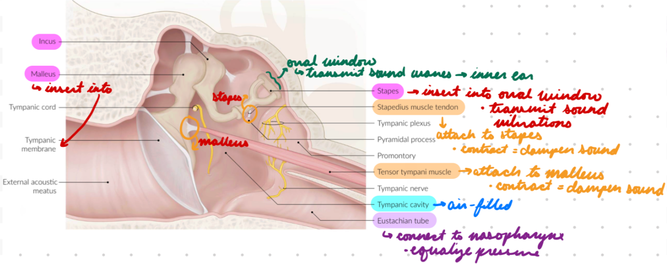
Middle Ear
Middle portion
Internal to tympanic membrane + external to oval window
Contains:
Tympanic cavity
Ossicles
Windows
Skeletal muscles
Mastoid process
Eustachian tube

Middle Ear: Tympanic Cavity
Air-filled space
Contain ossicles, muscles, nerves

Middle Ear: Ossicles
Small bones connecting tympanic window → Oval window
Malleus: Lateral
Insert into tympanic membrane
Incus: Middle
Stapes: Medial
Insert into oval window
Function:
Receive + amplify vibrations from tympanic membrane
Transmit vibrations to inner ear through oval window

Middle Ear: Windows
Oval: Opening at cochlea base
Covered by stapes
Function: Transmit sound waves to inner ear
Round: Membrane-covered opening below oval window
Perilymph transmit stapes vibrations from oval window = Vibrate in opposite phase
Middle Ear: Skeletal Muscles
Tensor Tympani: Attach to malleus
Contract = Pull malleus medially = Tense tympanic membrane = Dampen vibration + sound
Stapedius: Attach to stapes
Contract = Pull stapes laterally = Dampen stapes vibration = Dampen sound

Middle Ear: Mastoid Process
Temporal bone process behind ear
Middle Ear: Eustachian Tube
Connect tympanic cavity → Nasopharynx
Function: Equalize pressure around tympanic membrane
Middle ear pressure = External environment pressure
Open when chewing, swallowing, yawning
Inner Ear
Inner portion
Auditory section
Labyrinth
Cochlea
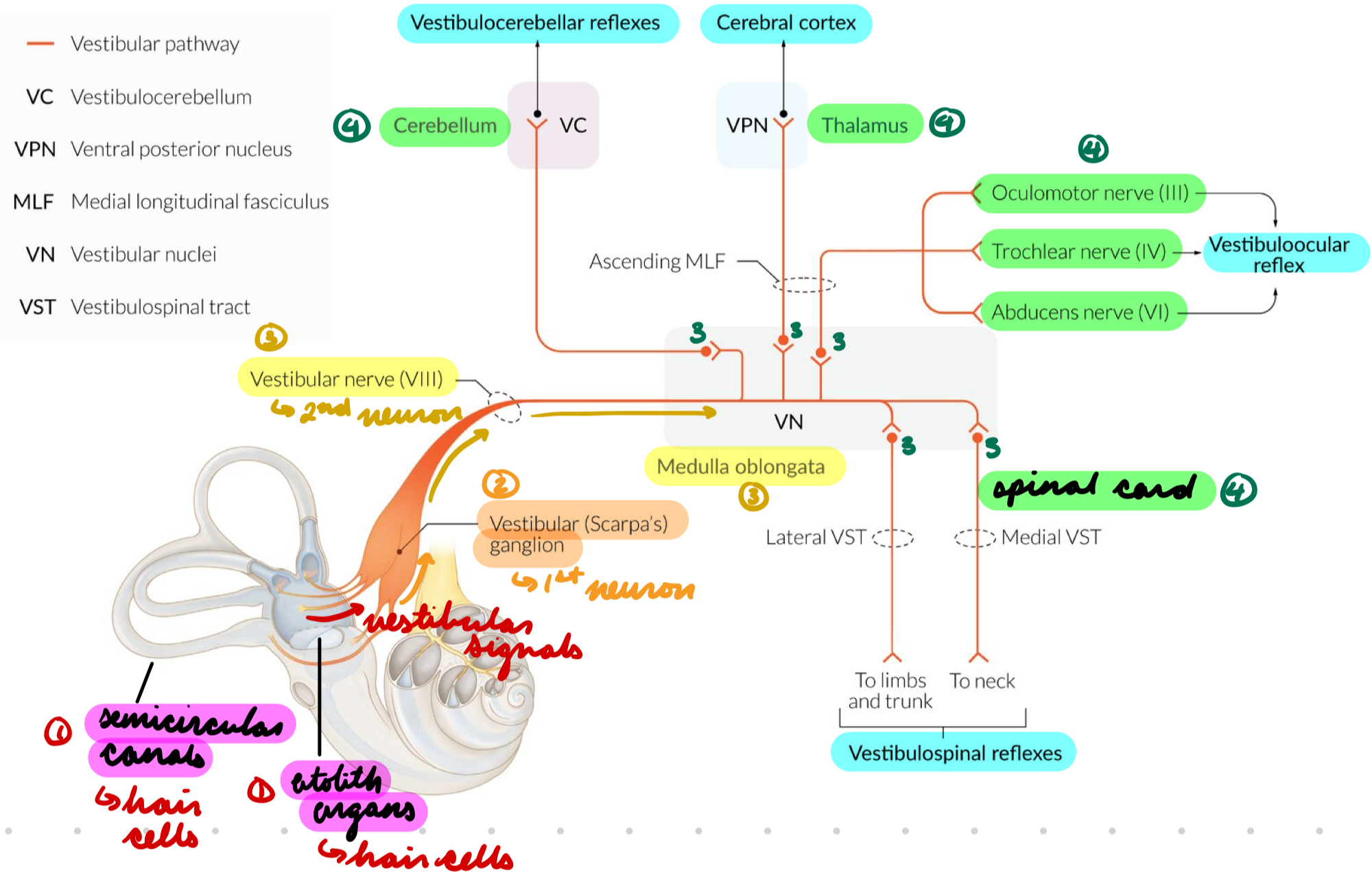
Vestibule section
Otolith organs
Semicircular canals
Vestibular hair cells

Inner Ear Auditory: Labyrinth
Osseous/Bony: Bony wall (temporal bone)
Filled with perilymph (low K+, high Na+)
Contain vestibule, semicircular canals, cochlea
Membranous: Inside osseous labyrinth
Filled with endolymph (high K+, low Na+)
Contain sensory organs
Function: Transmit vibrations from oval window to round window

Inner Ear Auditory: Cochlea
Fluid-filled cavity in osseous labyrinth
Layers:
Bone
Vestibular duct
Reissner membrane
Cochlear duct
Basilar membrane
Tympanic duct
Bone

Cochlea: Vestibular Duct
Filled with perilymph
Helicotrema (cochlea apex) connect oval window → Tympanic duct
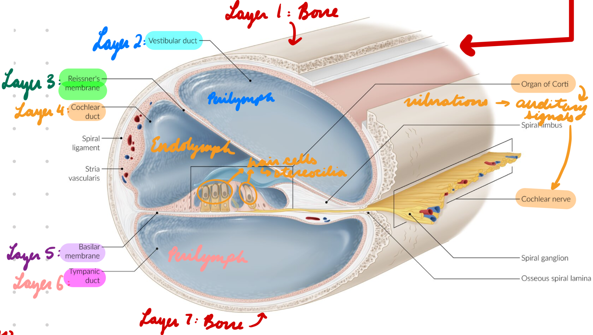
Cochlea: Cochlear Duct
Filled with endolymph
Organ of Corti: Transform vibrations → Auditory neural signals

Cochlea: Basilar Membrane
Support organ of Corti
Covered in auditory hair cells with stereocilia
High-Frequency: Stimulate hair cells at cochlea base = Thin + rigid basilar membrane
Low-Frequency: Stimulate hair cells at helicotrema = Wide + flexible basilar membrane

Cochlea: Tympanic Duct
Filled with perilymph
Connect to round window

Inner Ear Vestibule: Otolith Organs
Fluid-filled pouches with sensory epithelium
Utricle: Sense motion in horizontal plane
Forward-backward
Left-right
Saccule: Sense motion in sagittal plane
Up-down

Inner Ear Vestibule: Semicircular Canals
Sense rotary motion (angular acceleration)
Horizontal
Superior
Posterior
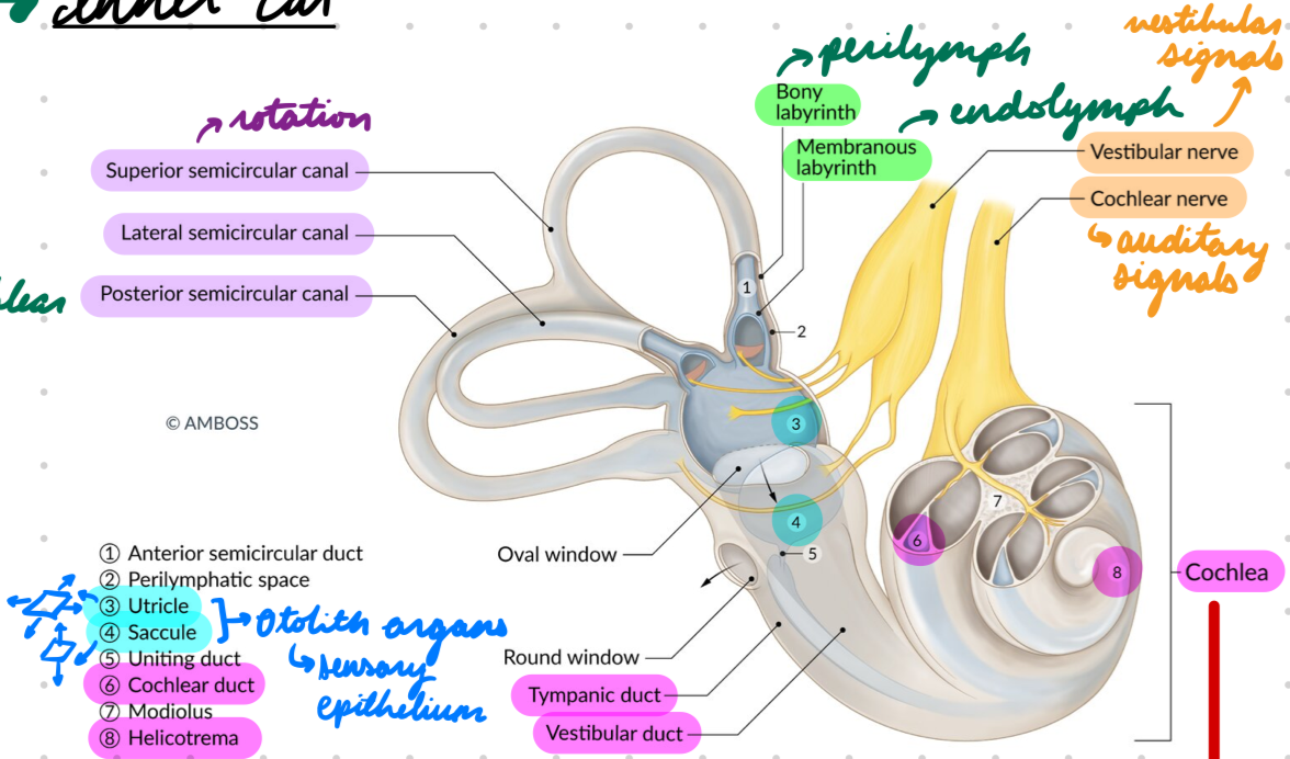
Inner Ear Vestibule: Vestibular Hair Cells
Transform displacement → Neural signals
In otolith organs + semicircular canals
Auditory Pathway
Sound waves collected into outer ear by pinna → Pass through external auditory meatus
Sound waves vibrate tympanic membrane = Transmit vibrations to ossicles (malleus → incus → stapes)
Ossicles amplify vibrations
Stapes transmit vibrations to oval window
Vibrations enter osseous labyrinth → Cochlea → Organ of Corti
Hair cells in organ of Corti vibrate = Transform into auditory neural signals
Signals transmitted to bipolar neurons (spiral ganglion)
1st order neuron
Bipolar neurons form cochlear nerve (part of vestibulocochlear nerve)
Synpase with ventral + dorsal cochlear nuclei (brainstem)
2nd order neuron
2 pathways:
Direct: Dorsal cochlear nucleus neurons join contralateral lateral lemniscus → Inferior colliculus
3rd order neuron
Indirect:
Ventral cochlear nucleus neurons synapse with ipsilateral + contralateral superior olivary nucleus
3rd order neuron
Superior olivary nucleus → Lateral lemniscus + synapse on inferior colliculus
4th order neuron
Inferior colliculus neurons synapse with medial geniculate body cells
4th (direct) + 5th (indirect) order neuron
Medial geniculate body neurons → Primary auditory cortex
Vestibular Pathway
Head movement = Move vestibular hair cells in otolith organs + semicircular canals = Transform into vestibular neural signals
Signals transmitted to bipolar neurons (vestibular ganglion)
1st order neuron
Bipolar neurons form vestibular nerve (part of vestibulocochlear nerve)
Synapse with vestibular nuclei complex (brainstem)
2nd order neuron
Vestibular nuclei complex transmit signal to cerebellum, CN III/IV/VI, reticular formation, spinal cord, thalamus → Vestibular cortex + reflexes
3rd order neuron
Hearing Loss: Description
Decreased auditory function
Hearing Loss: Types
Conductive: From impaired sound wave transmission to inner ear
Outer + middle ear dysfunction
Sensorineural: From impaired neuronal transmission to brain
Inner ear + cochlear nerve dysfunction
Mixed: Conductive + sensorineural components
Hearing Loss: Location
Central: Impaired brainstem + brain processing
No understanding of sound
Peripheral: Impaired ear (outer, middle, inner) + cochlear nerve
Decreased sound sensitivity
Hearing Loss: Epidemiology
20% in children
Conductive = Most common
Hearing Loss: Etiology
Pediatric:
Genetics
Perinatal complications
Premature birth
Increased brith weight
Hyperbilirubinemia
Trauma
Infection
Measles + mumps
Meningitis
Chronic otitis media
Adults:
Conductive:
Otosclerosis: Stapes overgrowth = Bone fixation = Decrease sound conduction
Otitis Media: Middle ear inflammation
Cerumen impaction
Sensorineural:
Idiopathic
Meniere disease: Impaired endolymph reabsorption
Older age
Presbycusis: Damage hair cells in organ of Corti (base first) = Lose high-frequency hearing
Trauma (noise-induced)
Neurologic
Acoustic neuroma: Schwannoma in vestibular nerve
Infection
Hearing Loss: Conductive Pathophysiology
Blockage (cerumen, inflammation) or dampening pathology (stapes fixing) = Decrease sound wave vibrations = Decrease sound transmission = Hearing loss
Hearing Loss: Sensorineural Pathophysiology
Decreased hair cells/movement or cochlear nerve pathology = Decrease auditory signal transmission = Hearing loss
Hearing Loss: Clinical Presentation
Conductive:
Hearing improves in noisy environments
Normal voice
No sound distortion
Sensorineural:
Hearing worsens in noisy environments
Loud voice
Sound distortion (lose high frequency)
Tinnitus
Pediatric:
Abnormal development
Delayed language + communication
Inappropriate response to sounds
Inattention
Symptoms of underyling cause
Facial asymmetry
Ear malformations
Microcephaly
Goiter
Hearing Loss: Investigations
Physical exam
Audiometry
Imaging
Hearing Loss Investigations: Physical Exam
Whispered voice test
Weber’s Exam: Tuning fork on forehead
Conductive: Louder in impaired ear
Detect vibration
Sensorineural: Louder in unimpaired ear
No sound transmission by damaged inner ear or auditory nerve
Rinne Exam: Tuning fork on mastoid bone until sound gone → In front of ear
Normal: Air conduction > bone conduction
Conductive: Bone > air
Vibrations bypass blockage in bone
Sensorineural: Air > bone (both decreased)
Inner ear + cochlear nerve cannot transmit sound to brain
Otoscopy:
Visualize tympanic membrane
Pneumatic: Apply pressure with pneumatic bulb = Tympanic membrane movement
Hearing Loss Investigations: Audiometry
Subjective:
Audiogram (pure tone testing)
Speech audiometry
Objective:
Impedance tympanometry
Otoacoustic emissions (OAE)
Audiometry: Audiogram
Pt listen to frequencies with headphones (air conduction) and bone oscillator (bone conduction) on mastoid bone
Determine threshold frequencies heard
Conduction: Threshold increase in air conduction + decrease in bone conduction
Sensorineural: Threshold increase in air + bone conduction
Audiometry: Speech Audiometry
Increasingly loud words played + pt repeat
Determine threshold level
Conductive: Louder = Increased comprehension
Sensorineural: Louder ≠ Increased comprehension
Audiometry: Impedance Tympanometry
Seal external auditory canal + emit frequencies inwards
Measure sound reflected from tympanic membrane as pressure
Record on tympanogram
Determine middle ear pathology
Eustachian tube dysfunction
Secretory otitis media
Audiometry: OAE
Measure sound emission (OAE) from cochlea in response to acoustic stimuli
Determine hair cell pathology
Normal cochlear function = Detectable
Cochlear hearing loss = Not detectable
Hearing Loss Investigations: Imaging
MRI/CT
Indication:
Unilateral
Gradual sensorineural
Acoustic neuroma
Hearing Loss: Investigations in Children
Hearing test
Otoscopy
Tympanometry
Hearing Loss Investigations in Children: Hearing Test
≤ 6 Months:
Automatic auditor brainstem response: Gold standard
Measure electrical activity along cochlear nerve + brainstem in response to sound
OAE
7 Months - 3 Years: OAE
≥ 4 Yeats:
Audiogram
OAE
Hearing Loss: Treatment
Treat underlying cause
Irreversible Causes:
Hearing aids: Bone/air-conduction aids
Cochlear implants
Hearing assistive technology
Hearing Loss Treatment: Hearing Aids
Amplify sound
Indications:
Conductive + sensorineural hearing loss
Mild to severe
Hearing Loss Treatment: Cochlear Implants
Surgically-implanted device electrically stimulate vestibulocochlear nerve
Improve speech discrimination
Indications:
Intact cochlea + cochlear nerve
Moderate to severe sensorineural hearing loss
Decreased speech recognition
Unsuccessful hearing aid treatment
Hearing Loss: Complications
Developmental delays
Language
Inattention
Cochlear implants: Increase meningitis risk
Prevention: Vaccinations