A + P Chapter 18
1/92
There's no tags or description
Looks like no tags are added yet.
Name | Mastery | Learn | Test | Matching | Spaced | Call with Kai |
|---|
No analytics yet
Send a link to your students to track their progress
93 Terms
What is the primary function of the plateau phase in cardiac myocyte action potentials?
to ensure full contraction and ejection of blood to chamber
During the Isovolumetric Relaxation Phase, ventricular volume:
stays the same
As with action potentials in other types of cells, the repolarization of cardiac myocytes involves the:
exit of potassium through voltage-gated channels.
Starting with the Superior Vena Cava, place the proper flow of blood in the correct order through the adult human heart and pulmonary circulation.
As a part of the parasympathetic system, the _______________ nerve innervates the heart acting on SA & AV nodes. When it stimulates these nodes, the resultant effect is to ________________ the heart rate.
vagus
decrease
An increase in the force of contraction of cardiomyocytes would ______ ESV and ______ SV.
decrease ESV
increase SV
Positive inotropic agents often work by increasing:
Calcium availability in cardiac muscle cells
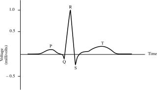
What phase on the graph represents atrial depolarization?
P wave

What phase on the graph represents ventricular depolarization?
QRS complex

What phase on the graph represents ventricular repolarization?
T wave

What phase on the graph represents the time it takes for electrical conduction to pass from the SA to the AV node
space between P wave and QRS complex

Atrial repolarization occurs during this part of the ECG waveform
QRS complex
What blood vessel is primarily responsible for dampening fluctuations in blood pressure due to its fiber content?
elastic arteries
Ventricular systole begins when the __________________________ and ends when the ________________________.
AV valves close
semilunar valves close
Smooth muscle cells would be found in which of the following layers of a typical blood vessel?
tunica media
How would an increase in afterload affect cardiac output?
decrease cardiac output
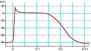
A typical Ventricular Action Potential is depicted below. The __________________ phase of the action potential allows for a sustained contraction of the cardiomyocytes.
plateau
What structure allows for ions to flow between adjacent cardiac myocytes?
gap junctions
The opening and closing of the valves of the heart is driven by:
differences in pressure across those valves
During the Isovolumetric Contraction phase of the cardiac cycle, the semilunar valves are ______________ and the AV valves are ________________.
closed
closed
One way cardiac myocytes differ from skeletal muscle cells?
have less developed sarcoplasmic reticulum
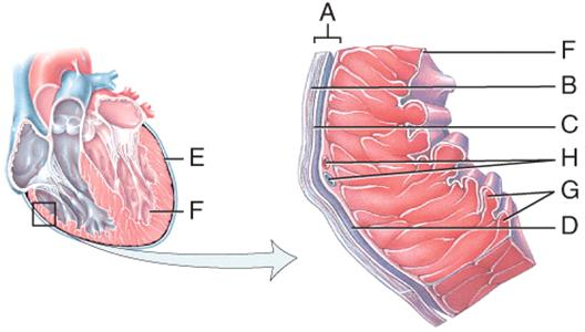
What letter is the innermost heart layer called the Endocardium? This layer is in direct contact with the blood contained within the chambers.
F
Right side of heart pumps through _______ circuit?
pulmonary
Left side of heart pumps through ______ circuit?
systemic

Identify structure 10
Right ventricle

Identify structure 13?
Pulmonary semilunar valves

Identify structure 9?
Left ventricle

Which structure represents the pulmonary trunk?
5

Which structure represents right + left pulmonary arteries?
5
What side of the heart receives oxygen-poor blood + pumps it to the lung?
Right
What side of the heart receives oxygenated blood from lungs + sends it throughout body?
Left
The heart has __ chambers?
4

Identify structure 1?
Right atrium
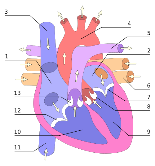
Identify structure 2?
Left atrium
Throughout the body, what type of blood vessels carry oxygen-rich blood away from the heart?
Arteries
Throughout the body, what type of blood vessels carry oxygen-depleted blood back TO the heart?
Veins (and venules)
Which blood vessels are typically depicted as blue?
Veins
Which blood vessels are typically depicted as red?
arteries
Unlike the rest of the body, what colors depict the pulmonary arteries + veins?
arteries= blue
veins= red
What does the color blue indicate about status of the blood in vessels?
nutrient poor
CO2 dense
O2 deficient
What does the color red indicate about status of the blood in vessels?
nutrient rich
O2 rich
CO2-less
The size of a heart can be compared to a?
fist
less than 1 lb.
What is the medial cavity of the thorax containing the heart called?
mediastinum
The heart is between what structures?
left and right lungs
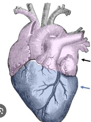
Identify the light purple area?
base of the heart
Identify the dark blue area?
apex of the heart
What side of the body is the majority of the heart positioned in?
Left
What is the inelastic, double-walled sac that encloses the heart called?
pericardium
The tough, outer, dense connective tissue layer of the pericardium?
fibrous pericardium
T/F: the pericardium is elastic
false
The thin, slippery two-layer tissue layer of the pericardium?
serous pericardium

What is A?
fibrous pericardium
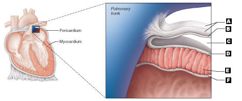
What is B?
parietal layer of serous pericardium
What two-layers comprise the serous pericardium?
parietal layer
visceral layer
What is the layer of the serous pericardium that lines the fibrous pericardium?
parietal layer
What is the layer of the serous pericardium that lines the fibrous pericardium?
visceral layer/epicardium
Between the parietal and visceral layers of the serous pericardium contains?
pericardial cavity, filled with serous fluid

What is C?
pericardial cavity

What is D?
epicardium

What is E?
myocardium

What is F?
endocardium
most superficial layer of the heart wall?
epicardium
What is the function of the epicardium?
supply blood to the heart with coronary blood vessels
What is the function of the myocardium?
contract the heart
The myocardium is comprised of interlacing bundles of ______ tissue fibers, arranged in ______ patterns.
connective
circular
Regarding the myocardium, the ____ side is significantly thicker as it pumps blood to the entirety of the body, not just to the lungs.
left
What is the function of the endocardium?
line heart chambers, covers valves
Which action leads to the closure of the right atrioventricular valve?
Contraction of the right ventricle
The four heart chambers are divided into top + bottom:
top: 2 atria
bottom: 2 ventricles
Internal partition of the heart that separates the atria?
interatrial septum
Internal partition of the heart that separates the ventricles?
interventricular septum
What is the structure + function of pectinate muscles?
comb-like ridges that assist in contraction of the atria
What is the structure + function of the papillary muscles?
pillar-like muscles ensuring valves in ventricles remain closed during systole (prevent backflow)
Which of the following is used to reduce friction between the layers of membranes surrounding the heart?
pericardial fluid
Within cardiac myocytes, which structure allows for ion flow between neighboring cells?
gap junctions
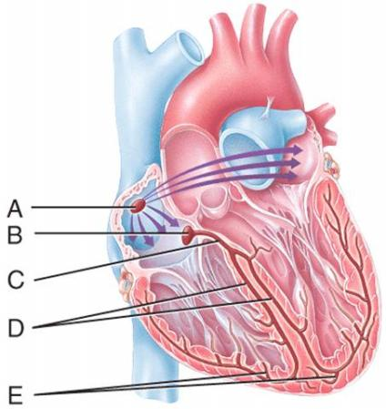
Which labeled structure in the figure is the AV node?
B
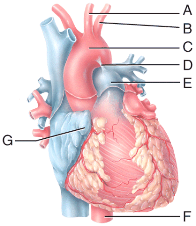
Which labeled blood vessel in the diagram is an artery carrying deoxygenated blood?
E - pulmonary artery
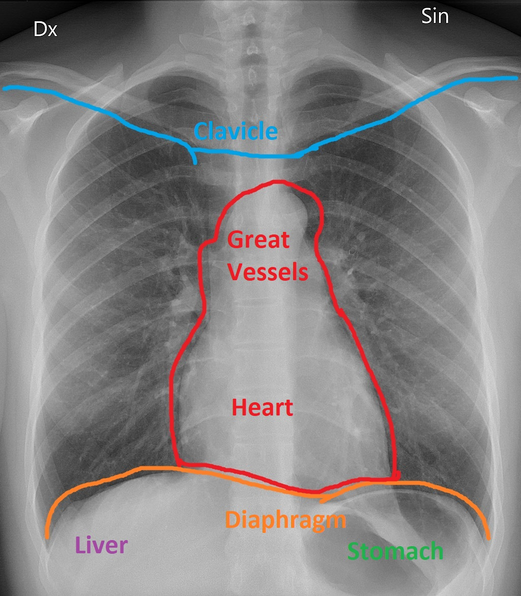
The accompanying image is a chest X-ray with the outline of the heart as it sits in the mediastinum resting atop the diaphragm. Which heart chamber forms the RIGHT border of human hearts?
right atrium
During which of the following cardiac cycle phases is ventricular pressure decreasing at the fastest rate?
isovolumetric relaxation
Blood leaving the left ventricle passes through which of the following structures?
aortic semilunar valve
In an ECG, the P wave is generated when the:
atria depolarizes
What of the following chambers of the heart contain deoxygenated blood?
right atrium + right ventricle
Which of the following occurs during the Isovolumetric phase of the cardiac cycle?
pressure within ventricles increases
Contraction of the atria of the heart leads to blood moving directly:
through atrioventricular valves

In the diagram, which labeled structure prevents blood flow from the right ventricle back into the right atrium?
B (tricuspid)
Which blood vessel carries oxygenated blood from the lungs to the heart?
pulmonary veins
The right coronary artery gives rise to which of the following arteries?
posterior interventricular artery

In the diagram, which labeled structure is the pulmonary semilunar valve?
A
Which of the following chambers of the heart is surrounded by the thickest layer of myocardium?
left ventricle
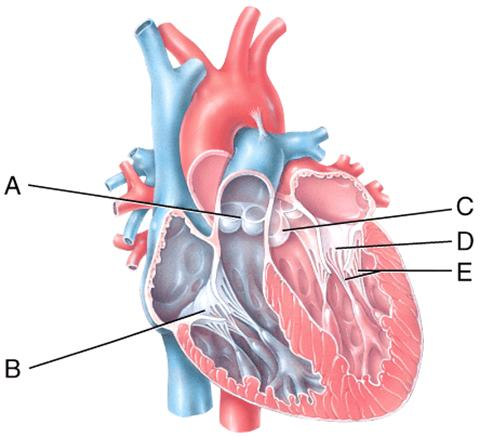
In the diagram, which labeled structures are atrioventricular valves?
B (tricuspid valve)
D (bicuspid valve)
As with action potentials in other types of cells, the repolarization of cardiac muscle cells involves the:
exit of potassium through voltage-gated channels
What are the functions of the pericardium?
prevents heart overfilling w/ blood, anchors, protects
What is the function of the pericardial cavity?
lubricate the heart