Cardiac cycle and heart murmurs
1/31
There's no tags or description
Looks like no tags are added yet.
Name | Mastery | Learn | Test | Matching | Spaced | Call with Kai |
|---|
No analytics yet
Send a link to your students to track their progress
32 Terms
Systole:
when the ventricles contract, blood is ejected into the circulation
During systole…
both AV valves are closed, thus preventing a back-flow into the atria. Both semilunar valves are open, allowing the blood to leave the ventricle
Diastole:
after ejection is completed, the ventricles relax and fill again
During diastole…
both semilunar valves are closed to prevent the ejected blood backflow into the ventricles.
Both AV valves are open to allow blood to flow from the large veins through the atria into the ventricles
CAVEAT: Atrial systole and diastole
the terms systole and diastole usually stand for the ventricles. Atria also have a systole and a diastole, but they are shorter and begin and end before the related ventricular actions
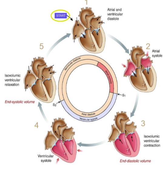
Diastole: Steps
5) Isovolumetric ventricular relaxation
1) Ventricular filling.
2) Atrial contraction or atrial systole
Systole: steps
3) Isovolumetric ventricular contraction
4) Ventricular ejection or systole
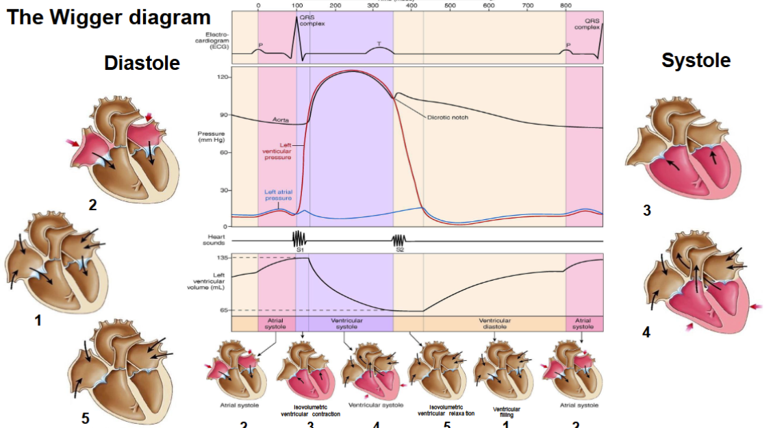
Cardiac cycle: phases
Phases and heart sound timing
Isovolumetric ventricular relaxation(transition from the previous systole: S2).
Ventricular filling (S3)
Atrial contraction or atrial systole (S4)
S2:
associated with the closure of the semilunar valves(pulmonic and aortic valves) (dub) and is typically loudest over these valve areas
Cardiac Cycle: Phases and heart sounds timing
S1
associated with the AV valves (lub) closure and is typically loudest over the mitral and tricuspid valve areas. The pulse occurs just after S1
Pressure-volume loop explained:
The pumping function of the heart.
A-B: diastole (ventricular filling and atrial systole).
B-C: systole (isovolumetric contraction).
C-D: systole (ventricular ejection).
D-A: diastole (isovolumetric relaxation).
EW = external work.
Cardiac Cycle: pressure-volume loop: allow us to explore the intricates of the heart
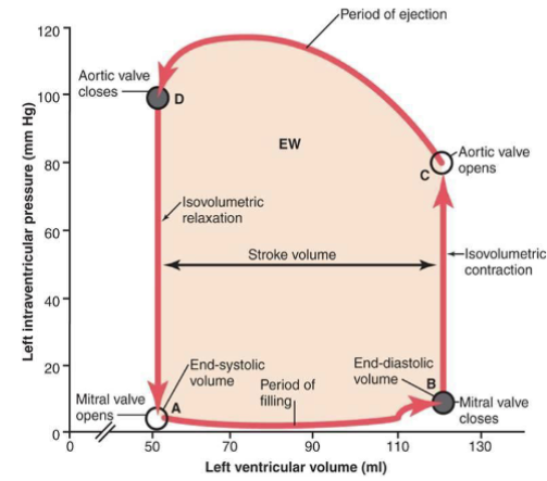
Preload(left ventricular end-diastolic pressure(LVEDP))
the degree of tension or stretch of the ventricular muscle when it begins to contract
Afterload
also known as systemic vascular resistance (SVR)
the amount of resistance the heart must overcome to open the aortic valve and push the blood volume into the systemic circulation.
Frank-starling mechanism
When the ventricular filling is increased, the myocardial fibers and their sarcomeres are stretched, optimizing the length-tension relationship of the sarcomere.
At the same time, troponin C becomes more sensitive to calcium as the sarcomeres stretch, accelerating the interaction between actin and myosis, leading to more force
In healthy hearts…
myofilaments become more sensitive to calcium as the myocardium is stretched within physiological limits.
Systolic-S1 sound:
associated with the closure of the AV valves (lub) and is typically loudest over the mitral and tricuspid valve areas. The pulse occurs just after S1
Diastolic-S2 sound:
associated with the closure of the semilunar valves (pulmonic and aortic valves)(dub) and is typically loudest over these valve areas
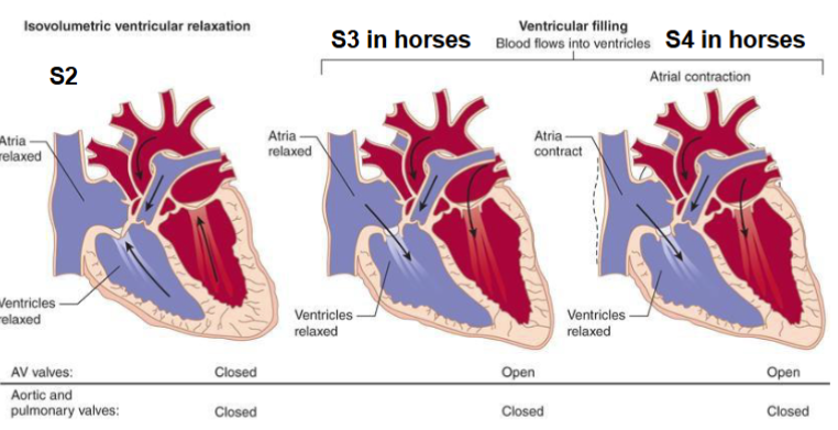
Diastolic-S3 sound:
low frequency, diastolic sound associated with rapid ventricular filling. Pathological in small animals
Diastolic-S4 sound:
low frequency, diastolic sound associated with atrial contraction.
Pathological in small animals: The pumping function creates a pressure gradient that guarantees blood flow
Heart murmurs
abnormal heart noises, either abnormalities of S1 and S2 heart sounds or additional abnormal noises
How are heart murmurs generated?
They are generated by turbulent blood flow through:
Altered valves.
Abnormal openings: between arteries and between heart chambers.
Benign murmurs: anemia
Mitral Valve Stenosis:
the mitral valve does not open as wide as it should, and blood flow from the left atrium to the left ventricle is partially restricted
Mitral Valve Insufficiency or regurgitation
the mitral valve leaks when the left ventricle contracts and some blood flows backward into the left atrium
Heart murmurs: mitral valve insufficiency
The murmur begins during the QRS complex (=systole) and ends during the T wave (end of systole). During diastole, the AV valves are open and consequently, the murmur dies out.
Patent Ductus Arteriosus
Heart murmur sounds “Machinery murmurs”.
Timing: systolic + diastolic murmur.
How common is patent ductus arteriosus?
PDA: is one of the three most common congenital heart defects identified in dogs.
■ PDA is most commonly hereditary in the dog, and it is characterized by ductal smooth muscle hypoplasia.
Causes of patent ductus arteriosus
Combination of stenosis and insufficiency
Mechanism of patent ductus arteriosus
The ductus arteriosus is a muscular blood vessel. Prostaglandins relax the ductus during fetal development.
At birth, lung expansion occurs, and oxygen tension in the systemic vasculature increases. The lung expansion and the vasodilatation of pulmonary blood vessels reduce the pulmonary resistance to the blood flow.
However, the ductus arteriosus contracts under a higher oxygen tension, and the smooth muscle then undergoes degeneration.
The wigger diagram