IB Biology - D2.1 Cell and Nuclear Division
1/65
There's no tags or description
Looks like no tags are added yet.
Name | Mastery | Learn | Test | Matching | Spaced | Call with Kai |
|---|
No analytics yet
Send a link to your students to track their progress
66 Terms
Parent Cell
original cell/mother cell before cell division and produces two daughter cells
Daughter Cell
The product of cell division.
Cytokenesis
The splitting of the cytoplasm of a parent cell to form two daughter cells. Each nucleur division is followed by cytokenesis.
Cleavage Furrow
The area of the cell membrane that pinches in and eventually separates the dividing cell using myosin and actin
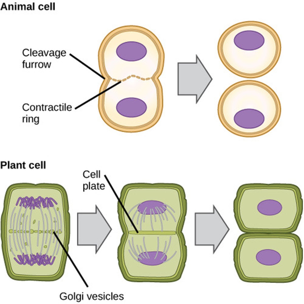
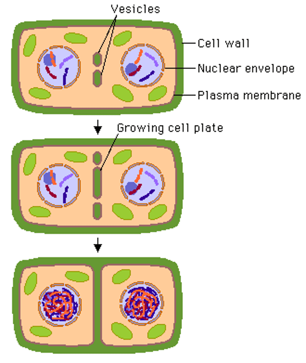
Cell plate
Cytokenesis in plants form vesicles of carbohydrates from the Golgi apparatus along the equator of the cell and fuse to form a cell plate and forms two separate cells, cellulse is secreted into the cell plate to form the cell wall of the new cells.
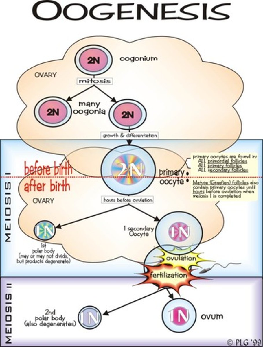
Oogenesis
Formation of eggs in females. Oogenesis involves unequal division of cells during meiosis to produce one large egg cell
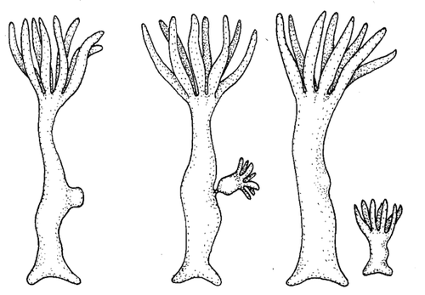
Budding
In yeast, it is a form of asexual reproduction where a small daughter cell buds off a larger cell by cytokenesis
Mitosis
Nuclear division which produces two genetically identical daughter nuclei. Parent cells undergoes Cytokenesis after mitosis.
Mitosis is required for?
growth and development (including embryological development)
Tissue repair as new cells are produced to replace dead cells
Asexual reproduction in unicellular (one cell) eukaryotic (with nucleus) organisms.
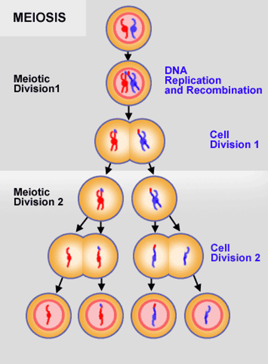
Meiosis
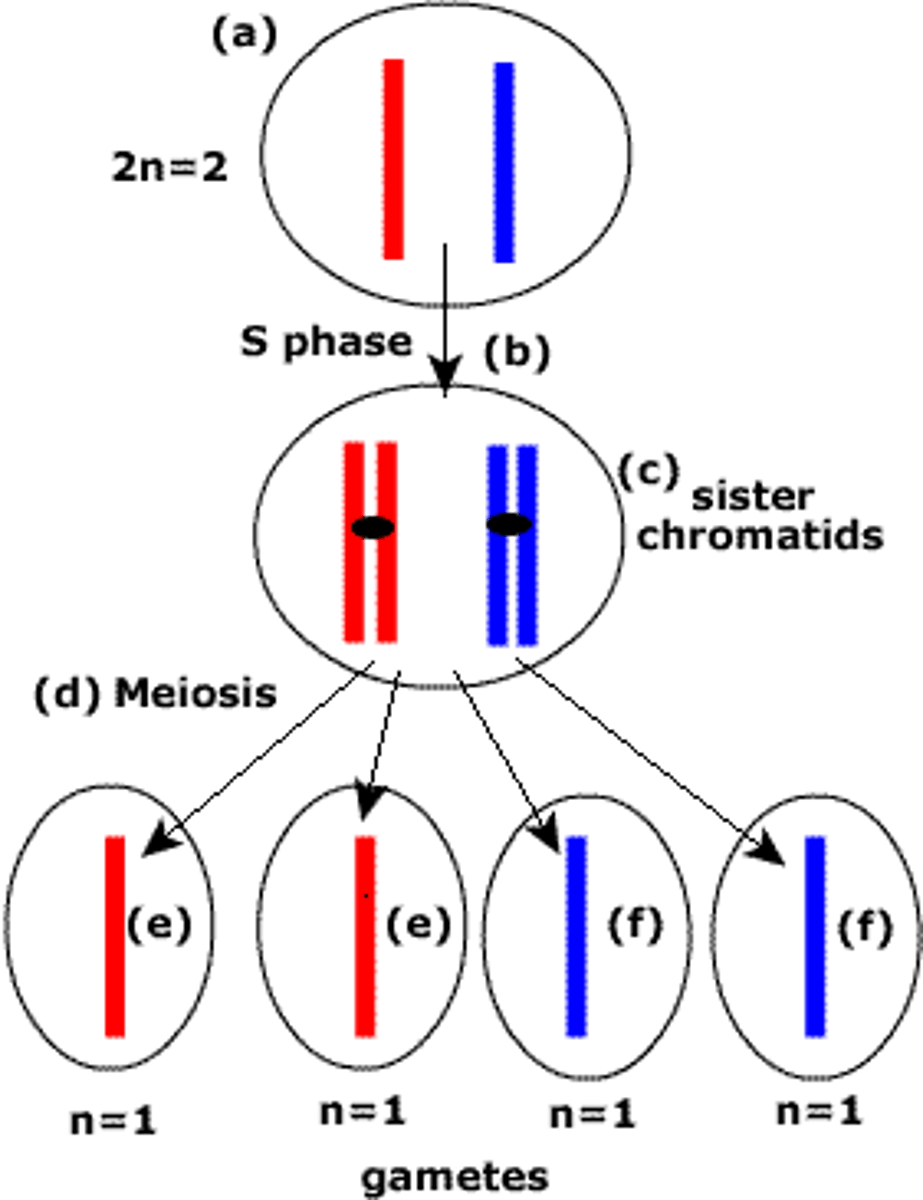
Nucleur division which produces four daughter cells, each with half the number of chromosomes as the parent cell. It is required for he production of gametes for sexual reproduction.
Gametes
Produced by meiosis, sex cells (egg and sperm)
Spores
Produced by meiosis. Grow into haploid organisms by mitosis.
Zygote
a diploid cell resulting from the fusion of two haploid gametes; a fertilized ovum.
Anucleate Cell
A cell without a nucleus
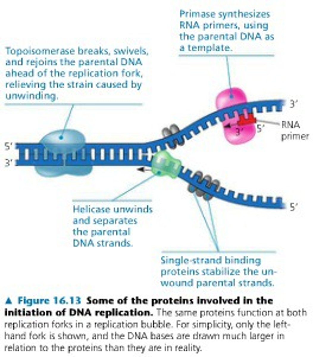
DNA Replication
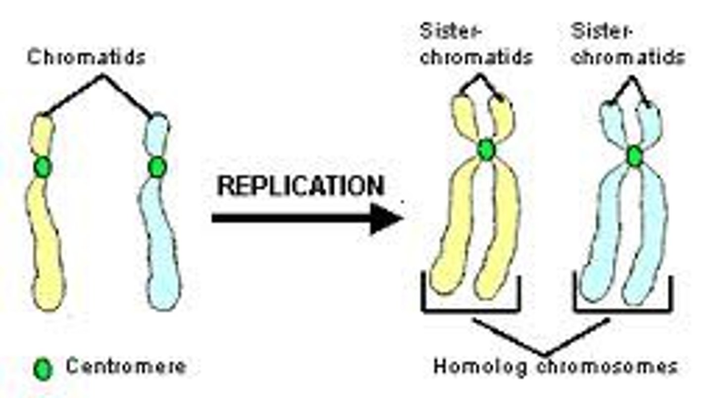
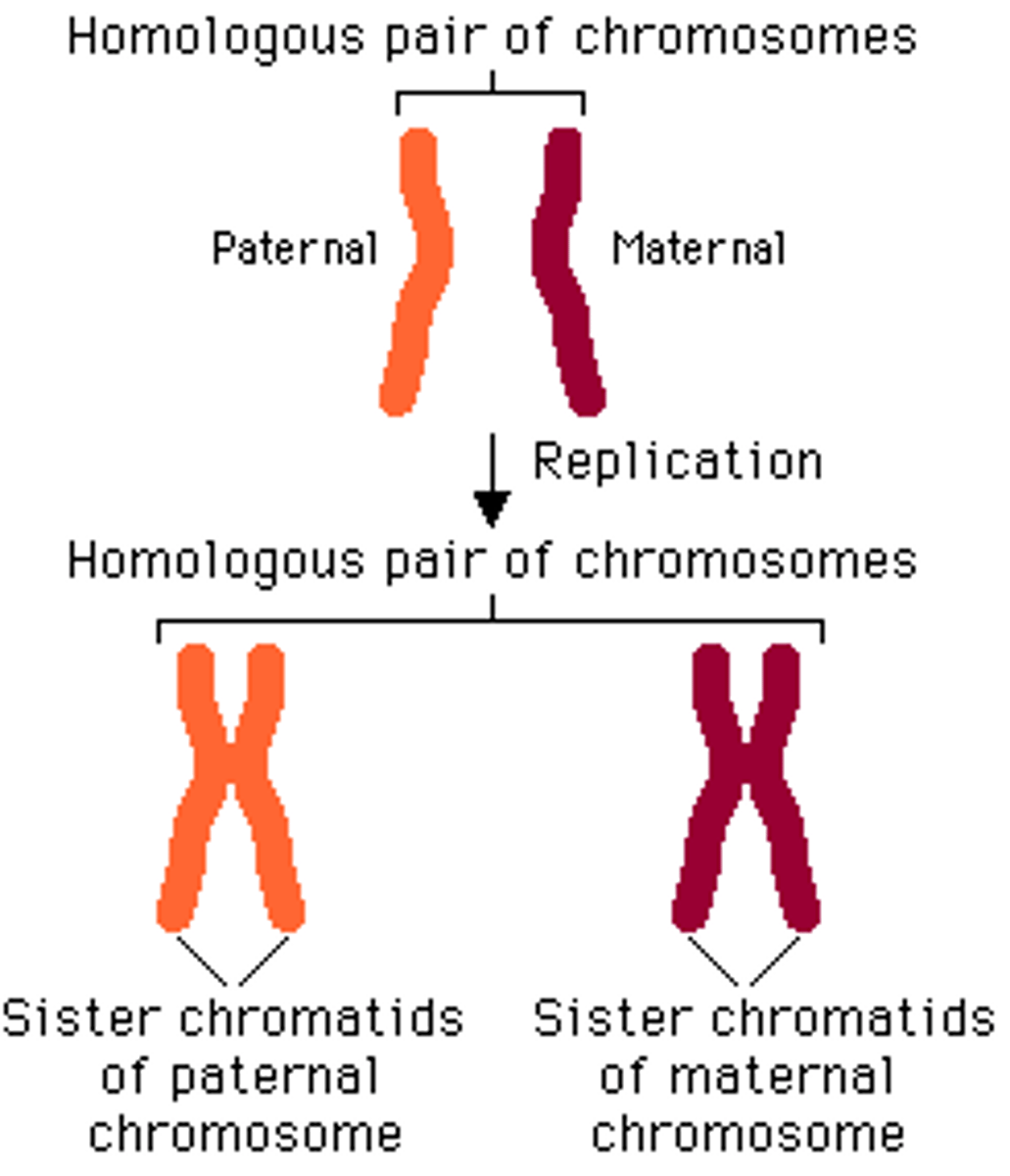
The production of two identical copies of a DNA molecule. Must occur before mitosis and meiosis and held together by a centromere.
Sister chromatids
Two identical copies of a duplicated chromosome; seperated during Anaphase of mitosis and Anaphase II of meiosis to form individual chromosomes
Condensation of Chromosomes
Chromatin condenses to form visible chromosomes, with sister chromatids during prophase of mitosis and prophase I and II of meiosis. During condensation, DNA coils around historic proteins to form nucleosomes. The nucleosomes coil around each other to form chromosomes with sister chromatids. Condensation of chromosomes allows for segregation of chromosomes during mitosis and meiosis
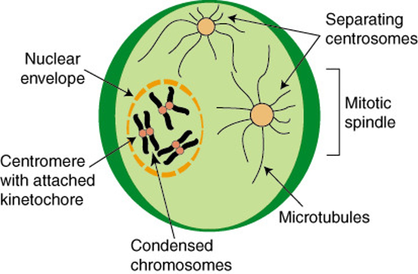
Microtubules
Spiral fibres of protein molecules that form a tubelike structure and attach to the centromeres of chromosomes by kinetochore during prophase of mitosis and meiosis.
Microtubule motors
on kinetochores and move the chromosomes to the poles during anaphase
Interphase
the resting phase between successive mitotic divisions of a cell, or between the first and second divisions of meiosis.
Nucleosome
repeating subunit of chromatin fibers, consisting of DNA coiled around histones
Histone protein
A simple protein bound to DNA, involved in the coiling of chromosomes
spindle fibers
Protein structures which move the chromosomes during cell division.
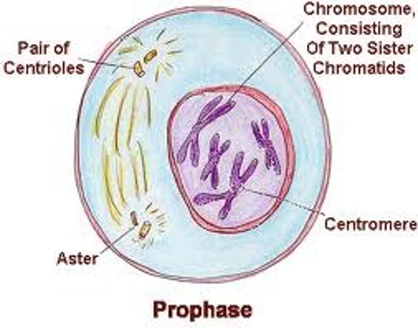
Prophase (mitosis)
Chromosomes condenses to form chromosomes which are visible under a light microscope. The chromosomes are composed of two identical sister chromatids attached to a centromere and the nucleur membrane breaks down. The centrioles move towards the poles producing spindle fibres, then the chromosomes attached to the spindle fibres at the centromeres.
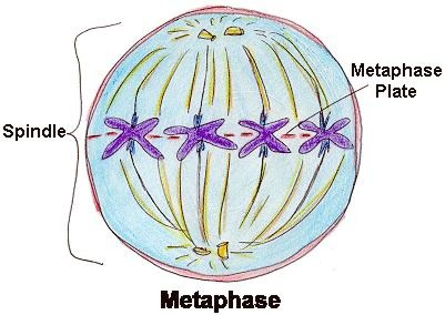
Metaphase (mitosis)
The chromosomes line up along the equator of the cell attached to spindle fibres
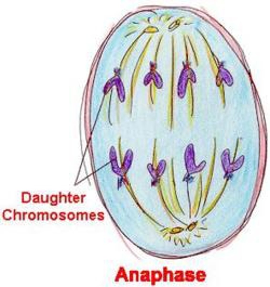
Anaphase (Mitosis)
The spindle fibers seperate the sister chromatids at the centromere to form single stranded chromosomes. The microtubule motors on the kinetochore, attached to centromeres, move chromosomes to opposite poles of the cell.
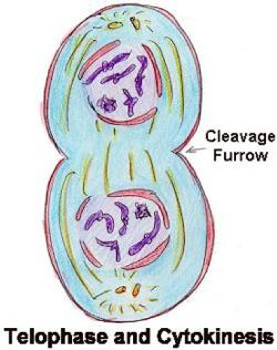
Telophase (mitosis)
Chromosomes arrive at the poils of the cell. Nucelur membranes form around the set of chromosomes at each pole. The chromosomes uncoil to form chromatin. Two genetically identical nuclei are produced. Cytokenesis begins during telophase to produce two genetically identical daughter cells.
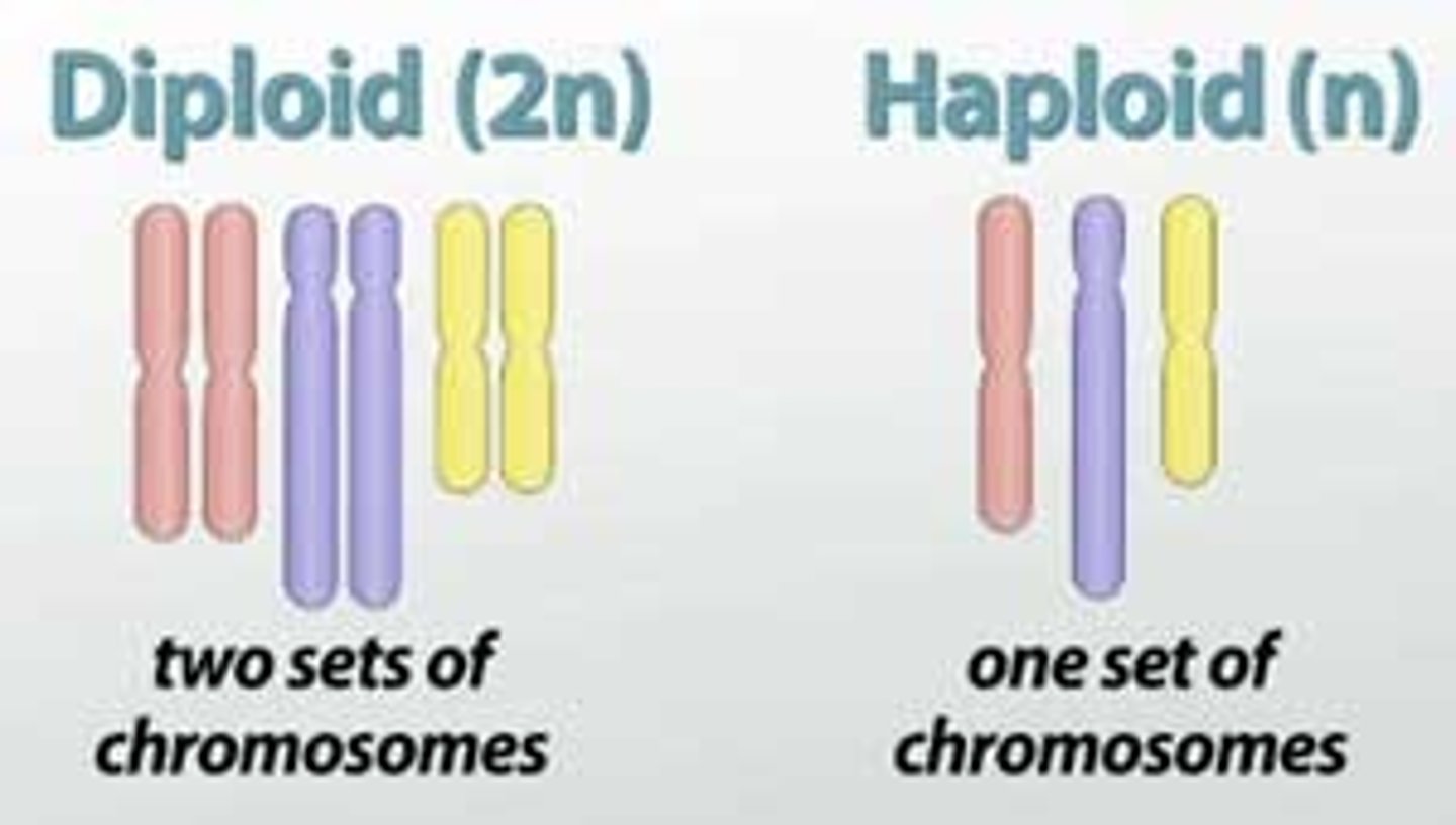
Diploid
2 sets of chromosomes (2n). A diploid cell has a nucleus with two sets of chromosomes. Each chromosomes will have a matching homolog (similar in position, structure, and evolutionary origin but not necessarily in function.)

Haploid
having a single set of unpaired chromosomes
Reduction division
Meiosis is known as a reduction division as the parent cell is diploid and the daughter cells are haploid and meiosis reduces the number by half, in the nucleus of the daughter cells.
Homologous chromosomes
Chromosomes that have the same sequence of genes and the same structure by may have different alleles
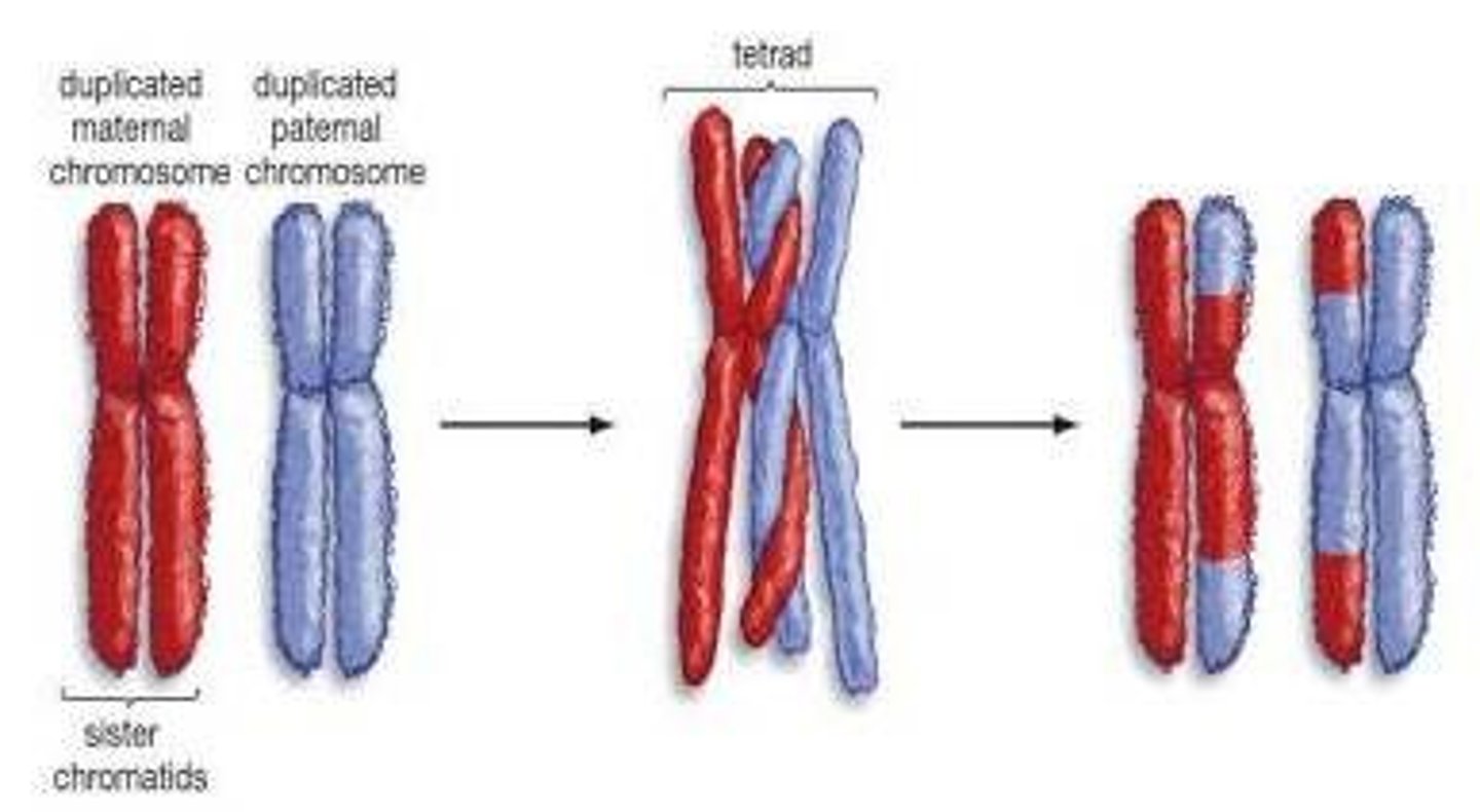
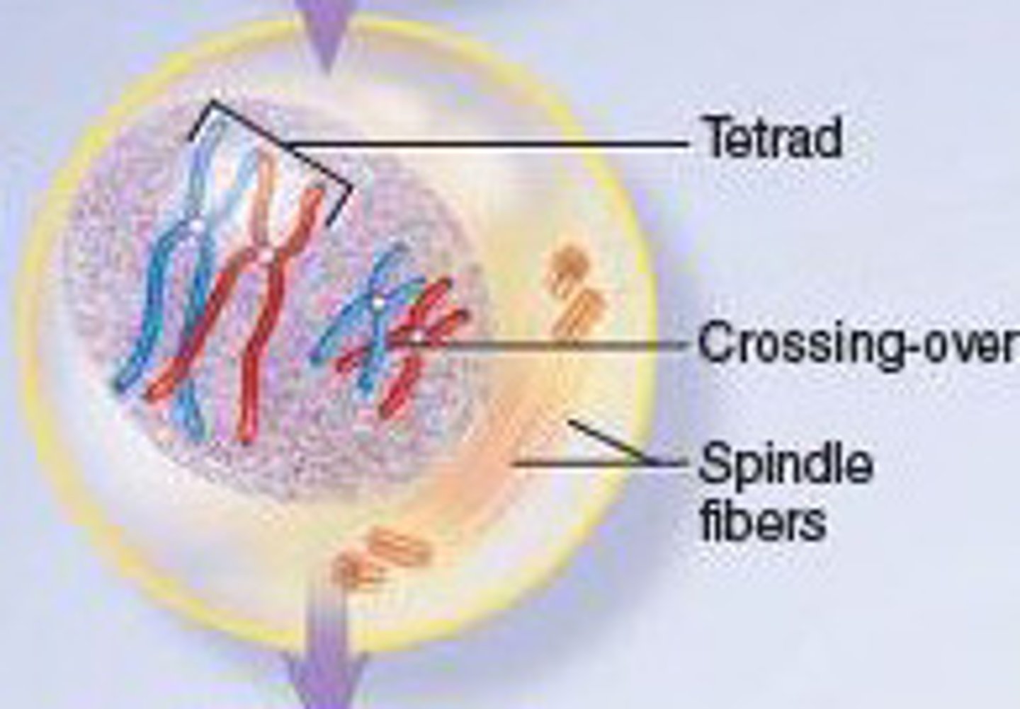
Bivalents
The paired chromosomes that result from the pairing of homologous pair. The non-sister chromatids cross over during the formation of bivalents, exchanging sections of non-sister chromatids.
Meiosis 1
Segregates the homologous chromosomes to produce two haploid cells
Meiosis II
Segregates the sister chromatids producing four haploid cells
Prophase I (meiosis 1)
Homologous chromosomes pair-up to form bivalents. Crossing over occurs where alleles switch between non-sister chromatids. Chromatin condenses to form sister chromatids (after crossing over). The centrioles move towards the poles ad start to produce spindle fibres. The nuclear membrane breaks down.
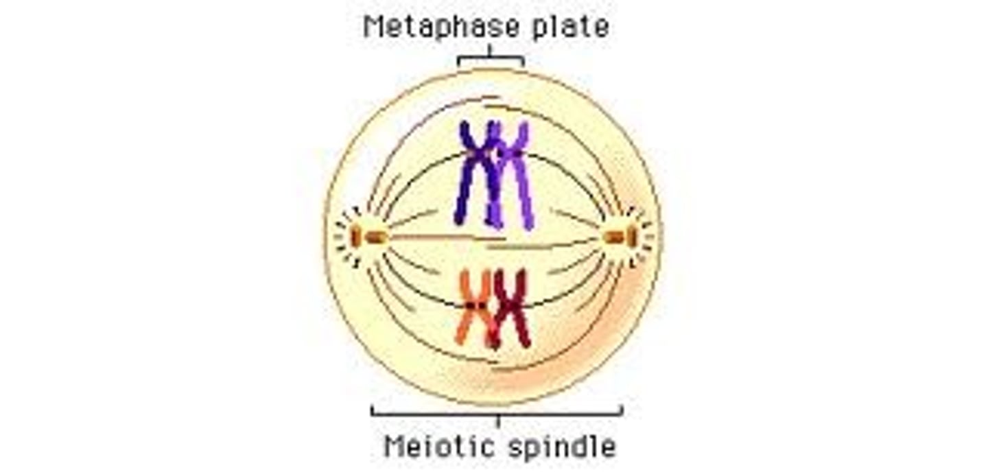
Metaphase I (meiosis I)
Spindle fibers move the bivalents to the equator of the cell. Sister chromatids are attached to the spindle fibres at the centromere. The maternal and paternal homologous chromosomes are randomly assorted as they line up along the equator of the cell.
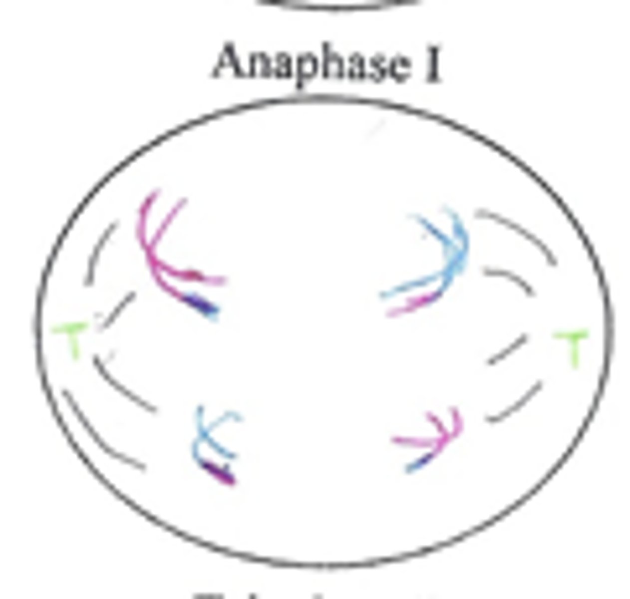
Anaphase I (meiosis I)
Homologous chromosomes from the bivalents separate and are pulled towards the poles of the cell by the spindle fibers. The microtubule motors on the kinetochore, attached to centromeres, move chromosomes to opposite poles of the cell.
Telophase I (meiosis I)
The chromosomes, as sister chromatids, arrive at the poles and uncoil. A nuclear membrane forms around the sister chromatids at each pole, producing two haploid nuclei. Cytokensis occurs to produce two haploid cells.
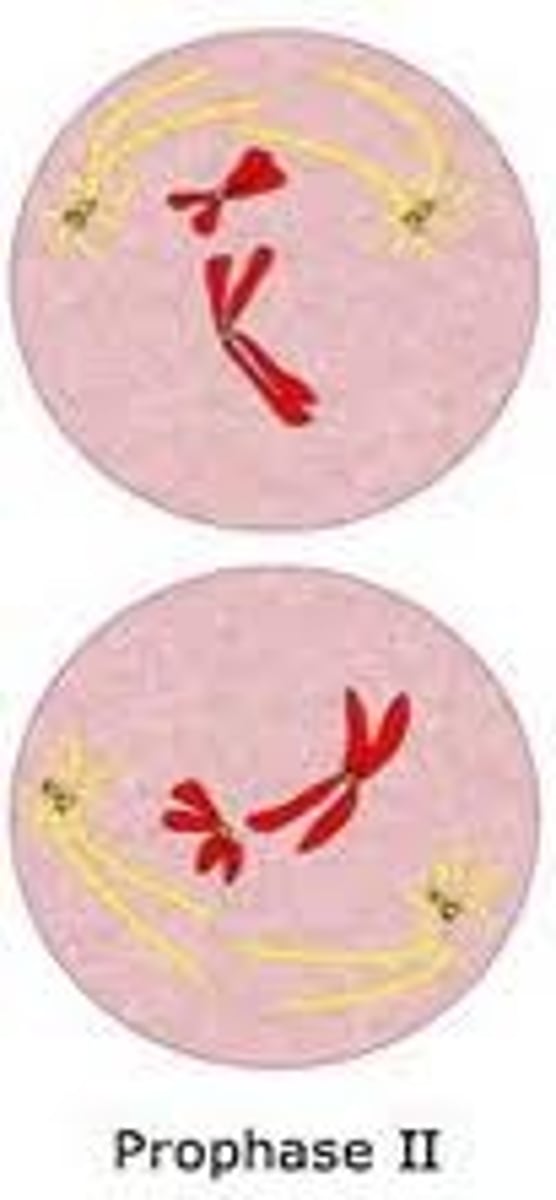
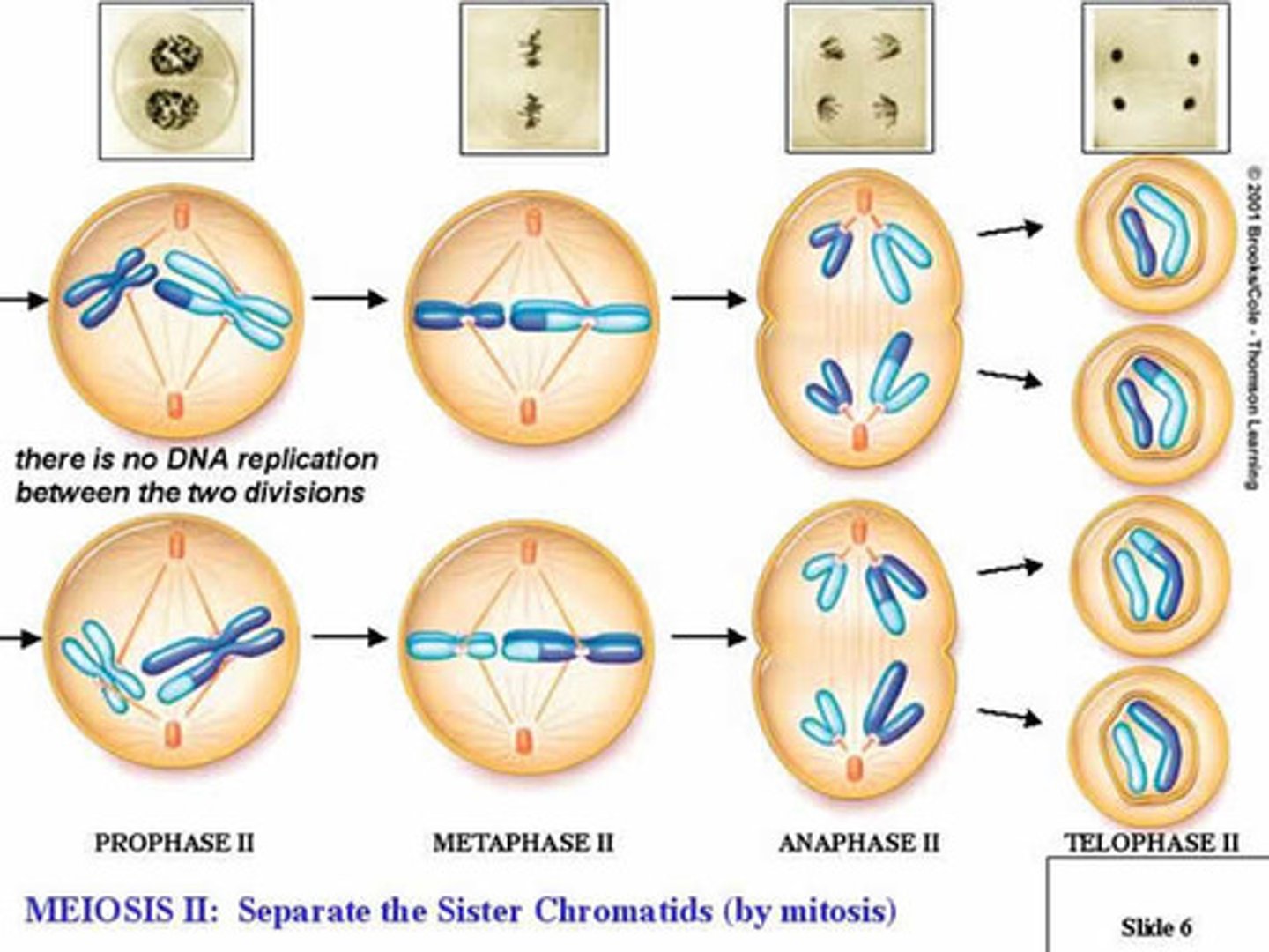
Prophase II (Meiosis II)
Chromosomes, as sister chromatids, super coil and appear in both haploid cells. The centrioles move towards the poles, producing the spindle fibres microtubules. The chromosomes (sister chromatids attach to the spindle fibres at the centromere.
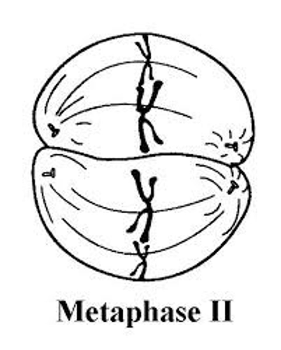
Metaphase II (Meiosis II)
Chromosomes line up along the equator of the cells, attached to the spindle fibre by the centromere. Bivalents dont line up only single chromosomes.
Anaphase II (Meiosis II)
Sister Chromatids are pulled apart (at the centromere) producing single stranded chromosomes. Those chromosomes are moved towards the poles by the microtubule motors on the kinetochore
Telophase II (Meiosis II)
The chromosomes reach the poles of each cell. The chromosomes uncoil. A nuclear membrane forms around each set of chromosomes. Cytokenesis occurs forming 4 cells with haploid nuclei with are not identical.
Non-disjunction
The failure of one or more pairs of homologous chromosomes or sister chromatids to separate fully during nuclear division. Can occur during Anaphase I if homologous chromosomes dont separate correctly, or Anaphase II of meiosis if sister chromatids dont seperate correctly or if it produces gametes with an extra chromosome or a missing chromosome.
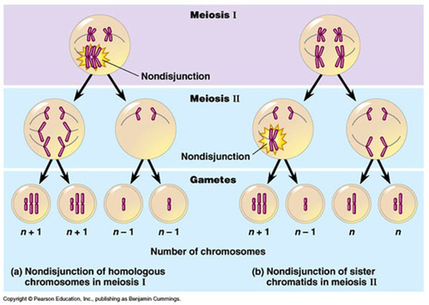
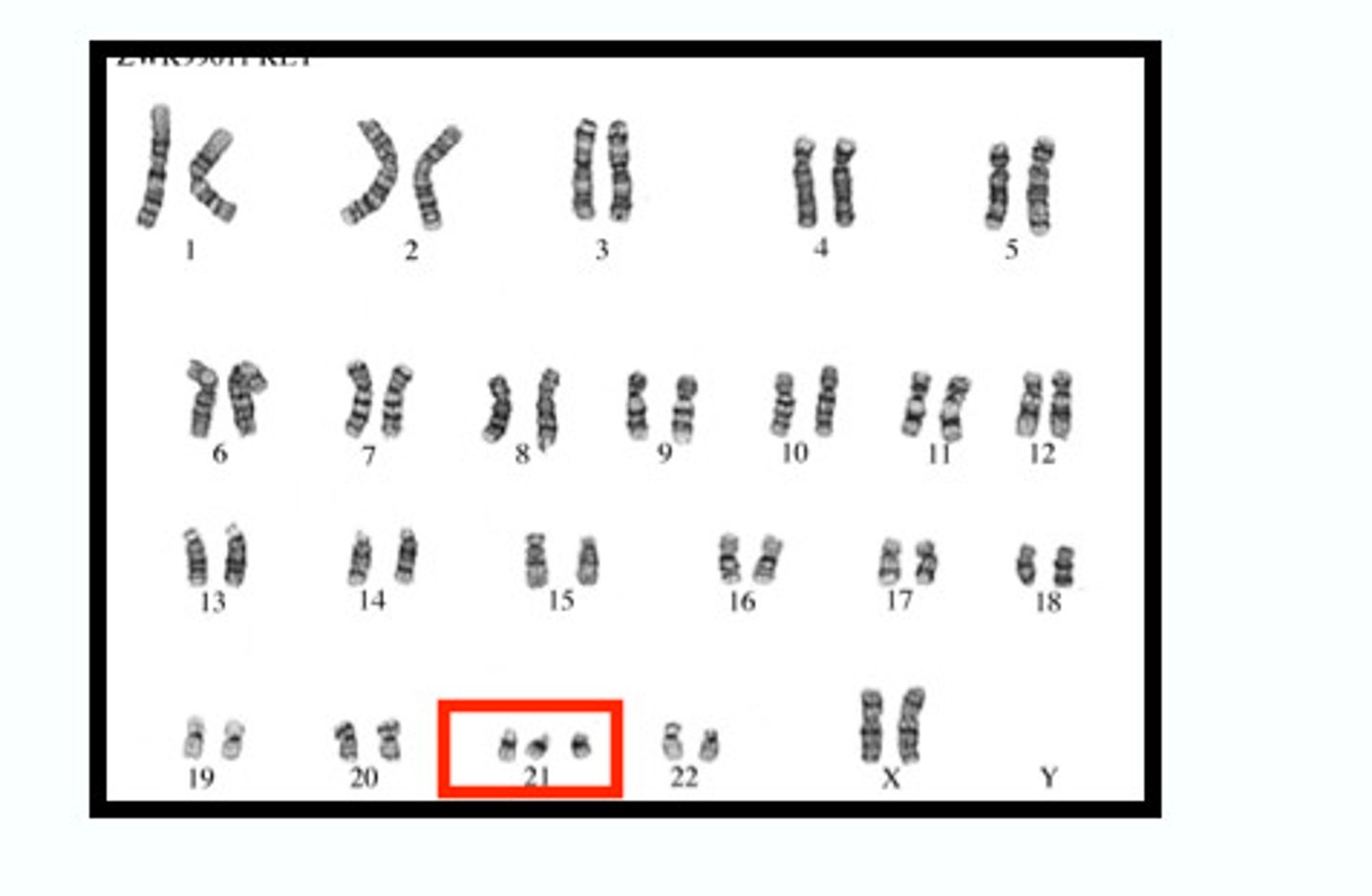
Down Syndrome (Trisomy 21)
A genetic disorder caused by the presence of all or part of a third copy of chromosome 21. Often caused by non disjunction during meiosis. The zygote will have 47 chromosomes with three copies of chromosome 21.
Cell Proliferation
The process by which a cell grows and divides to produce two daughter cells. Cell proliferation leads to an exponential increase in the number of cells is a rapid mechanism for tissue growth and repair
What are examples of Cell proliferation?
Embryological development: early embryos grow by cell proliferation through mitosis and cytokenesis
Plant meristems: Meristems are groups of undifferentiated cells which reproduce and differentiate to produce plant issues and organs.
Tissue replacement and healing: Dead and damaged tissue is replaced by cell proliferation. Skin cells are lost and replaced throughout an animal's life
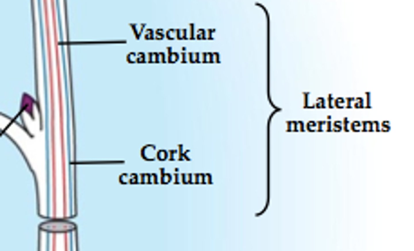
Meristems
Meristems are groups of undifferentiated cells which reproduce and differentiate to produce plant issues and organs.
Cell Cycle
interphase, mitosis, cytokinesis
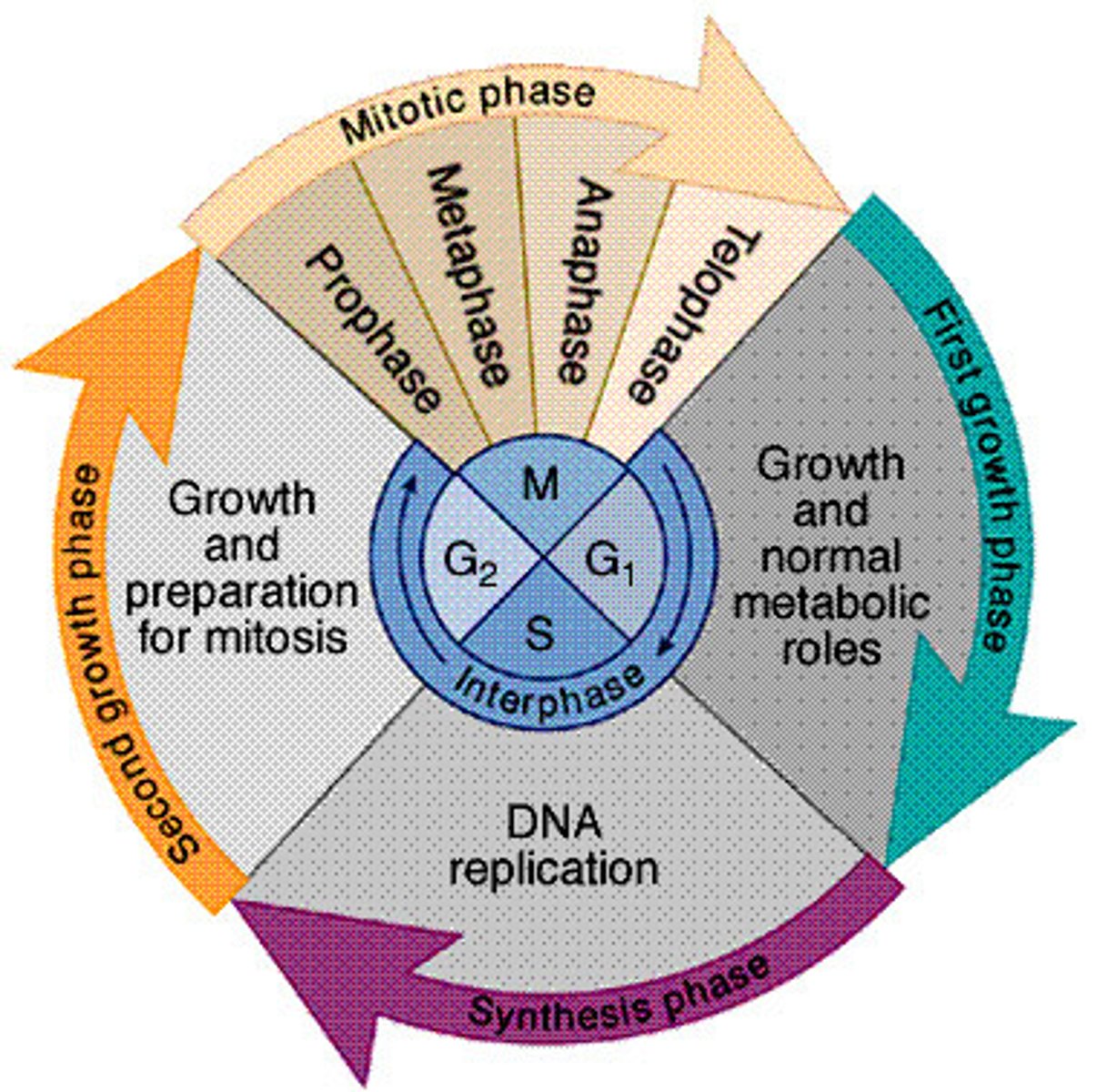
Interphase (HL)
Cell spends most of its life cycle in interphase. Transcription and translation occurs during interphase. The numbers of chloroplasts and mitochondria increase as the cell grows and during G1 and G2 phases of interphase.
G1 Phase
First stage of interphase. Cell grows after mitosis and cytokinesis. There is a lot of protein synthesis allowing the cell to grow and build new organelles.
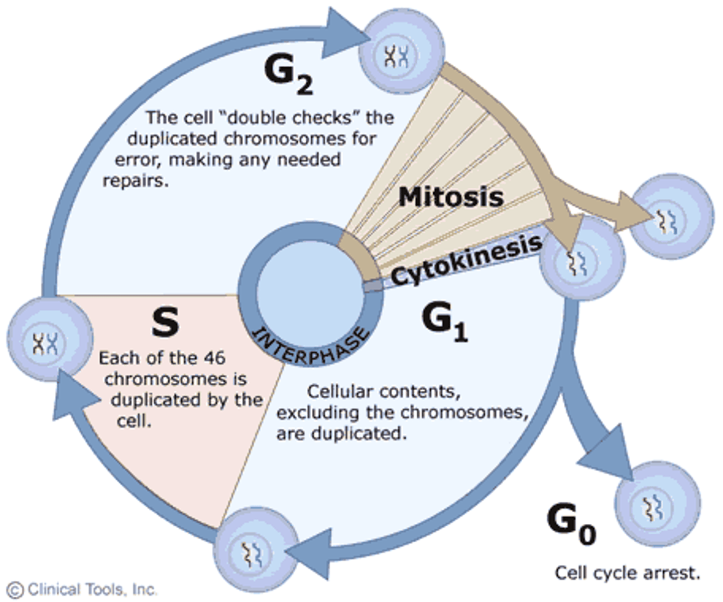
S Phase
2nd stage of interphase. DNA replication occurs to produce chromosomes with sister chromatids.
G2 Phase
Last stage of interphase. Cell prepares for mitosis by growing and replicating organelles.
Cyclins
A group of four proteins which control the movement of a cell through the cell cycle. A treshold level of a specific cyclin is required to pass each checkpoint in the cycle. Concentrations of different cyclins increase and decrease during the cell cycle.
Cyclin-dependent kinases (CDKs)
enzyme to which cyclin binds during interphase and mitosis, triggering and controlling activities during the cell cycle
Mutations
Any change to the DNA or RNA base sequence of a cell or virus.
Tumour suppressor genes
Tumour suppressor genes regulate cell division, as they normally inhibit cell proliferation and tumor development. Mutations to a tumour suppressor gene will cause the genes functions to be lost and uncontrolled cell division may occur.
Proto=oncogenes
Genes involved in regulating normal cell growth. Mutations to a proto-oncogene may cause it to become an oncogene
Oncogenes are genes that change a cell into a tumour cell which can lead to cancer
Tumours
abnormal proliferation of cells, either benign or malignant.
Primary tumour
first tumour produced throughout the body
Metastasis
The spread of cancer cells beyond throughout the body to form secondary tumours
secondary tumours
result from the metastasis of cancer cells throughout the body
cancer
uncontrolled cell division
Benign tumours (tumour by abnormal cell growth)
Non-cancerous and grow slowly and do not invade neighbouring tissue or undergo metastasis
Malignant Tumours (tumour by abnormal cell growth)
Cancerous, grow quickly and do undergo metastasis to spread to other parts of the body.
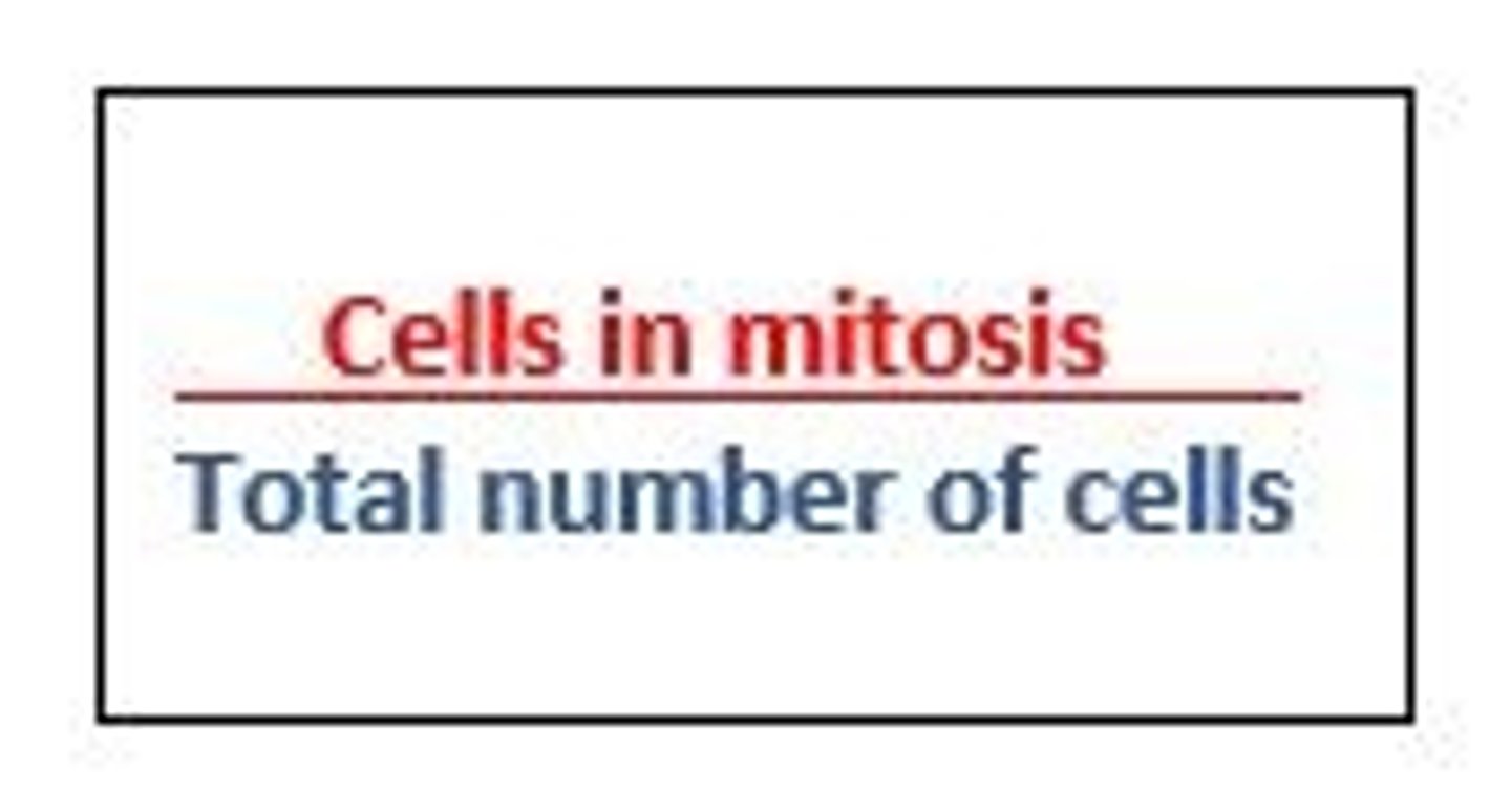
Mitotic Index
Ratio of cells in mitosis to the total number of cells and can be expressed as a percentage. A high mitotic index strongly suggests cancer is present in the tissue.