Cellular pathology (EXAM 1- MODS)

0.0(0)
Studied by 0 people
call kaiCall Kai
learnLearn
examPractice Test
spaced repetitionSpaced Repetition
heart puzzleMatch
flashcardsFlashcards
GameKnowt Play
Card Sorting

1/41

encourage image

There's no tags or description

Looks like no tags are added yet.

Last updated 9:35 PM on 6/8/26
Name
Mastery
Learn
Test
Matching
Spaced
Call with Kai

No analytics yet

Send a link to your students to track their progress

42 Terms

1
New cards

What are the mechanisms of the four basic types of cellular adaptation ?

  • Hyperplasia

  • Hypertrophy

  • Atrophy

  • Metaplasia

2
New cards

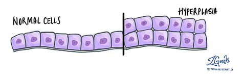
Describe Hyperplasia in cellular adaptation

An increase in the number of cells in an organ or tissue, which may increase its volume.

<p>An<span style="color: red;"> increase in the number of cells </span>in an organ or tissue, which may increase its volume.</p>
3
New cards

What are physiological adaptations for hyperplasia

occurs due to normal stressor:

  • Hormonal: ^# of glandular epithelial breast cells during pregnancy → enlargement of breasts→ preparation for lactation

  • Increased functional demand: Living at high altitude leads to hyperplasia of erythrocyte precursors in bone marrow

  • Compensatory: Regeneration of liver following partial hepatectomy. Regeneration of epidermis after abrasion

4
New cards

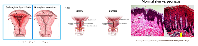
What are pathologic adaptations for hyperplasia

Occurs due to an abnormal stressor: ( ex; excessive stimulation of hormones)

  • Endometrial hyperplasia (increased estrogen)

  • Benign prostatic hyperplasia (androgens)

  • Epidermal hyperplasia in psoriasis

<p>Occurs due to an <span style="color: red;">abnormal stressor:</span> ( ex; excessive stimulation of hormones)</p><ul><li><p><span style="color: red;">Endometrial hyperplasia</span><span style="color: rgb(250, 249, 249);"> (increased estrogen)</span></p></li><li><p><span style="color: red;">Benign prostatic hyperplasia </span><span style="color: rgb(255, 247, 247);">(androgens)</span></p></li><li><p><span style="color: red;">Epidermal hyperplasia in psoriasis</span></p></li></ul><p></p>
5
New cards

Describe Hypertrophy in cellular adaptation

An increase in the size of a cell, resulting in an increased organ size.

<p>An increase in the size of a cell, resulting in an increased organ size.</p><p></p>
6
New cards

What are pathologic adaptations for hypertrophy

increase in size of heart due to aortic stenosis

7
New cards

What are physiological adaptations for hypertrophy

caused by functional demand/ hormones

  • Enlargement of skeletal muscle with exercise (increased functional demand)

  • Physiological growth of uterus during pregnancy (Hyperplasia + Hypertrophy)

8
New cards

Describe Atrophy in cellular adaptation

decrease in the size of a cell (organ)

  • May be due to loss of blood supply, loss of endocrine stimulus, disuse, decreased workload, aging etc.

9
New cards

What are physiological adaptations for atrophy

caused by normal aging or hormonal drops

  • Atrophy of brain with aging

  • Atrophy of gonads after menopause (decreased hormones)

  • Decrease in the size of the uterus after pregnancy

10
New cards

What are pathologic adaptations for atrophy

Caused by disuse

  • Immobilization of a limb after fx (disuse)

  • Cachexia (starvation, insufficient nutrients)

  • Ischemic process (inadequate supply of oxygen)

11
New cards

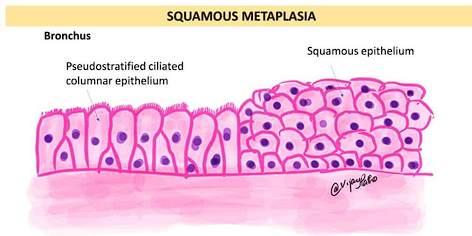
Describe metaplasia in cellular adaptation

change of epithelium at a site, or location, from one type to another (change in cell type)

  • epithelium normally present at a site cannot handle the new environment so it converts to a type of epithelium that can adapt.

<p>change of epithelium at a site, or location, from one type to another <span style="color: red;">(change in cell type)</span></p><ul><li><p>epithelium normally present at a site cannot handle the new environment  so it converts to a type of epithelium that can adapt.</p></li></ul><p></p>
12
New cards

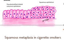
What are pathologic adaptations for metaplasia

abnormal changes

  • Cigarette smoking

  • Barrett’s esophagus

<p>abnormal changes</p><ul><li><p>Cigarette smoking</p></li><li><p>Barrett’s esophagus</p></li></ul><p></p>
13
New cards

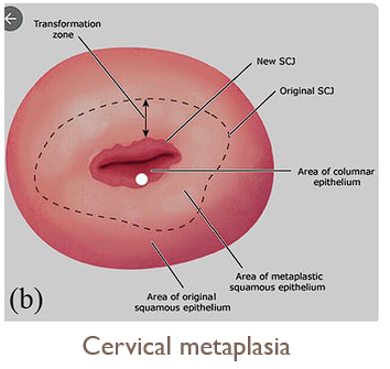
What are physiological adaptations for metaplasia

Normal cervical changes at the transformation zone

<p>Normal cervical changes at the transformation zone</p><p></p>
14
New cards

describe/ list the causes of cell injury

  • occurs when the cells cannot adapt to their new environment

  • Causes:

    • Hypoxia (common)

    • Ischemia (common)

    • Physical agents

    • chemical agents

    • infectious agents

    • radiation and toxins

    • metabolic abnormalities

    • immune dysfunction

    • Nutritional imbalances

    • Aging

15
New cards

List the mechanisms of cellular injury?

  1. Hypoxia

  2. Free radicals

  3. Chemical injury

  4. Increased mitochondrial cytosolic calcium (mitochondrial damage)

16
New cards

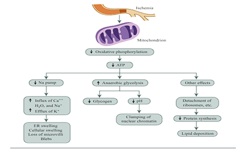
Describe the mechanism of hypoxia in cellular injury

  • No Blood No Oxygen→ No ATP

  • Results in: Swelling (pump fails), Acid/Clumping (backup power fails), and Fat Build-up (protein factories break).

  • refer to diagram

<ul><li><p><strong>No Blood </strong><span><strong>→</strong></span><strong> No Oxygen→ No ATP</strong></p></li><li><p><strong>Results in: </strong><span style="color: red;"><strong>Swelling</strong> (pump fails),</span> <span style="color: red;"><strong>Acid/Clumping</strong></span> (backup power fails), and<span style="color: red;"> <strong>Fat Build-up</strong></span> (protein factories break).</p></li><li><p>refer to diagram</p></li></ul><p></p>
17
New cards

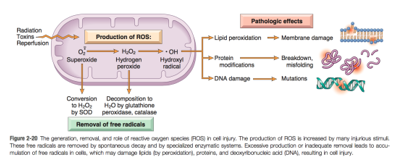
Describe the mechanism of generation of oxygen-derived free radicals in cellular injury

  • chemically unstable and react with other molecules damage

  • Produced by physiologic oxidation-reduction reactions, UV light, ionizing radiation, metals, chemicals (smoking, pollution), inflammation, stress

<ul><li><p><span style="font-family: &quot;Gill Sans MT&quot;;"> </span><span style="font-family: &quot;Gill Sans MT&quot;; color: red;">chemically unstable</span><span style="font-family: &quot;Gill Sans MT&quot;;"> and react with other molecules </span>→ <span style="font-family: &quot;Gill Sans MT&quot;; color: red;">damage</span></p></li><li><p><span style="font-family: &quot;Gill Sans MT&quot;;">Produced by </span><span style="font-family: &quot;Gill Sans MT&quot;; color: red;">physiologic oxidation-reduction reactions</span><span style="font-family: &quot;Gill Sans MT&quot;;">, UV light, ionizing radiation, metals, chemicals (smoking, pollution), inflammation, stress</span></p></li></ul><p></p>
18
New cards

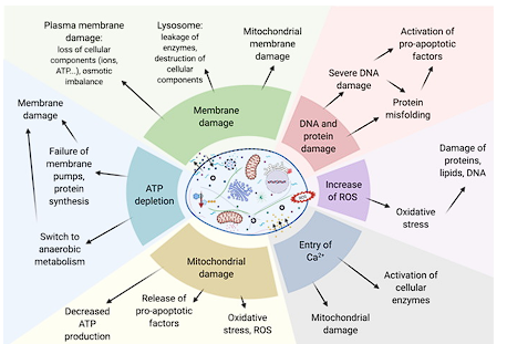
Describe the mechanism of chemical injury in cellular injury

  • can affect any of the processes listed on the chart

    • membrane damage

    • DNA and protein damage

    • ATP depletion

    • Mitochondrial damage

    • Entry of Ca 2+

    • Increase of ROS

<ul><li><p>can affect any of the processes listed on the chart</p><ul><li><p>membrane damage</p></li><li><p>DNA and protein damage</p></li><li><p> ATP depletion</p></li><li><p>Mitochondrial damage</p></li><li><p>Entry of Ca 2+</p></li><li><p>Increase of ROS</p></li></ul></li></ul><p></p>
19
New cards

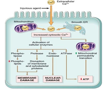
Describe the mechanism of Increased mitochondrial cytosolic calcium in cellular injury

  • Results in:

    • Lipid peroxidation

    • Mitochondrial injury

    • Loss of calcium homeostasis

    • ↓ ATP production

    • Apoptosis

<ul><li><p><span style="font-family: &quot;Gill Sans MT&quot;;">Results in:</span></p><ul><li><p><span style="font-family: &quot;Gill Sans MT&quot;;">Lipid peroxidation</span></p></li><li><p><span style="font-family: &quot;Gill Sans MT&quot;;">Mitochondrial injury</span></p></li><li><p><span style="font-family: &quot;Gill Sans MT&quot;;">Loss of calcium homeostasis</span></p></li><li><p><span style="font-family: &quot;Gill Sans MT&quot;;">↓ ATP production</span></p></li><li><p><span style="font-family: &quot;Gill Sans MT&quot;;">Apoptosis</span></p></li></ul></li></ul><p></p>
20
New cards

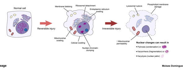
Define and explain the process of reversible cell injury

  • Stress is mild → moderate

  • injured cell may recover

  • caused by hypoxia, decreased ATP, sodium potassium pump failures, or failures of calcium channels

  • Result= cellular swelling, Mitochondrial swelling, ER swelling → blebbing of those membranes

  • Ca enters cell→ ribosomal detachment→ decreased protein synthesis and alteration of lipids → clumping of chromatins

  • Leads to protein denaturation and changes in the structure of DNA

  • Goes back into a state of homeostasis and becomes a normal cell

<ul><li><p>Stress is mild → moderate</p></li><li><p>injured cell may recover</p></li><li><p>caused by hypoxia, decreased ATP, sodium potassium pump failures, or failures of calcium channels</p></li><li><p>Result= <span style="color: red;">cellular swelling, Mitochondrial swelling, ER swelling → blebbing of those membranes</span></p></li><li><p><span style="color: rgb(255, 255, 255);">Ca enters cell→ ribosomal detachment→ decreased protein synthesis and alteration of lipids → clumping of chromatins</span></p></li><li><p><span style="color: rgb(255, 255, 255);">Leads to protein denaturation and </span><span style="color: red;">changes in the structure of DNA</span></p></li><li><p><span style="color: red;">Goes back into a state of homeostasis and becomes a normal cell</span></p></li></ul><p></p>
21
New cards

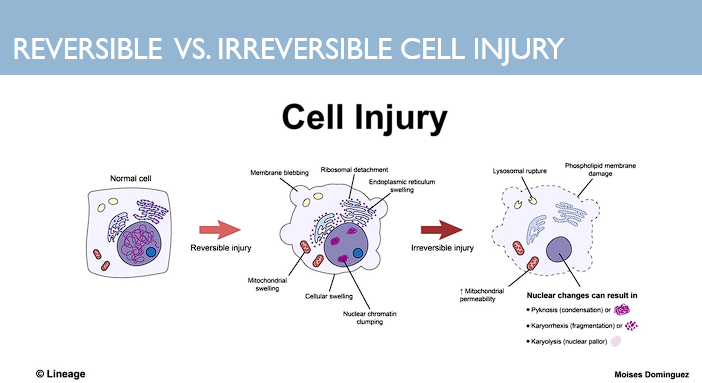
Define and explain the process of irreversible cell injury

  • Rupturing of the organelles → overall rupturing of those cells

  • Depositions/ densities in mitochondria

  • myelin fissures → nuclear degradation/ nuclear changes can result in:

    • Pyknosis (condensation)

    • Karyorrhexis (fragmentation)

    • Karyolysis (nuclear pallor)
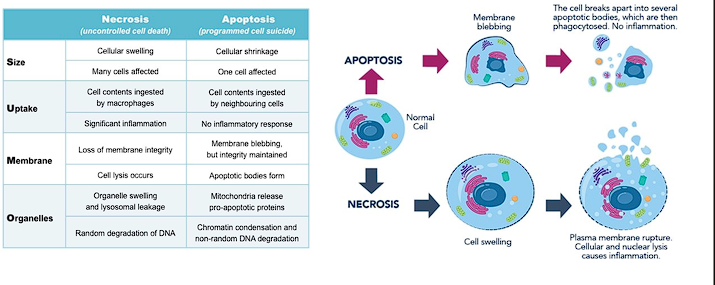
<ul><li><p><span style="color: red;">Rupturing of the organelles </span><span style="color: rgb(255, 255, 255);">→ overall rupturing of those cells</span></p></li><li><p><span style="color: rgb(255, 255, 255);">Depositions/ densities in mitochondria</span></p></li><li><p><span style="color: rgb(255, 255, 255);">myelin fissures → nuclear degradation/ </span><span style="color: red;">nuclear changes can result in:</span></p><ul><li><p><span style="color: red;">Pyknosis</span> (condensation)</p></li><li><p><span style="color: red;">Karyorrhexis</span> (fragmentation)</p></li><li><p><span style="color: red;">Karyolysis</span> (nuclear pallor)</p></li></ul></li></ul><p></p>
22
New cards

Define apoptosis

controlled (programed) cell death/breakdown of cells occurring in response to irreversible cellular damage or as part of normal growth and development.

Phases:

  • Initiation: caspases become catalytically active

  • Execution: action of caspases causes cell death

<p><span style="color: red;">controlled (programed) </span>cell death/breakdown of cells occurring in response to irreversible cellular damage or as part of normal growth and development.</p><p>Phases:</p><ul><li><p><span style="color: red;">Initiation:</span> caspases become catalytically active</p></li><li><p><span style="color: red;">Execution:</span> action of caspases causes cell death</p></li></ul><p></p>
23
New cards

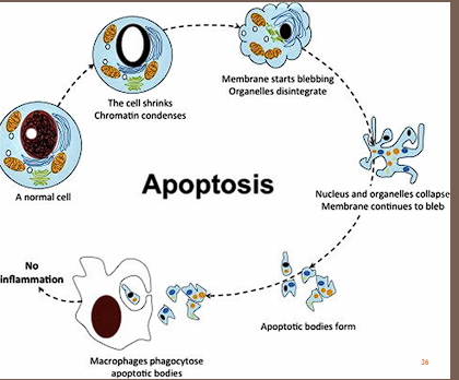
What is the microscopic morphology of Apoptosis?

Chromatin condensation and fragmentation

24
New cards

What is the microscopic and gross morphology of Necrosis?

Gross:

  • softening and discoloration of the organ

Micro:

  • Coagulative necrosis

  • Liquefactive necrosis

  • Fat necrosis

  • Caseous necrosis

25
New cards

Define necrosis

uncontrolled breakdown of cells/cell death in response to irreversible cellular injury; triggers inflammatory response

26
New cards

Compare and contrast apoptosis and necrosis

  • KEY DIFFERENCE:

    • Apoptosis does not generate an inflammatory response, where as necrosis does

  • Refer to chart

<ul><li><p>KEY DIFFERENCE:</p><ul><li><p>Apoptosis does not generate an<span style="color: red;"> inflammatory response</span>, where as necrosis does</p></li></ul></li><li><p>Refer to chart</p></li></ul><p></p>
27
New cards

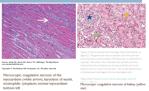
Define coagulative necrosis and describe the microscopic morphology

  • Due to ischemia, infarction

  • Increased eosinophilia of the cytoplasm (denatured proteins) and decreased basophilia of the nucleus (loss of DNA/RNA).

  • General cellular architecture is initially preserved.

<ul><li><p>Due to ischemia, infarction</p></li></ul><p></p><ul><li><p><span style="color: red;">Increased eosinophilia</span> of the cytoplasm (denatured proteins) and <span style="color: red;">decreased basophilia</span> of the nucleus (loss of DNA/RNA).</p></li></ul><ul><li><p>General cellular architecture is initially preserved.</p></li></ul><p></p>
28
New cards

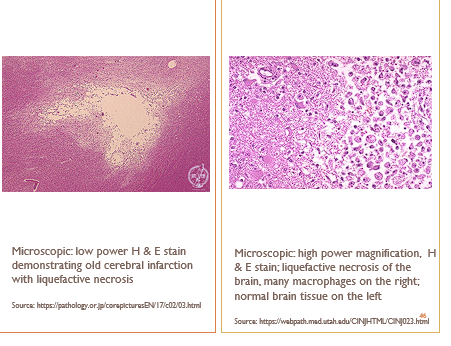
Define liquefactive necrosis and describe the microscopic morphology

  • enzyme breakdown in lipid-rich organs

  • loss of organ cellular architecture

  • lipid-laden macrophages replace the dead tissue

<ul><li><p>enzyme breakdown in lipid-rich organs</p></li></ul><p></p><ul><li><p>loss of organ cellular architecture</p></li><li><p>lipid-laden macrophages replace the dead tissue</p></li></ul><p></p>
29
New cards

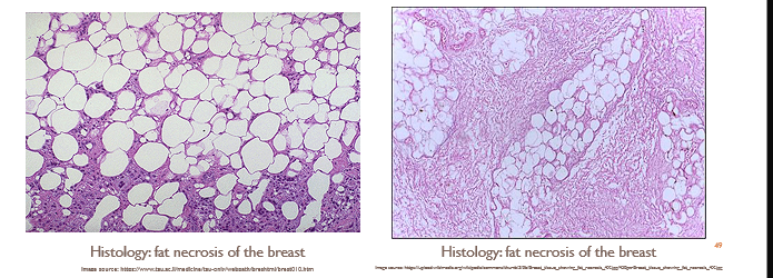
Define fat necrosis and describe the microscopic morphology.

  • Change in adipose tissue due to trauma or the release of enzyme from adjacent organs

  • Large, lipid filled vacuoles

<ul><li><p>Change in adipose tissue due to trauma or the release of enzyme from adjacent organs</p></li></ul><p></p><ul><li><p>Large, lipid filled vacuoles</p></li></ul><p></p>
30
New cards

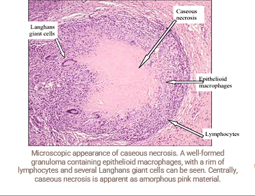
Define caseous necrosis and describe the microscopic morphology.

  • Immune system cannot successfully remove a foreign stimuli (e.g. tuberculosis). Forms granuloma

  • granuloma with central necrosis, eosinophilia

<ul><li><p>Immune system cannot successfully remove a foreign stimuli (e.g. tuberculosis). Forms granuloma </p></li></ul><p></p><ul><li><p><span style="color: red;">granuloma</span> with central necrosis, eosinophilia</p></li></ul><p></p>
31
New cards

List the substances that can accumulate

  • Lipofuscin

  • Calcium

  • Protein

  • Iron

  • Fat

  • Cholesterol

  • Glycogen

  • Pigments

32
New cards

Describe the mechanism of formation, sites of accumulation, and gross/ microscopic morphology of Lipofuscin.

  • Mechanism of formation: product of lipid peroxidation which accumulates in lysosomes as cells ages; cells cannot get rid of it

  • Organs affected: commonly in the heart, liver, skin

  • Gross morphology: brown discoloration to organs

  • Microscopic morphology: finely granular, yellow-brown pigment, which often surrounds the nucleus

<ul><li><p><span style="font-family: &quot;Gill Sans MT&quot;;"><strong>Mechanism of formation: </strong>product of</span><span style="font-family: &quot;Gill Sans MT&quot;; color: red;"> lipid peroxidation</span><span style="font-family: &quot;Gill Sans MT&quot;;"> which accumulates in lysosomes as cells ages; cells cannot get rid of it</span></p></li><li><p><span style="font-family: &quot;Gill Sans MT&quot;;"><strong>Organs affected: </strong>commonly in the heart, liver, skin</span></p></li><li><p><span style="font-family: &quot;Gill Sans MT&quot;;"><strong>Gross morphology: </strong>brown discoloration to organs</span></p></li><li><p><span style="font-family: &quot;Gill Sans MT&quot;;"><strong>Microscopic morphology</strong></span><span style="font-family: &quot;Gill Sans MT&quot;; color: red;"><strong>: </strong>finely granular, yellow-brown pigment</span><span style="font-family: &quot;Gill Sans MT&quot;;">, which often surrounds the nucleus</span></p></li></ul><p></p>
33
New cards

Describe the mechanism of formation, sites of accumulation, and gross/ microscopic morphology of Calcium

2 types of mechanism

  • Mechanism of metastatic calcification: patients who have hypercalcemia have deposition of calcium within normal or abnormal tissue

  • Mechanism of dystrophic calcification: patients who have normal levels of calcium have deposition of calcium only within abnormal tissue (necrosis or damage)

  • Organs affected:  vasculature, kidneys, lungs

  • Gross morphology: hard, yellow nodules

  • Microscopic morphology:  chunky, smooth, purple granules

<p>2 types of mechanism</p><ul><li><p><span style="font-family: &quot;Gill Sans MT&quot;;">Mechanism of </span><span style="font-family: &quot;Gill Sans MT&quot;; color: red;"><u>metastatic calcification</u></span><span style="font-family: &quot;Gill Sans MT&quot;;">: patients who have <u>hypercalcemia </u>have deposition of calcium within </span>normal <span style="font-family: &quot;Gill Sans MT&quot;;">or abnormal tissue</span></p></li><li><p><span style="font-family: &quot;Gill Sans MT&quot;;">Mechanism of </span><span style="font-family: &quot;Gill Sans MT&quot;; color: red;"><u>dystrophic calcification</u></span><span style="font-family: &quot;Gill Sans MT&quot;;">: patients who have normal levels of calcium have deposition of calcium only within abnormal tissue (necrosis or damage)</span></p></li><li><p><span style="font-family: &quot;Gill Sans MT&quot;;">Organs affected: &nbsp;vasculature, kidneys, lungs</span></p></li><li><p><span style="font-family: &quot;Gill Sans MT&quot;;">Gross morphology: hard, yellow nodules</span></p></li><li><p><span style="font-family: &quot;Gill Sans MT&quot;;">Microscopic morphology:&nbsp; chunky, smooth, purple granules</span></p></li></ul><p></p>
34
New cards

Describe the mechanism of formation, sites of accumulation, and gross/ microscopic morphology of protein

  • Mechanism of formation: Increased protein absorption in proximal renal tubule (renal disease), Increased cytoskeletal proteins, increased protein production, amyloidosis, defect in intracellular transport and protein secretion.

  • Organs affected: liver and kidneys

  • Gross morphology: blue-black foci on tissue once stained

  • Microscopic morphology:

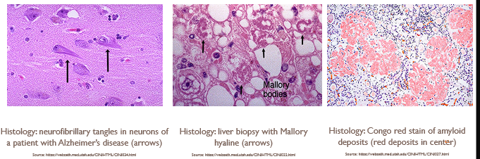
    • Amyloidosis: amyloid deposits stain eosinophilic/pink-pale; pink or red on Congo red stain

    • Alzheimer’s disease: neurofibrillary tangles

    • Fatty liver: Mallory hyaline-eosinophilic, pink, rope-like deposits

<ul><li><p><span style="font-family: &quot;Gill Sans MT&quot;;"><strong>Mechanism of formation: </strong>Increased protein absorption in </span><span style="font-family: &quot;Gill Sans MT&quot;; color: red;">proximal renal tubule </span><span style="font-family: &quot;Gill Sans MT&quot;;">(renal disease), Increased cytoskeletal proteins, increased protein production, amyloidosis, defect in intracellular transport and protein secretion.</span></p></li><li><p><span style="font-family: &quot;Gill Sans MT&quot;;"><strong>Organs affected:</strong> liver and kidneys</span></p></li><li><p><span style="font-family: &quot;Gill Sans MT&quot;;"><strong>Gross morphology</strong>: blue-black foci on tissue once stained</span></p></li><li><p><span style="font-family: &quot;Gill Sans MT&quot;;"><strong>Microscopic morphology:</strong></span></p><ul><li><p><span style="font-family: &quot;Gill Sans MT&quot;; color: red;">Amyloidosis:</span><span style="font-family: &quot;Gill Sans MT&quot;;"> amyloid deposits stain eosinophilic/pink-pale; pink or red on Congo red stain</span></p></li><li><p><span style="font-family: &quot;Gill Sans MT&quot;;">Alzheimer’s disease: neurofibrillary tangles</span></p></li><li><p><span style="font-family: &quot;Gill Sans MT&quot;;">Fatty liver: Mallory hyaline-eosinophilic, pink, rope-like deposits</span></p></li></ul></li></ul><p></p>
35
New cards

Describe the mechanism of formation, sites of accumulation, and gross/ microscopic morphology of iron

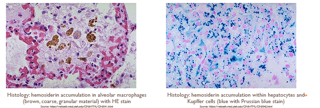
  • Mechanism: accumulation of hemosiderin due to iron

    • Hemosiderosis: accum. of iron in organs w/o resultant side effects

    • Hemochromatosis: accum. of iron in parenchymal cells resulting in side effects

  • Organs affected by hemochromatosis: liver, skin, pancreas, heart

  • Gross morphology: dark brown color

  • Microscopic morphology: chunky, yellow-brown granules on H & E stain; blue on Prussian blue stain

<ul><li><p><strong>Mechanism: </strong>accumulation of<span style="color: red;"> hemosiderin</span> due to <span style="font-family: &quot;Cambria Math&quot;;">↑</span><span style="font-family: &quot;Gill Sans MT&quot;;"><strong> </strong>iron</span></p><ul><li><p><span style="color: red;">Hemosiderosis</span>: accum. of iron in organs w/o resultant side effects</p></li><li><p><span style="color: red;">Hemochromatosis:</span> accum. of iron in parenchymal cells resulting in side effects</p></li></ul></li><li><p><span style="font-family: &quot;Gill Sans MT&quot;;"><strong>Organs affected by hemochromatosis: </strong></span>liver, skin, pancreas, heart</p></li><li><p><span style="font-family: &quot;Gill Sans MT&quot;;"><strong>Gross morphology: </strong>dark brown color</span></p></li><li><p><span style="font-family: &quot;Gill Sans MT&quot;;"><strong>Microscopic morphology: </strong></span><span style="font-family: &quot;Gill Sans MT&quot;; color: red;">chunky, yellow-brown granules</span><span style="font-family: &quot;Gill Sans MT&quot;;"> on H &amp; E stain; blue on Prussian blue stain</span></p></li></ul><p></p>
36
New cards

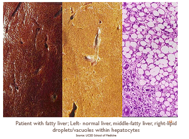
Describe the mechanism of formation, sites of accumulation, and gross/ microscopic morphology of fat (steatosis)

  • Mechanism: can indicate reversible damage or may be a sign of intrinsic abnormality in fat metabolism

    • Seen in fatty liver disease (alcohol use, nonalcoholic), diabetes mellitus, obesity

  • Organs affected: liver, kidney, heart, skeletal muscle

  • Gross morphology:  yellow discoloration of an organ

  • Microscopic morphology: one or several clear vacuoles within the cell

<ul><li><p><strong>Mechanism:</strong> <span style="font-family: &quot;Gill Sans MT&quot;;">can indicate reversible damage or may be a sign of intrinsic abnormality in fat metabolism</span></p><ul><li><p><span style="font-family: &quot;Gill Sans MT&quot;;">Seen in fatty liver disease (alcohol use, nonalcoholic), diabetes mellitus, obesity</span></p></li></ul></li><li><p><span style="font-family: &quot;Gill Sans MT&quot;;"><strong>Organs affected: </strong>liver, kidney, heart, skeletal muscle</span></p></li><li><p><span style="font-family: &quot;Gill Sans MT&quot;;"><strong>Gross morphology: </strong>&nbsp;yellow discoloration of an organ</span></p></li><li><p><span style="font-family: &quot;Gill Sans MT&quot;;"><strong>Microscopic morphology: </strong>one or several clear vacuoles within the cell</span></p></li></ul><p></p>
37
New cards

Describe the mechanism of formation, sites of accumulation, and gross/ microscopic morphology of cholesterol

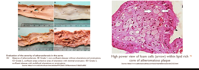
  • Mechanism: Hypercholesterolemia; elevated blood cholesterol levels lead to disruption in cellular function, oxidative stress, inflammation

  • Organs affected: blood vessels (atherosclerosis)

  • Gross morphology:  yellow discoloration of an organ

  • Microscopic morphology: foam cells (lipid-laden macrophages)

<ul><li><p><strong>Mechanism:</strong> <span style="font-family: &quot;Gill Sans MT&quot;; color: red;"><strong>Hypercholesterolemia</strong></span><span style="font-family: &quot;Gill Sans MT&quot;;"><strong>;</strong>&nbsp;elevated blood cholesterol levels lead to disruption in cellular function, oxidative stress, inflammation</span></p></li><li><p><span style="font-family: &quot;Gill Sans MT&quot;;"><strong>Organs affected: </strong>blood vessels (atherosclerosis)</span></p></li><li><p><span style="font-family: &quot;Gill Sans MT&quot;;"><strong>Gross morphology: </strong>&nbsp;yellow discoloration of an organ</span></p></li></ul><ul><li><p><span style="font-family: &quot;Gill Sans MT&quot;;"><strong>Microscopic morphology: </strong>foam cells (lipid-laden macrophages)</span></p></li></ul><p></p>
38
New cards

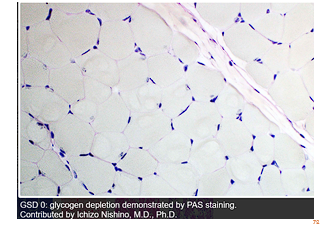
Describe the mechanism of formation, sites of accumulation, and gross/ microscopic morphology of Glycogen

  • Mechanism: accumulates due to glycogen storage disorders or disease of glucose metabolism

  • Organs affected: liver and skeletal muscle

  • Microscopic morphology: clear vacuoles in the cytoplasm

<ul><li><p><strong>Mechanism:</strong> <span style="font-family: &quot;Gill Sans MT&quot;;">accumulates due to glycogen storage disorders or disease of glucose metabolism</span></p></li><li><p><span style="font-family: &quot;Gill Sans MT&quot;;"><strong>Organs affected: </strong>liver and skeletal muscle</span></p></li><li><p><span style="font-family: &quot;Gill Sans MT&quot;;"><strong>Microscopic morphology: </strong>clear vacuoles in the cytoplasm</span></p></li></ul><p></p>
39
New cards

what are the types of pigments

  • Exogenous pigments (produced outside of the body): Melanin

  • Endogenous pigments (produced inside of the body): Bilirubin

40
New cards

Describe the mechanism of formation, sites of accumulation, and gross/ microscopic morphology of bilirubin

  • Mechanism: produced from the breakdown of RBCs and excreted through bile and urine

    • Hyperbilirubinemia (hemolysis, obstruction, genetic disorders, drugs)

  • Organs affected:

    • Jaundice- yellowing of skin, eyes

    • Kernicterus-brain

  • Gross morphology: yellow pigmentation

  • Microscopic morphology: brown deposits

  • bilirubin is a component of bile

41
New cards

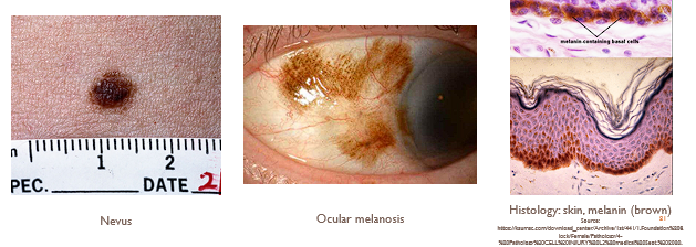
Describe the mechanism of formation, sites of accumulation, and gross/ microscopic morphology of mealnin

  • Mechanism: brown-black pigment formed by melanocytes and transferred to keratinocytes

    • Function is to prevent the harmful effects of ultraviolet (UV) light

  • Organs affected: skin, eye

  • Gross & microscopic morphology: brown–black pigment

<ul><li><p>Mechanism<span style="font-family: &quot;Gill Sans MT&quot;;"><strong>: </strong>brown-black pigment formed by melanocytes and transferred to keratinocytes</span></p><ul><li><p><span style="font-family: &quot;Gill Sans MT&quot;;">Function is to prevent the harmful effects of ultraviolet (UV) light</span></p></li></ul></li><li><p><span style="font-family: &quot;Gill Sans MT&quot;;"><strong>Organs affected: </strong>skin, eye</span></p></li><li><p><strong>Gross &amp; microscopic morphology: </strong>brown–black pigment</p></li></ul><p></p>
42
New cards

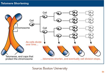
Describe the role of the telomere in relation to cellular aging

As a cell divides, its telomeres shorten. Eventually, the telomeres become so short that the cell can no longer divide, which leads to cellular aging (senescence).

<p>As a cell divides, its <strong>telomeres shorten</strong>. Eventually, the telomeres <span style="color: red;">become so short that the cell can no longer divide</span>, which leads to cellular aging (senescence).</p>