Porcine MSK ICVA
1/26
There's no tags or description
Looks like no tags are added yet.
Name | Mastery | Learn | Test | Matching | Spaced | Call with Kai |
|---|
No analytics yet
Send a link to your students to track their progress
27 Terms
Hernias: D.X
Physical Examination:
A soft, reducible, or irreducible swelling at the umbilical or inguinal/scrotal regions.
In cases of strangulation, the hernia may become firm and painful on palpation
Ultrasound: In complicated or unclear cases, ultrasound can be used to evaluate the contents of the hernia sac, assess the involvement of abdominal organs, and check for signs of strangulation or torsion, particularly in scrotal hernias.
Differential Diagnosis: Other conditions that may mimic hernias include abscesses, hematomas, or neoplasms. Therefore, differentiating these conditions through palpation, aspiration, or imaging may be necessary.
Infectious Causes of Lameness
Mycoplasma hyorhinis:
Causes polyserositis and arthritis.
Mycoplasma hyosynoviae:
Leads to arthritis in older pigs.
Erysipelothrix rhusiopathiae:
Causes erysipelas,
leading to swollen joints and skin lesions.
Streptococcus suis:
Can cause septicemia and arthritis,
especially in young pigs.
Nutritional causes of Lameness
Calcium and phosphorus:
Essential for bone formation;
deficiencies lead to rickets or osteomalacia.
Vitamin D:
Necessary for calcium absorption;
deficiency results in poor bone mineralization.
Zinc:
Important for skin integrity and enzyme function;
deficiency leads to parakeratosis and lameness.
Iron:
Essential for red blood cell production;
Deficiency causes anemia and general weakness.
Developmental Causes of Lameness
Angular limb deformities:
Abnormal bone growth leading to crooked legs.
Osteochondrosis:
Defects in cartilage and bone development
Leading to joint pain and lameness.
Hip dysplasia:
Malformation of the hip joint,
Causing instability and lameness.
Mycoplasma hyorinis infection what age is commonly affected
In young pigs, typically between 3 to 10 weeks of age. (Nursery)
It is a cause of significant economic losses in pig production due to morbidity and impaired growth rates, though mortality is generally low.
Mycoplasma hyorinis infection:C.S
Respiratory Signs: Early infection can present with coughing, nasal discharge, and mild respiratory distress, particularly in piglets.
Polyserositis: The hallmark of systemic Mycoplasma hyorhinis infection is polyserositis. This results in inflammation of multiple serous membranes, including pleuritis, peritonitis, and pericarditis.
Pleural Involvement: Signs include tachypnea, respiratory distress, and reluctance to move due to thoracic pain.
Pericardial Involvement: Can lead to muffled heart sounds and signs of congestive heart failure if severe.
Peritoneal Involvement: Abdominal distension and discomfort, often accompanied by poor feed intake and a hunched posture.
Arthritis: In the chronic phase, polyarthritis is common. Affected pigs may exhibit:
Swollen joints
Lameness
Reluctance to rise or move
Pain on palpation of affected joints
In some cases, chronic arthritis may lead to reduced weight gain and secondary infections due to decreased mobility and poor welfare. Severe cases of polyserositis can cause sudden death, though this is uncommon.
Mycoplasma hyorinis infection: D.X
Clinical Observation:
Polyserositis and arthritis in young pigs are clinical indicators.
Respiratory signs in combination with poor growth rates in affected animals.
Necropsy Findings:
Fibrinous Polyserositis: This is a hallmark finding during necropsy. Affected animals will show fibrinous pleuritis, peritonitis, and pericarditis.
Polyarthritis: Synovial fluid will be increased in volume, and joints will often be swollen with fibrinous exudate. Chronic cases may show fibrosis in the joints.
Laboratory Diagnostics:
PCR (Polymerase Chain Reaction): This is the preferred method for diagnosing Mycoplasma hyorhinis from nasal swabs, joint fluid, or serous membranes. PCR is sensitive and specific.
Culture: While Mycoplasma can be cultured, it is fastidious, and culture is not routinely performed due to the slow-growing nature of the organism.
Serology: Antibody detection may help in identifying exposure but does not differentiate between active and past infection.
Differential Diagnosis:
Other Causes of Polyserositis/Arthritis: Conditions like Haemophilus parasuis (Glässer’s disease), Streptococcus suis, or Erysipelothrix rhusiopathiae can present with similar signs.
Respiratory Diseases: Co-infections with viral or bacterial respiratory pathogens must be considered
Mycoplasma hyorinis infection:T.X
Antimicrobials:
Tetracyclines (e.g., oxytetracycline or doxycycline):
Macrolides (e.g., tylosin, tilmicosin):
Lincosamides (e.g., lincomycin):
Anti-inflammatory Treatment:
NSAIDs (flunixin meglumine or meloxicam)
Control pain + reduce inflammation in polyarthritis and polyserositis cases.
Supportive Care:
Ensure adequate hydration and nutrition to support growth in affected pigs.
In severe cases, segregating sick pigs to avoid further stress and secondary infections may be necessary.
Prognosis:
The prognosis for pigs infected with Mycoplasma hyorhinis depends on the severity of clinical signs and the timeliness of intervention. While mortality is generally low, chronic polyarthritis can result in long-term mobility issues and reduced productivity in affected pigs. With appropriate antimicrobial therapy and supportive care, many pigs recover fully.
Hallmark of Mycoplasma hyorinis infection
Hallmark Clinical Signs and Diagnosis:
Fibrinous Polyserositis: The most characteristic postmortem finding, often seen with pleuritis, pericarditis, and peritonitis.
Arthritis: Synovial swelling and lameness are hallmark clinical signs of joint involvement, particularly in the chronic phase.
PCR Testing: The definitive diagnostic tool for confirming Mycoplasma hyorhinis infection, especially when combined with clinical and necropsy findings
What age are most commonly affected by Mycoplasma Hyosynoviae Arthritis
Pigs are most commonly affected between 3 to 6 months of age, coinciding with the period of rapid growth and stress due to changes in housing or feeding.
Mycoplasma Hyosynoviae Arthritis:C.S
Clinical Signs
The hallmark clinical sign of M. hyosynoviae infection is lameness, typically appearing suddenly in pigs aged 3 to 6 months. The lameness is often bilateral but may affect multiple joints, including the stifles, elbows, and hocks. Affected pigs may exhibit:
Reluctance to stand or move
Often Bilateral lameness
Swollen, painful joints
Stiffness and abnormal gait
Decreased feed intake and reduced growth rates
Weight shifting between legs to avoid pressure on painful joints
In severe cases, pigs may be recumbent due to intense pain, leading to secondary issues such as pressure sores.
The absence of systemic signs, such as fever or weight loss, distinguishes M. hyosynoviae from other joint infections like those caused by Erysipelothrix rhusiopathiae.
Mycoplasma Hyosynoviae Arthritis:D.X
Diagnostics
Accurate diagnosis of M. hyosynoviae relies on clinical signs, history, and confirmatory laboratory testing:
Joint fluid analysis: Affected joints often contain cloudy synovial fluid with increased white blood cell counts (predominantly neutrophils) but typically lack visible bacteria.
PCR testing: Polymerase Chain Reaction (PCR) is the most reliable method for detecting M. hyosynoviae in joint fluid, tonsillar swabs, or nasal secretions.
Culture: While possible, culturing M. hyosynoviae is challenging due to its fastidious nature and slow growth. PCR is preferred for speed and sensitivity.
Serology: Antibody detection via ELISA may help identify past exposure, but it is not definitive for diagnosing active disease.
Postmortem findings: In chronic cases, joint necropsy may reveal synovial thickening, cartilage erosion, and joint capsule fibrosis, though these findings are non-specific.
Mycoplasma Hyosynoviae Arthritis:T.X
Treatment
Treatment is focused on controlling inflammation and managing pain:
Antibiotics: Although M. hyosynoviae is resistant to beta-lactams like penicillin so macrolides (such as tylosin or tiamulin), tetracyclines, or fluoroquinolones are typically effective. Antibiotic treatment is most effective when administered early in the disease course to prevent chronic joint damage.
Anti-inflammatory drugs: Non-steroidal anti-inflammatory drugs (NSAIDs) such as flunixin meglumine or meloxicam can help reduce inflammation and pain.
Supportive care: Rest and reduced stress (e.g., minimizing handling and crowding) are important in recovery, especially for severely affected pigs.
Mycoplasma Hyosynoviae Arthritis Image

Typical lesions of Mycoplasma hyorhinis infection. (A) Joint with excess synovial fluid. (B) Serosanguinous synovial fluid. (C) Serofibrinous pericarditis. (D) Serofibrinous pleuritis.
Prognosis
The prognosis for M. hyosynoviae infections depends on the timing of treatment and the severity of joint damage. Early intervention with appropriate antibiotics and NSAIDs can result in full recovery, with pigs returning to normal growth rates. However, delayed treatment can lead to chronic joint changes, reducing the animal's productivity and longevity.
Osteochondrosis:C.S
Clinical Signs
Osteochondrosis in pigs manifests primarily as lameness and joint swelling. Clinical signs can vary depending on the severity and location of the lesions. Common clinical signs include:
Lameness: Often bilateral, affecting the front or hind limbs.
Joint effusion: Swelling of the affected joint, typically seen in weight-bearing joints.
Reduced mobility: Reluctance to move or bear weight on the affected limb.
Stiff gait: Pigs may develop an abnormal, stiff-legged gait due to joint pain.
Joint deformities: In advanced cases, joints may become visibly misshapen or enlarged.
Recumbency: In severe cases, affected pigs may become recumbent due to pain and immobility.
Lameness can significantly affect growth rates, feed conversion efficiency, and overall production performance.
Genetic predisposition
Osteochondrosis:D.X
Diagnostics
Diagnosing osteochondrosis involves a combination of clinical evaluation, radiographic imaging, and post-mortem examination.
Clinical Examination: A thorough physical examination reveals signs of joint swelling, lameness, and reluctance to move. A detailed history, including growth rate and housing conditions, should also be taken into account.
Radiography: X-rays are the primary diagnostic tool for osteochondrosis. Radiographs reveal irregularities in the epiphyseal cartilage, such as cartilage flaps, fissures, or retained cartilage islands. Osteochondritis dissecans (OCD) may be identified as visible cartilage or bone fragments within the joint space.
Computed Tomography (CT): In research or valuable breeding animals, CT scans provide detailed images of cartilage and bone architecture and are more sensitive than radiographs.
Post-mortem Examination: In culling cases, post-mortem evaluation allows for direct examination of the cartilage lesions, confirming the diagnosis.
Osteochondrosis:T.X
Weight Management: Reducing the growth rate in affected pigs by adjusting the diet and controlling feed intake can decrease joint stress and slow the progression of the disease.
Pain Management: Non-steroidal anti-inflammatory drugs (NSAIDs), such as meloxicam or flunixin meglumine, can provide temporary relief from pain and inflammation. However, long-term use is generally avoided due to withdrawal times and potential side effects.
Corrective Surgery: In valuable breeding stock, surgical intervention may be considered, especially for osteochondritis dissecans. Surgery involves removing loose cartilage fragments and debriding the affected joint. This is rarely performed in commercial pigs due to cost and practicality.
Environmental Modifications: Providing soft bedding, reducing trauma to joints by improving flooring, and optimizing space can alleviate biomechanical stress on the joints.
Prevention
Prevention of osteochondrosis in pigs centers on genetic selection, nutritional management, and environmental modifications.
Genetic Selection: Breeding for animals with slower growth rates and sound skeletal conformation can reduce the incidence of osteochondrosis.
Nutritional Management: Balanced diets that provide adequate calcium, phosphorus, and trace minerals are essential for proper bone and cartilage development. Avoiding excessive energy intake that promotes rapid growth can reduce the risk.
Environmental Management: Flooring that reduces trauma to joints, such as straw or rubber mats, is beneficial in preventing joint damage. Housing designs that promote adequate space for movement and reducing stocking density are also important.
Streptococcal synovitis:C.S
Clinical Signs
Streptococcal synovitis typically presents in weaned piglets and growing pigs. Clinical signs can vary in severity but are primarily related to arthritis and joint inflammation. Key clinical signs include:
Lameness: Affected pigs often exhibit lameness, favoring one or more limbs.
Joint Swelling: Swollen, warm joints, especially in the limbs, are common. Knees, hocks, and elbows are frequently affected.
Stiff Gait: Due to joint pain, pigs may show a stiff gait or reluctance to move.
Recumbency: Severely affected pigs may remain recumbent due to pain, with difficulty standing or walking.
Fever: Systemic infection may cause fever, although this is more common in severe or generalized infections.
Reduced Appetite and Growth: Chronically affected pigs may have poor weight gain and reduced feed intake.
In some cases, septicemia can develop, leading to multisystemic signs such as lethargy, depression, and even death if left untreated.
Streptococcal synovitis:D.X
Diagnostics
Diagnosis of streptococcal synovitis is based on a combination of clinical signs, bacterial culture, and post-mortem examination. Diagnostic steps include:
Clinical Examination: Identification of swollen, painful joints and lameness in pigs, especially in conjunction with a history of poor housing conditions or recent trauma.
Joint Fluid Analysis: Synovial fluid aspirates may reveal increased cell counts (primarily neutrophils), elevated protein levels, and evidence of bacterial infection.
Bacterial Culture: Isolation of Streptococcus dysgalactiae equisimilis from joint fluid, blood, or tissue samples is definitive. Joint fluid or affected tissue should be submitted for culture and sensitivity testing.
Histopathology: Histological examination of affected joints can reveal severe synovial inflammation, with infiltrates of neutrophils and macrophages, and erosion of articular cartilage.
Radiography: X-rays of affected joints may reveal soft tissue swelling, synovial effusion, and, in chronic cases, joint destruction or osteolysis.
Streptococcal synovitis:T.X
Antibiotics:
Penicillin is the antibiotic of choice, as Streptococcus species are typically highly susceptible. Injectable penicillin G or amoxicillin can be administered based on weight and severity.
In cases where penicillin resistance is suspected, ceftiofur or florfenicol may be used, guided by sensitivity testing.
Treatment should be continued for at least 7-10 days to ensure eradication of the infection.
Non-Steroidal Anti-Inflammatory Drugs (NSAIDs): NSAIDs like flunixin meglumine or meloxicam help reduce inflammation and relieve pain, improving mobility and comfort for the animal.
Supportive Care: Severely affected pigs may benefit from supportive measures, such as isolation in a dry, clean, and comfortable environment, with access to soft bedding to minimize further trauma to the joints.
Drainage of Joint Fluid: In cases of severe synovial effusion, joint aspiration or drainage may help relieve pain and reduce bacterial load within the joint.
Hallmark Clinical Signs and Diagnosis
The hallmark of streptococcal synovitis is the combination of lameness, joint swelling, and pain. The rapid onset of symptoms in young, weaned pigs, often in conjunction with other risk factors such as overcrowding or tail biting, strongly suggests a streptococcal etiology. Definitive diagnosis requires bacterial culture of synovial fluid, which isolates the causative pathogen.
Prevention
Preventative measures are critical in reducing the incidence of streptococcal synovitis. Strategies include:
Improving Hygiene: Maintaining clean, dry pens with proper waste removal reduces the risk of skin abrasions and infections.
Minimizing Stress: Reducing overcrowding, providing adequate space for movement, and preventing tail biting or aggressive behaviors can lower the incidence of trauma and subsequent infection.
Tail Docking Management: If tail docking is necessary, ensure proper hygiene and technique to minimize infection risk.
Vaccination: Although no specific vaccine exists for Streptococcus dysgalactiae equisimilis, general herd health practices, including vaccination against other common diseases, can reduce stress and the risk of opportunistic infections.
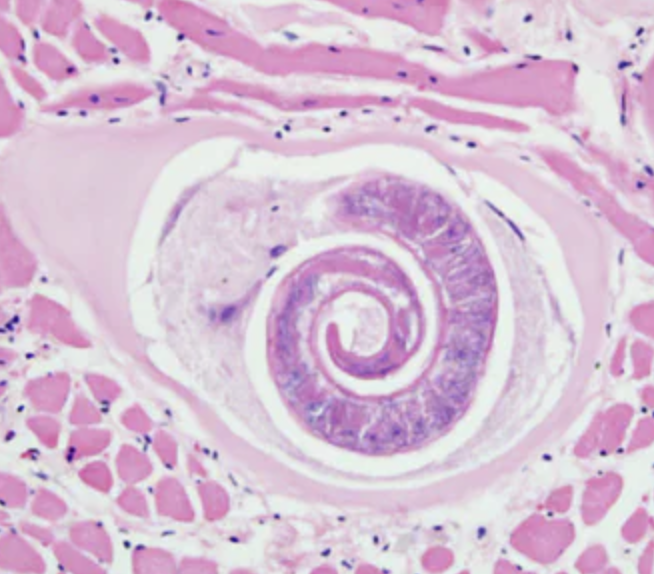
Trichinosis:C.S
HX of eating raw meat, carcasses, or exposure to wildlife
Clinical Signs
Trichinosis is often asymptomatic in pigs, but when clinical signs do occur, they typically manifest during the larval migration phase and in heavy infections. Clinical signs may include:
Gastrointestinal signs during the intestinal phase (early infection, 1-2 weeks post-infection):
Diarrhea
Abdominal pain
Vomiting
Anorexia
Muscle-related signs during the migration and encystment phase (2-4 weeks post-infection):
Muscle pain and stiffness
Lameness or reluctance to move
Swollen, tender muscles
Weakness
Fever
In severe cases, pigs may show significant debilitation and reduced weight gain. Respiratory difficulties may arise from diaphragm muscle involvement, while involvement of ocular and tongue muscles can lead to ocular swelling and dysphagia.
Trichinosis: D.X
Diagnostics
Trichinosis diagnosis in pigs can be challenging, particularly in asymptomatic animals. The following diagnostic methods are most commonly used:
Histopathology: Examination of muscle tissue, particularly the diaphragm, tongue, or masseter muscles, may reveal encysted Trichinella larvae. These larvae can be visualized under a microscope.
Serology: ELISA (Enzyme-Linked Immunosorbent Assay) tests can detect antibodies against Trichinella spp. Serological testing is more effective in detecting chronic infections but may not be useful in acute stages.
Artificial digestion test: Muscle samples are digested using pepsin-HCl, and the presence of larvae can be examined under a microscope. This method is commonly used in slaughterhouses to detect infections in pork.
Polymerase Chain Reaction (PCR): PCR can be used to detect the presence of Trichinella DNA in muscle tissues or blood. This is a more sensitive and specific test but may not be routinely available in all clinical settings.
Clinical examination and necropsy: In cases where pigs present with clinical signs such as muscle pain or stiffness, a thorough physical examination followed by necropsy (with examination of diaphragm and muscle tissue) can confirm the presence of larvae.
Trichinosis:T.X
Currently, there are no specific treatments for trichinosis in pigs. Management strategies focus on prevention. However, in clinical settings where treatment is attempted, anthelmintics such as benzimidazoles (e.g., albendazole, mebendazole) have shown some efficacy in reducing larval migration and intestinal infections. High doses may be needed, and the treatment must be administered early in infection before larvae have encysted in muscle tissue.
For pigs in the later stages of infection, treatment is often less effective because encysted larvae are difficult to eliminate. Supportive care, including anti-inflammatory drugs for muscle pain, may be provided for symptomatic animals.
Prevention
Proper feeding practices: Preventing access to raw or undercooked meat, carcasses, or wildlife sources of infection is critical. Cook all food scraps and meat-based feed thoroughly before offering them to pigs.
Rodent control: Implement strict rodent control programs to minimize exposure to potential reservoirs of Trichinella.
Farm hygiene and biosecurity: Maintaining good farm hygiene, proper waste management, and preventing contact with wildlife can reduce the risk of infection.
Meat inspection programs: Regular meat inspection and post-mortem muscle examination in slaughterhouses is essential to prevent the spread of Trichinella through the food chain. The artificial digestion test is frequently used for routine inspection in endemic areas.
Hallmark Clinical Signs and Diagnosis for Trichinosis
The hallmark sign of trichinosis in pigs is the presence of encysted larvae in muscle tissues, particularly in the diaphragm, tongue, and masseter muscles. Infections are often subclinical, but when clinical signs are present, muscle pain, stiffness, and swelling are key indicators during the larval migration phase.
Definitive diagnosis is typically achieved through the artificial digestion test or muscle biopsy with histological examination for encysted larvae. Serological tests may be used to confirm exposure, especially in chronic infections, but are not useful in early infections.
Vitamin/Mineral deficiency
Vitamin and mineral deficiencies in pigs can lead to a wide range of health problems, affecting growth, reproduction, and overall animal welfare. These deficiencies often go unnoticed until clinical signs become evident, and by then, production losses may already be significant. Early detection and prevention are crucial in ensuring the health and productivity of pigs. This article will explore the pathophysiology, clinical signs, risk factors, diagnostics, and treatment of key vitamin and mineral deficiencies in pigs.
Pathophysiology
Vitamins and minerals are essential nutrients that support various metabolic processes in pigs. Deficiencies occur when the intake of these nutrients is insufficient to meet the metabolic demands of the animal. The most common deficiencies in pigs involve calcium (Ca), phosphorus (P), vitamin D, zinc (Zn), iron (Fe), selenium (Se), and vitamin E, although other vitamins and minerals may also be involved.
1. Calcium, Phosphorus, and Vitamin D Deficiency
Calcium and Phosphorus are critical for bone mineralization, muscle function, and metabolic processes. A deficiency in either mineral, or an imbalance in their ratio, can lead to poor bone development and metabolic disorders.
Vitamin D plays a key role in calcium and phosphorus absorption from the intestines and their deposition into bone.
Pathophysiology: Calcium and phosphorus deficiencies result in impaired bone mineralization, leading to conditions such as rickets in young pigs and osteomalacia in older animals. Vitamin D deficiency exacerbates this by limiting calcium absorption, leading to hypocalcemia and secondary hyperparathyroidism.
2. Zinc Deficiency
Zinc is involved in enzyme function, protein synthesis, and immune response. Zinc deficiency can impair keratinization and wound healing.
Pathophysiology: Zinc deficiency leads to reduced cellular proliferation and impaired immune function, affecting skin and mucosal barrier integrity, resulting in a condition known as parakeratosis.
3. Iron Deficiency
Iron is necessary for hemoglobin formation and oxygen transport. Suckling piglets are particularly susceptible to iron deficiency anemia due to their rapid growth and the low iron content in sow’s milk.
Pathophysiology: Iron deficiency leads to reduced hemoglobin synthesis, resulting in anemia. This limits the oxygen-carrying capacity of blood, leading to fatigue, poor growth, and susceptibility to infections.
4. Selenium and Vitamin E Deficiency
Selenium and vitamin E are antioxidants that protect cell membranes from oxidative damage. They are also crucial for muscle function and immune response.
Pathophysiology: Deficiencies result in oxidative damage to cell membranes, particularly in muscles, leading to white muscle disease, a condition characterized by degeneration of skeletal and cardiac muscle.
5. Other Vitamins and Minerals
B Vitamins: Involved in metabolic pathways such as energy production and nervous system function. Deficiencies can lead to neurologic symptoms, poor growth, and anemia.
Magnesium: Essential for enzyme function and neuromuscular activity. Deficiencies are rare but can cause nervousness, tremors, and convulsions.
Clinical Signs1. Calcium, Phosphorus, and Vitamin D Deficiency
Rickets in growing pigs: lameness, swollen joints, bowed legs, and poor growth.
Osteomalacia in adult pigs: lameness, fractures, and decreased productivity.
Reduced feed efficiency and poor reproductive performance.
2. Zinc Deficiency
Parakeratosis: Thickened, scaly skin with crusts, particularly on the legs, abdomen, and face.
Reduced feed intake and poor growth rates.
Delayed wound healing and increased susceptibility to skin infections.
3. Iron Deficiency
Anemia: Pale skin and mucous membranes, lethargy, reduced growth, and difficulty breathing in severe cases.
Increased susceptibility to infections due to reduced immune function.
Poor growth and general unthriftiness in piglets.
4. Selenium and Vitamin E Deficiency
White Muscle Disease: Stiffness, muscle weakness, and sudden death due to cardiac failure.
Subcutaneous edema and difficulty standing.
Sudden death in piglets due to heart muscle degeneration.
5. Other Deficiencies
B Vitamin Deficiencies: Poor growth, nervous signs such as incoordination or paralysis, rough hair coat, and anemia.
Magnesium Deficiency: Tremors, convulsions, hyperexcitability, and sudden death in severe cases.
Prevention
Balanced Diets: Ensure all feed rations are properly formulated to meet the nutritional requirements of the herd, particularly during critical growth and reproductive stages.
Soil and Forage Testing: Regularly test soil and forage for mineral content and supplement accordingly.
Routine Blood Testing: Periodic testing of blood nutrient levels in high-risk groups (e.g., fast-growing piglets, breeding sows) can help identify deficiencies early.
Iron Supplementation for Piglets: Routine administration of iron injections to piglets born indoors.
Selenium Supplementation in Deficient Areas: Regions with known selenium-deficient soils should incorporate selenium supplements into feed.
Conclusion
Vitamin and mineral deficiencies in pigs are common but can be effectively managed with proper nutritional management. Early diagnosis and treatment are critical to avoid production losses and ensure the welfare of pigs. By understanding the specific roles of vitamins and minerals in pig physiology, veterinarians can help implement preventative strategies and provide effective treatments when deficiencies arise.
Vitamin/Mineral deficiency:D.X
Diagnostics1. Blood Tests
Serum calcium, phosphorus, zinc, iron, selenium, and vitamin levels can be measured to detect deficiencies.
Hematology: Low hemoglobin and hematocrit in iron deficiency anemia.
Serum alkaline phosphatase: Elevated in calcium or vitamin D deficiency due to bone turnover.
Glutathione peroxidase activity: Low in selenium deficiency.
2. Bone Analysis
Radiographs: In rickets or osteomalacia, radiographs may reveal reduced bone density, bone deformities, or fractures.
Bone ash analysis can determine the calcium and phosphorus content of bones post-mortem.
3. Histopathology
Muscle biopsies can reveal the characteristic lesions of white muscle disease in selenium/vitamin E deficiency, including muscle fiber degeneration and necrosis.
4. Clinical Observation
Diagnosis often relies on correlating clinical signs with potential risk factors such as dietary history, environmental management, and herd health.
Calcium, Phosphorus, and Vitamin D Deficiency Treatment
Treatment1. Calcium, Phosphorus, and Vitamin D Deficiency
Dietary Correction: Ensure an adequate and balanced supply of calcium and phosphorus, with a Ca
ratio between 1.5:1 and 2:1.
Vitamin D Supplementation: Administering vitamin D3 via feed or injection to support calcium absorption.
Supportive Care: In severe cases, parenteral calcium and phosphorus may be required.
2. Zinc Deficiency
Zinc Supplementation: Oral or injectable zinc preparations are used to correct deficiencies. Zinc oxide or sulfate is commonly added to feed.
Topical Treatments: Skin lesions can be managed with zinc-based ointments to promote healing.
3. Iron Deficiency
Iron Dextran Injection: Typically administered to piglets within the first few days of life to prevent anemia.
Oral Iron Supplements: Can be added to feed or water for ongoing supplementation.
4. Selenium and Vitamin E Deficiency
Selenium Supplementation: Injectable selenium compounds (e.g., sodium selenite) or dietary selenium additives.
Vitamin E Supplementation: Vitamin E should be provided through feed or injections, particularly during periods of stress or high metabolic demand.
5. Other Deficiencies
B vitamin and magnesium deficiencies are typically treated through dietary supplementation of premixes that include the missing vitamins or minerals. In acute cases, injectable forms may be used.