Animal Sensory Systems
1/41
There's no tags or description
Looks like no tags are added yet.
Name | Mastery | Learn | Test | Matching | Spaced | Call with Kai |
|---|
No analytics yet
Send a link to your students to track their progress
42 Terms
sensory receptor cells
transduce external stimuli into changes in membrane potentials- may either depolarize or hyperpolarize in response to the stimulus
if the changes in membrane potential are sufficient to induce action potentials → transduced to the nervous system (efferent division of PNS to CNS)
sensory receptor cells can be either:
specialized neurons (receptor cell is a neuron)
specialized sensory cells which synapse with neurons (receptor secretes neurotransmitters to a neuron)
different types:
mechanoreceptors
photoreceptors
chemoreceptors
nociceptors
thermoreceptors
stimuli are integrated in the ___ and responses are sent to appropriate body systems via the ___
stimuli are integrated in the CNS and responses are sent to appropriate body systems via the PNS
3 stages of sensory system
sensory input → integration → motor output
(afferent neurons → interneurons → efferent neurons)
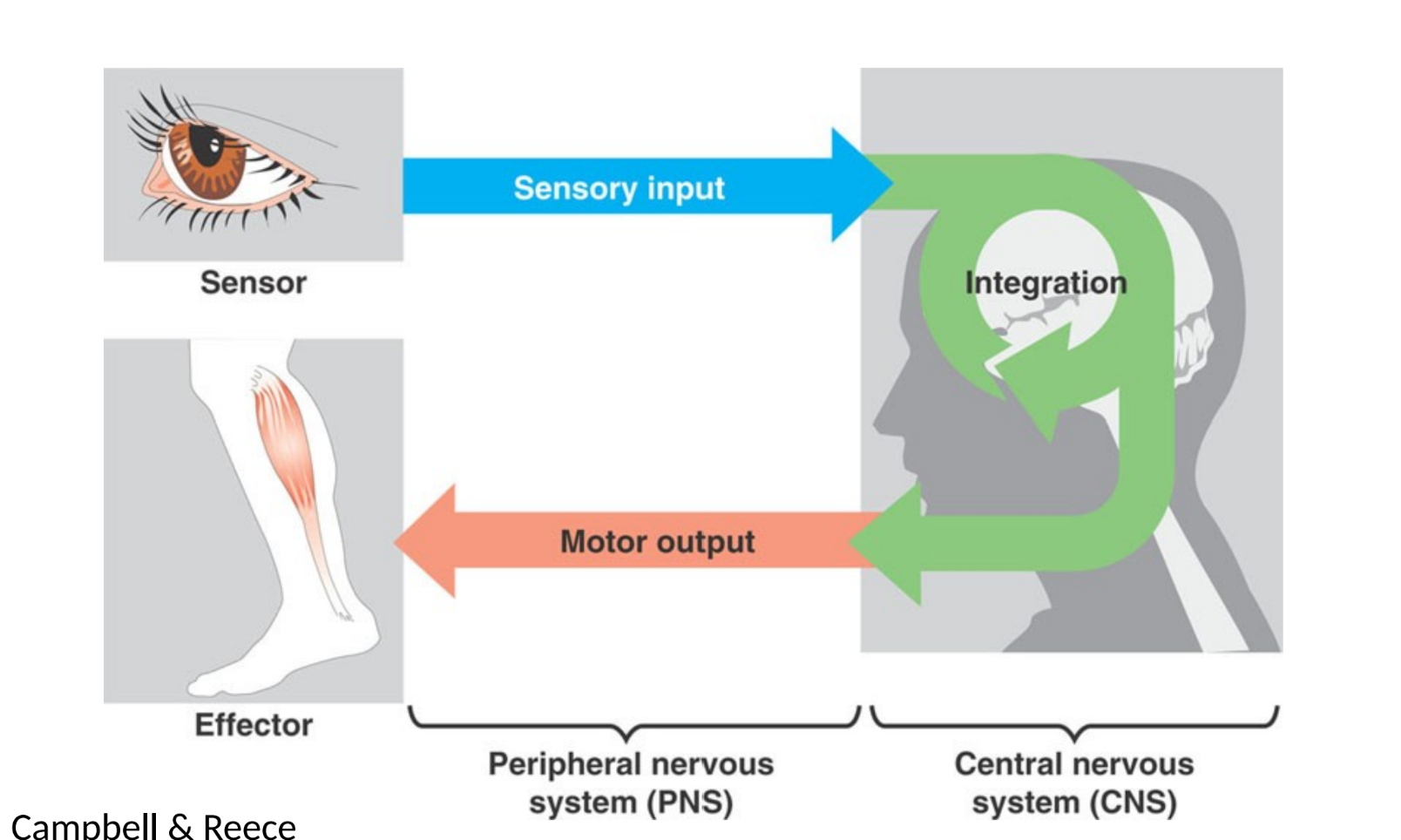
processing sensory information
transduction within the PNS
receptor cells may depolarize or hyperpolarize
transmission to the CNS via the PNS
signals from different sensory systems transmit to different parts of the brain
integration in the CNS
sensory adaptation
incoming signals integrated at axon hillock via summation of potentials from many receptors
response via the PNS (usu motor output)
mechanoreceptors
detect pressure
touch, sound, balance
some types of mechanoreceptors are located near the upper layers of the skin, and thus are more sensitive to lighter touch and are able to precisely localize gentle touch
mechanoreceptors located deeper in the skin are only activated by stronger pressure and are not as highly sensitive to identify the precise location of the touch
photoreceptors
respond to light
chemoreceptors
respond to taste/smell
oldest of all sensory receptors
nociceptors
detect tissue damage- which our brains interpret as pain
thermoreceptors
respond to heat or cold
the intensity/degree of a stimulus is encoded in three different ways:
rate/frequency of action potentials produced by the sensory receptor
number of receptors activated
which specific receptors are activated
frequency (sound)
number of waves per unit of time
pitch
high-frequency sounds are higher-pitched and shorter wavelength than low-frequency, long-wavelength sounds
amplitude (sound)
dimension of a wave from peak to trough
volume
sound waves of louder sounds have a greater amplitude than those of softer sounds
structure of the human ear
outer ear collects and amplifies signal which hits the tympanic membrane → ossicles vibrate to transmit signal to fluid-filled cochlea → soundwaves transduced into action potentials by hair cells between basilar and tectorial membranes within the cochlea
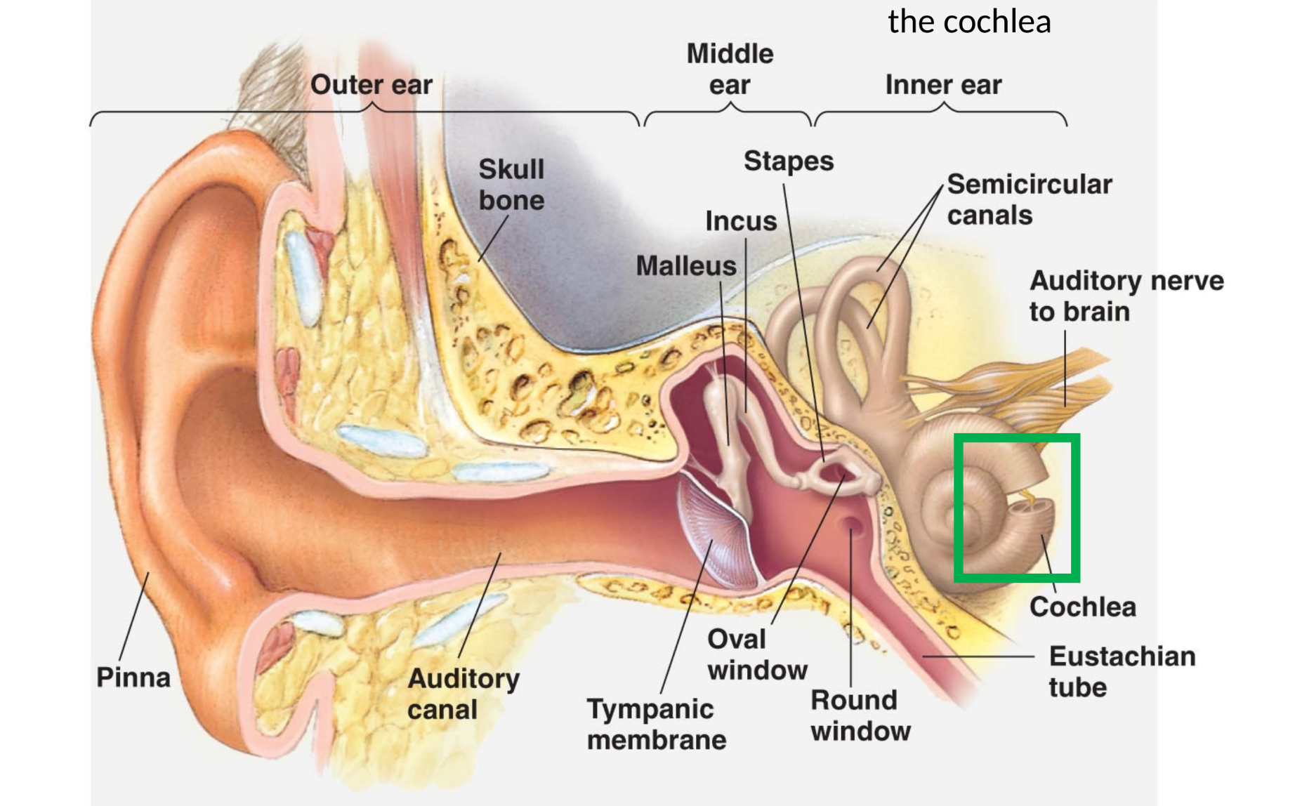
cochlea
fluid-filled whorled structure that contains the auditory mechanoreceptors that allow us to perceive pressure waves in the air as sound
contains the basilar membrane
basilar membrane
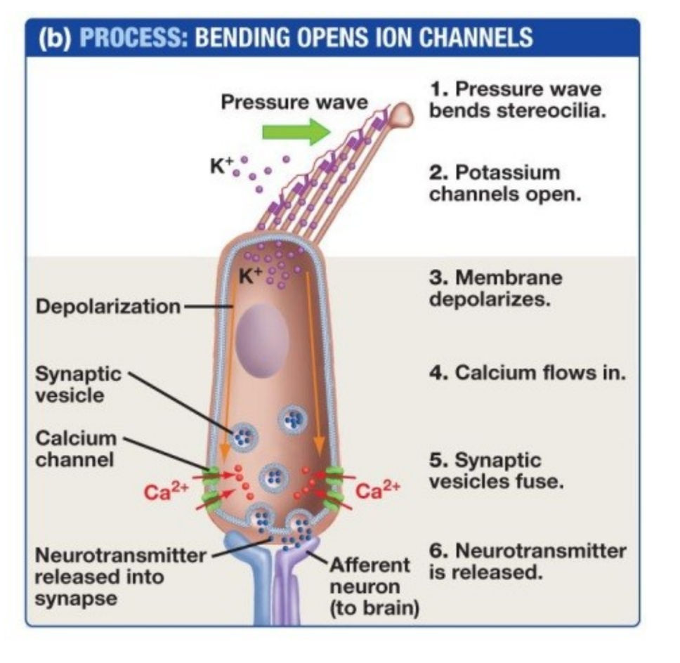
flexible membrane that runs the length of the cochlea and contains the mechanoreceptors called hair cells- transduce sound waves into action potentials
stereocilia: tiny hair-like protrusions on hair cells- pressed against the tectorial membrane when basilar vibrates- stereocilia bend → intitiates action potentials
mechanoreceptors bend and open ion channels in response to pressure
we perceive volume based on:
how many hair cells are activated
we perceive pitch based on:
which hair cells are activated (which region of the basilar membrane vibrates)
stiffer region of the basilar membrane (narrow part)
vibrates in response to high frequency (higher-pitched) sounds
flexible region of the basilar membrane (wider part)
vibrates in response to low frequency (lower-pitched sounds)
vestibular system
detects positions and movement of our head in space
stimuli:
linear acceleration (gravity)
angular acceleration and deceleration
vestibular labyrinth
contain vestibular hair cells
adjacent to cochlea
hair cells in the vestibular labyrinth detect stimuli in two ways:
detect head position and movement through gelatin shifting and stereocilia bending caused by movement of calcium carbonate crystals (ear stones) in a gelatinous layer in response to the head tilting or accelerating/decelerating- bending signals to the brain for balance
some hair cells project into a gelatinous cap called the cupula. When the head turns, the fluid in the canals shift → bending stereocilia and sending signals to the brain; when movement stops → movement of the fluid within the canals slows or stops.
to detect pressure changes, bony fish use:
lateral line systems comprised of hair cells in a cupula
statocysts
organs with dense statoliths adjacent to hair cells
how invertebrates detect balance
wavelength (light)
detected as hue/color
varies inversely with frequency
light at red end of the visible spectrum has longer wavelengths; light at violet end has shorter wavelengths
amplitude (light)
perceived as brightness
eye cups
in flatworms
dimple-shaped; detect direction of a light source
compound eyes
arthropods
contain multiple lenses and detect shapes, patterns, and movements
pinhole eyes
in the nautilus
contain no lens and form simple, low-resolution images
simple eyes
cephalopods and vertebrates
contain a single lens and form high-resolution images
all photoreceptors contain:
a combination of a protein and a pigment molecule
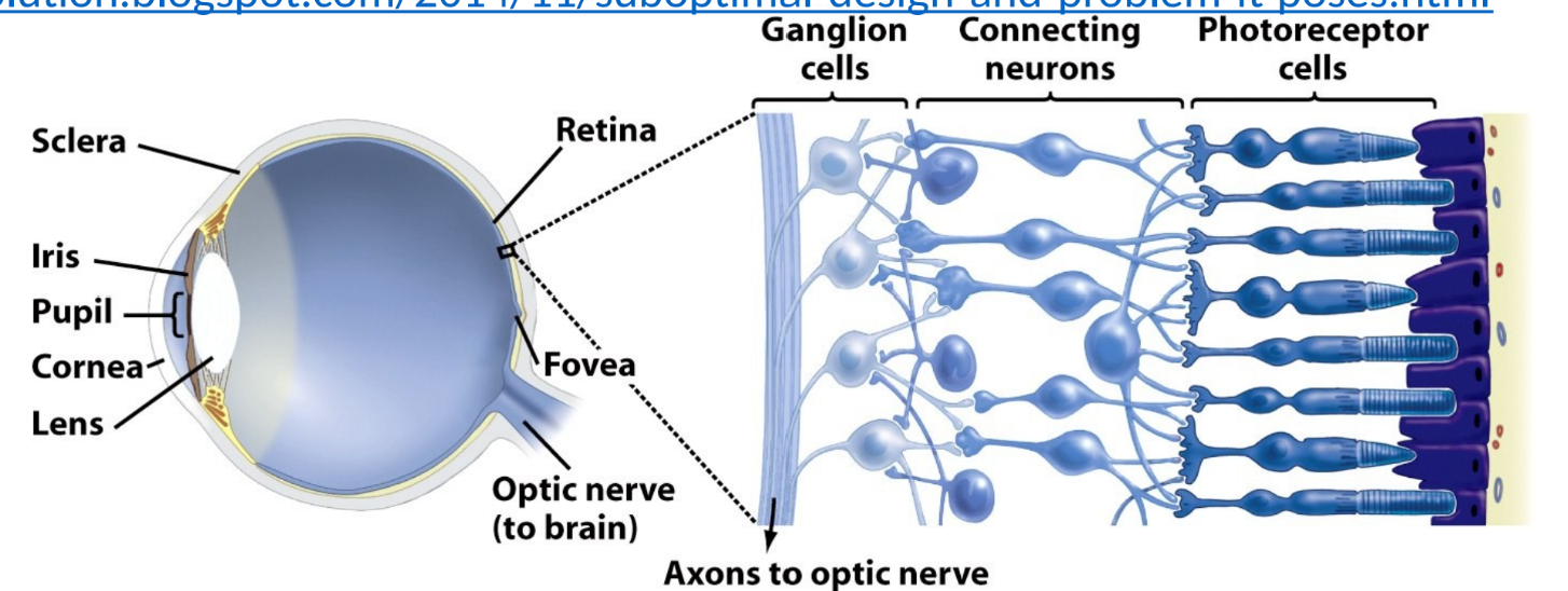
the vertebrate eye contains:
cornea: transparent sheet of connective tissue- functions with the lens to focus light on the retina
iris: pigmented ring of muscle that controls amount of light entering eye
pupil: hole in center of iris
lens: curved structure that focuses light on retina (by bending) in conjunction with cornea
retina: thin layer of photoreceptor cells and neurons
photoreceptor cells: light-detecting sensory cells
fovea: site of retina w/ only cones- area of highest visual resolution
optic nerve: axons of the ganglion cells
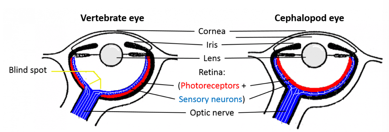
cephalopod vs. vertebrate eye
cephalopod eyes move the lens to focus rather than changing shape
vertebrate eyes have an inverted retina, where blood vessels and nerves are in front of the photoreceptor cells instead of behind
results in vertebrates having blind spot as well as age-related macular degeneration, and increased chance of a detached retina
retinal
pigment contained in opsin (protein)
reversibly changes shape when it is hit by a photon of light
opsin
protein that holds the retinal pigment and changes shape/activity when the retinal changes shape in response to absorption of light
responsible for ability to perceive differences in color or hue
each different version is capable of absorbing a different wavelength of light:
s opsin (short-wavelength opsin); m opsin (medium-wavelength opsin); l opsin (long-wavelength opsin)
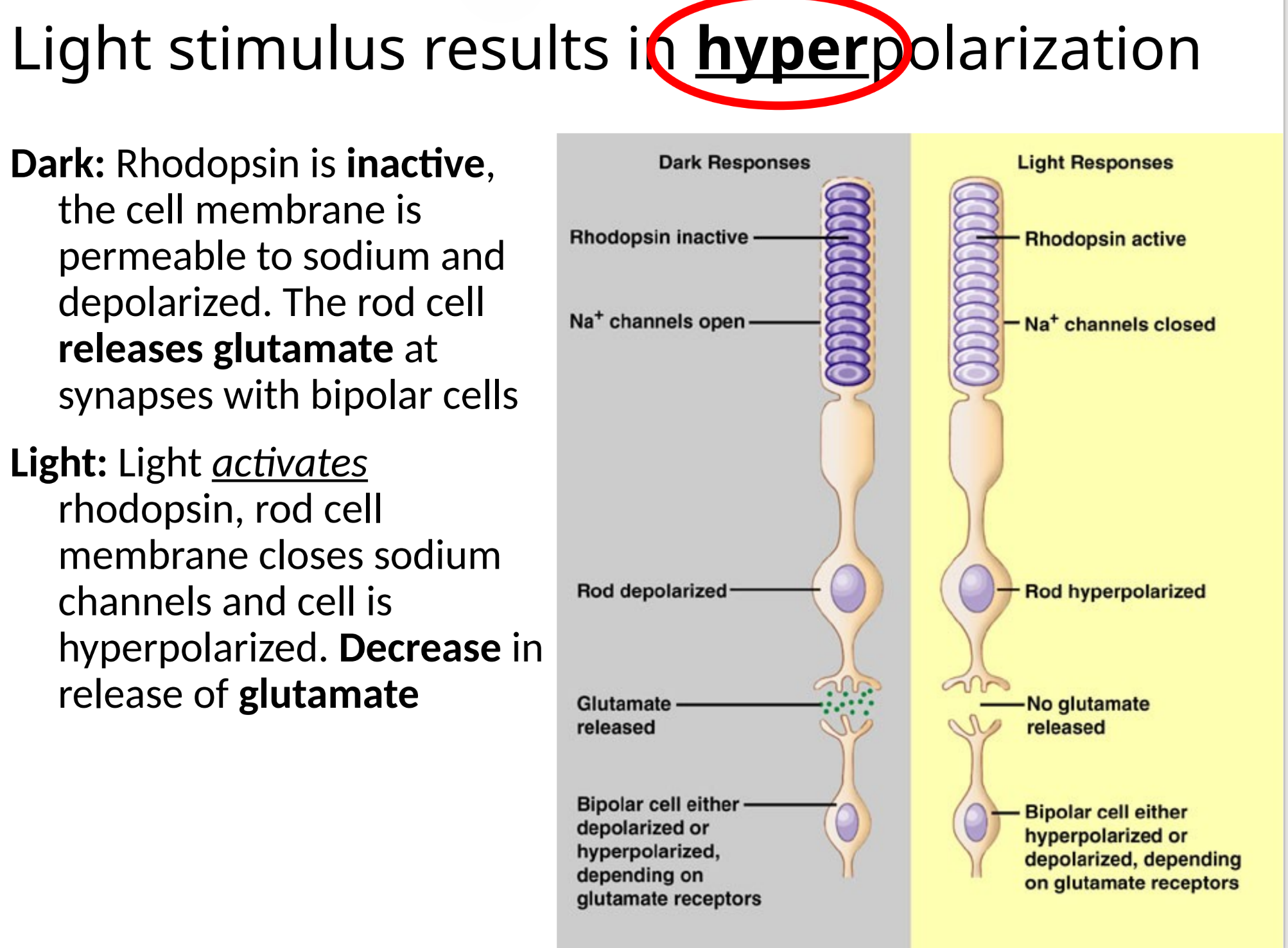
rhodopsin
complex made up of retinal and opsin- allows us to detect light and color
inactive in the dark
activated by light
light stimulus results in:
hyperpolarization
cones
contain a single type of color-sensitive opsin (so 3 types of opsin = 3 types of cones)
require high levels of light to work
use for color vision
good for detail
heavily concentrated at the fovea
rods
contain a fourth type of opsin called rod opsin- activated by an intermediate wavelengths of light
active in low light
not color-sensitive
good for detecting movement in field of vision
heavily concentrated at the periphery (outer edges) of retina
rods and cones in the dark vs in the light
in the dark: rods and cones are depolarized → releasing neurotransmitters to their synapsed bipolar cells
in the light: rods and cones hyperpolarize → stop releasing neurotransmitter