ALL Dimorphic fungi
1/44
There's no tags or description
Looks like no tags are added yet.
Name | Mastery | Learn | Test | Matching | Spaced | Call with Kai |
|---|
No analytics yet
Send a link to your students to track their progress
45 Terms
2 morphological states
Moulds - filamentous - in environment at low temperatures
Yeast - unicellular - form in host tissues at body temperature
Dimorphism is dependent on what
Temperature
How do conidia end up in body
By inhalation
Why does fungus switch to yeast
To resist phagocytosis
List off dimorphic fungi (5)
Blastomycetes dermatitidis, histoplasma capsulatum, histoplasma farciminosum, coccidioides immitis, sporothric schenckii
What temps for mould and yeast forms
Mould 25
Yeast 37
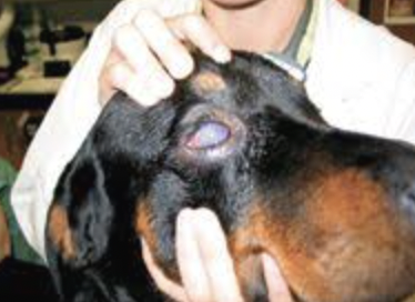
What is the name of the infection by blastomyces dermatitidis
Blastomycosis
Which host does blastomycosis affect
Dogs and humans
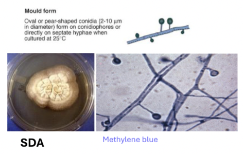
Mould form of blastomyces dermatitidis
Septate hyphae with single conidia
Culture on SDA - methyl blue
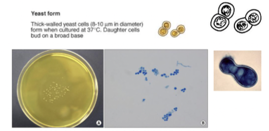
Yeast form blastomyces dermatitidis
Thick walled, broad based budding cells
Culture on BHI agar - gram or methyl blue
Where do blastomyces dermatitidis convert to yeast
Convert in lungs when reach alveoli
What kind of inflammation is caused by blastomyces dermatitidis
Pyogranulomatous inflammation
Blastomyces dermatitidis virulence factor
BAD1 (blastomyces adhesin 1)
Promote adhesion to cell, suppresses host response
Blastomycosis clinical signs
Chronic cough, fever, weight loss, draining skin lesions, lameness, ocular lesions
What is the disease name of histoplasma capsulatum
Histoplasmosis
Which hosts does histoplasmosis affect
Dogs, cats and humans
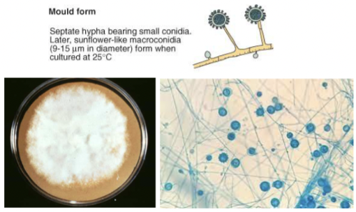
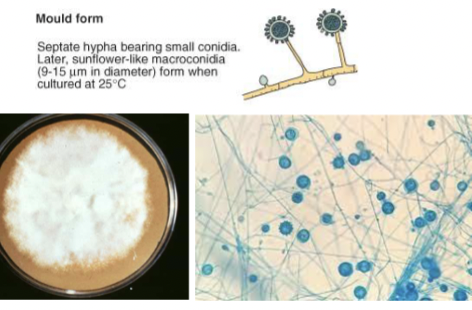
Histoplasma capsulatum mould form
Septate hyphae with small conidia and sunflower like macroconidia when cultured
Culture on SDA - methyl blue
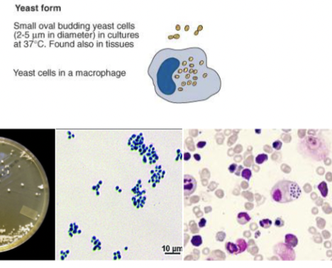
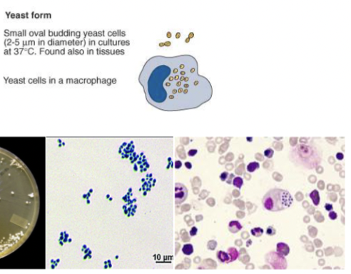
Histoplasma capsulatum yeast form
Small oval budding least cells, can live in macrophages
Where do histoplasma capsulatum turn into yeast
Alveolar macrophages phagocytose macroconidia
Spores convert to yeast and multiply inside macrophages
How does histoplasma capsulatum spread
Via lymphatics and blood and cause granulomatous inflammation
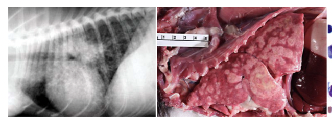
Histoplasmosis clinical signs
Chronic cough, dyspnea, depression, loss of weight, diarrhea, intestinal ulcers
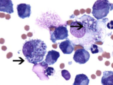
Histoplasma capsulatum diagnosis
Cytology will show macrophages filled with small yeast with clear halos
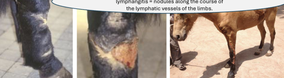
What is the disease name of Histoplasma farciminosum
Equine histoplasmosis or Epizootic lymphangitis
What does equine histoplasmosis affect
Horses, mules and donkeys
Mould form of histoplasma farciminosum
Septate hyphae with small conidia and sunflower like macroconidia when cultured
Culture on SDA at 25
Yeast form of Histoplasma farciminosum
Small oval budding yeast cells, can live in macrophages
How is histoplasma farciminosum transmitted
NOT INHALED
Through skin wounds or abrasions
What does histoplasma farciminosum target
Lymphatic vessels
What does Histoplasma farciminosum cause
Nodules along lymphatics on limbs, discharge pus, lymphocutaneous lesions
What is the name of the infection caused by Coccidioides immitis
Coccidioidomycosis
What does coccidioides immitis affect
Dogs, horses
What is special about the dimorphism in coccidioides immitis
Not dimorphic but biphasic as it doesn’t transform into true yeast cells
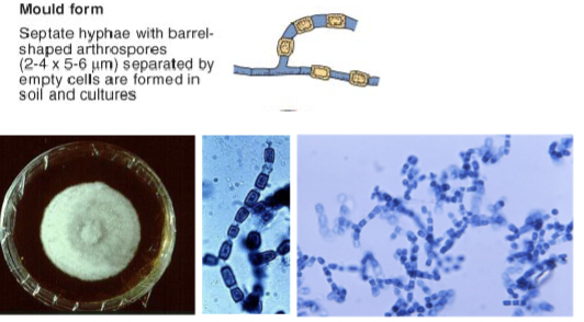
Mould form Coccidioides immitis
Septate hyphae which fragments into arthroconidia with barrel shaped arthrospores
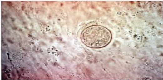
Spherules in coccidioides immitis
Mature spherules are filled with endospores and are found in tissue
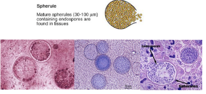
Coccidioides immitis pathogenesis
Inhalation arthroconidia which transform into spherules in lungs
They release endospores and new spherules form
Coccidioidomycosis clinical signs
Dogs - cough, fever, weight loss, lameness, skin lesions
Horses - pulmonary and cutaneous forms, nasal granulomas
Diagnosis of coccidioides immitis
Microscopy to identify spherules in tissues
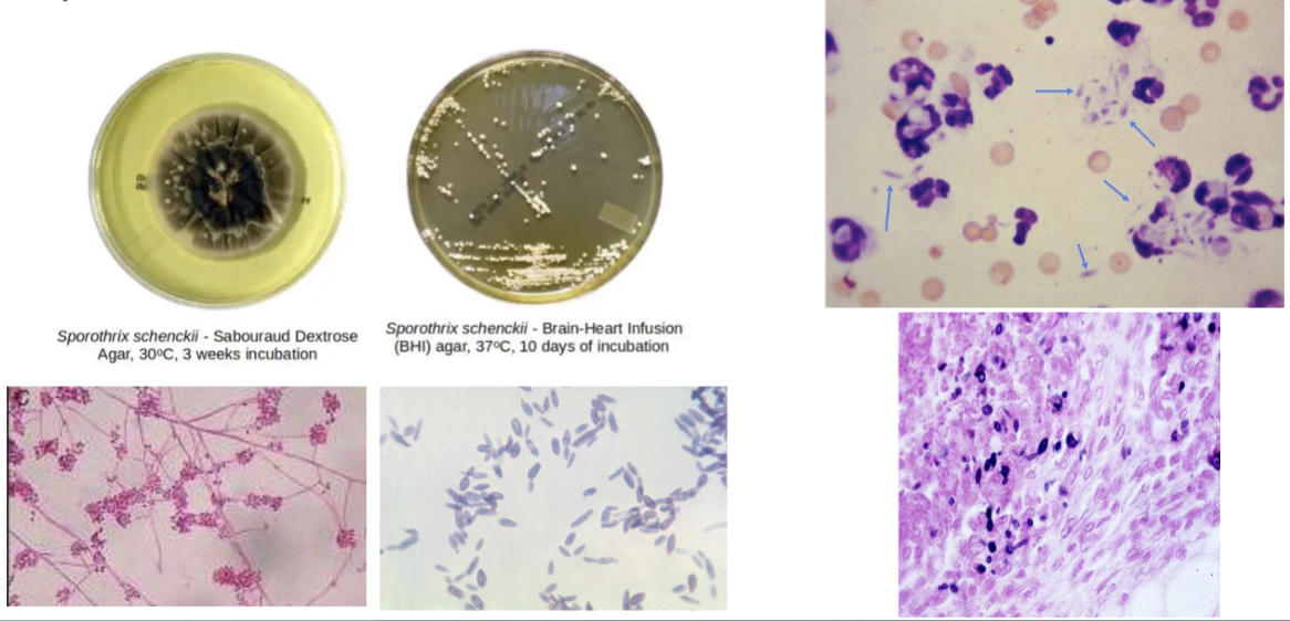
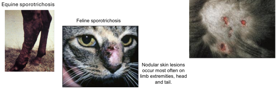
What is the name of the infection caused by Sporothric schenckii
Sporotrichosis
Who does Sporothric schenckii affect
Dogs, cats, horses and humans
How does Sporothric schenckii enter body
Traumatic inoculation of spores (splinters, bites, thorns…)
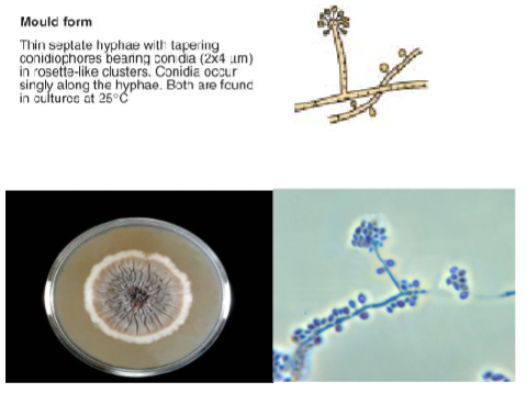
Mould form Sporothric schenckii
Septate hyphae with conidiophores bearing conidia
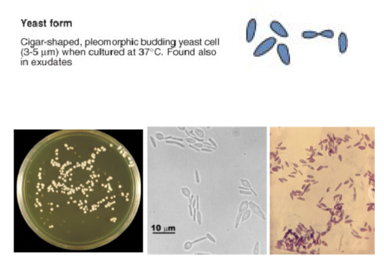
Yeast form Sporothric schenckii
Cigar shaped pleomorphic budding yeast cells
Where does Sporothric schenckii transform to yeast
Beneath the skin after inoculation
Nodules spread along lymphatic vessels
Clinical signs of sporotrichosis in horses, cats and dogs
Horses - nodules ulcerating yellow discharge, subcutaneous edemas
Cats - nodular skin lesion (often on limb extremities, head and tail)
Dogs - ulcerated, crusted and alopecic cutaneous lesions over head or trunk
Microscopy and culture Sporothric schenckii