Ear Final Exam
1/106
There's no tags or description
Looks like no tags are added yet.
Name | Mastery | Learn | Test | Matching | Spaced | Call with Kai |
|---|
No analytics yet
Send a link to your students to track their progress
107 Terms
Peripheral Auditory System
Function: Collect the sound waves and transform them from mechanical to electrochemical energy into the brain
Central Auditory system
interpret the electrochemical signals and differenciate the sounds
microtia
being born with a small pinna
anotia
complete absence of the pinna (fails to develop)
What is unique about the external auditory meatus in children
Their EAM is more horizontal- makes it easier to get things stuck
In adults, what is helpful about the structure and celia of the EAM?
It slopes downward to help drain fluid and keep dust, insects, and moisture out of the middle ear.
What is one reason why ear infections so painful?
Epithelial and connective tissue is tightly bound to the cartilage and bone of the ear
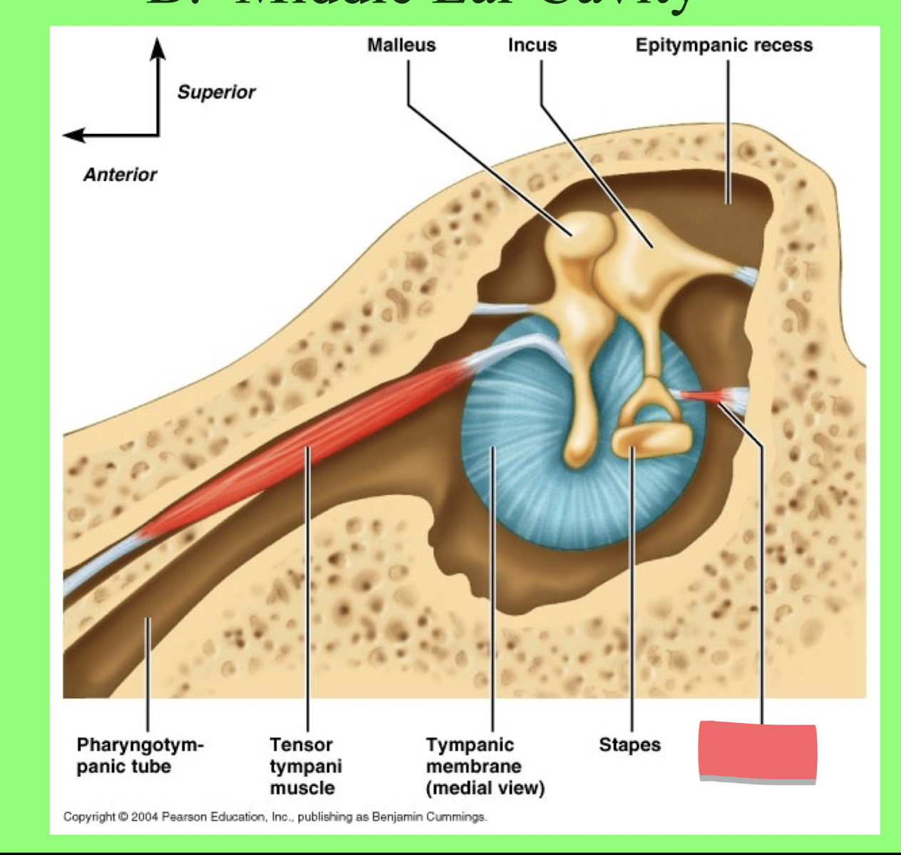
What 5 components make up the Middle Ear?
Tympanic membrane
Middle ear cavity
Auditory Ossicles (Ear bones)
Middle ear muscles
Eustachian tube
How many layers of tissue are in the tympanic membrane?
3 layers (two in the middle makes 4)
What are the different layers of tissue in the tympanic membrane
Outer Cuticular Layer (continuous with the outside tissue)
Intermediate Fibrous Layer (Made up fo 2 layers)
a. Superficial layer = center-outward fibers
b. deeper layer = circular
Inner mucous layer continuous with inside
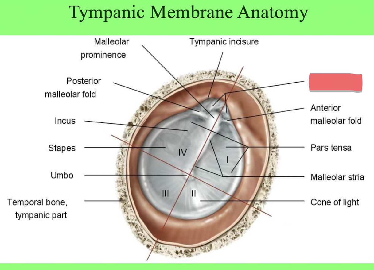
Pars flaccida
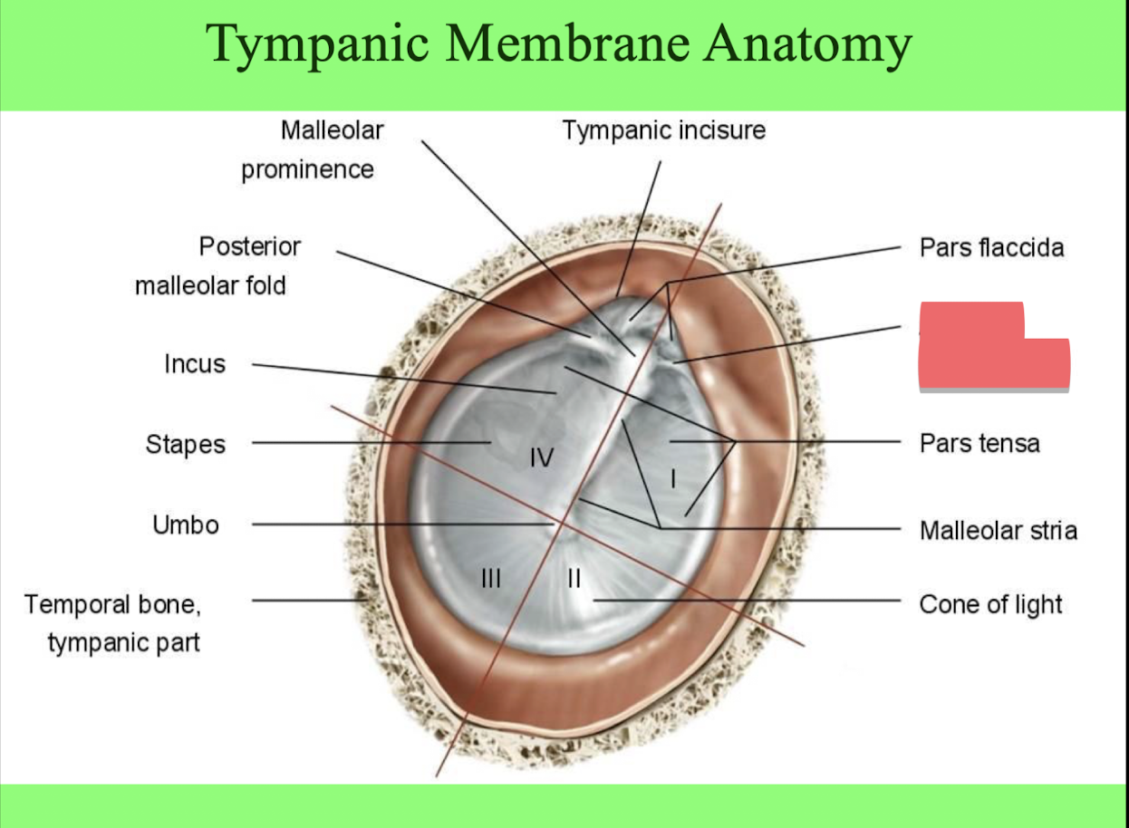
anterior malleolar fold
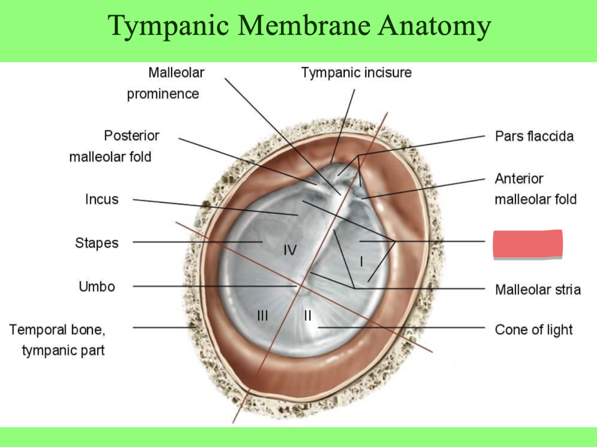
pars tensa
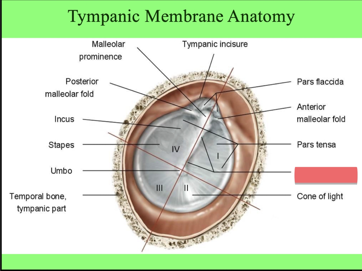
malleolar stria
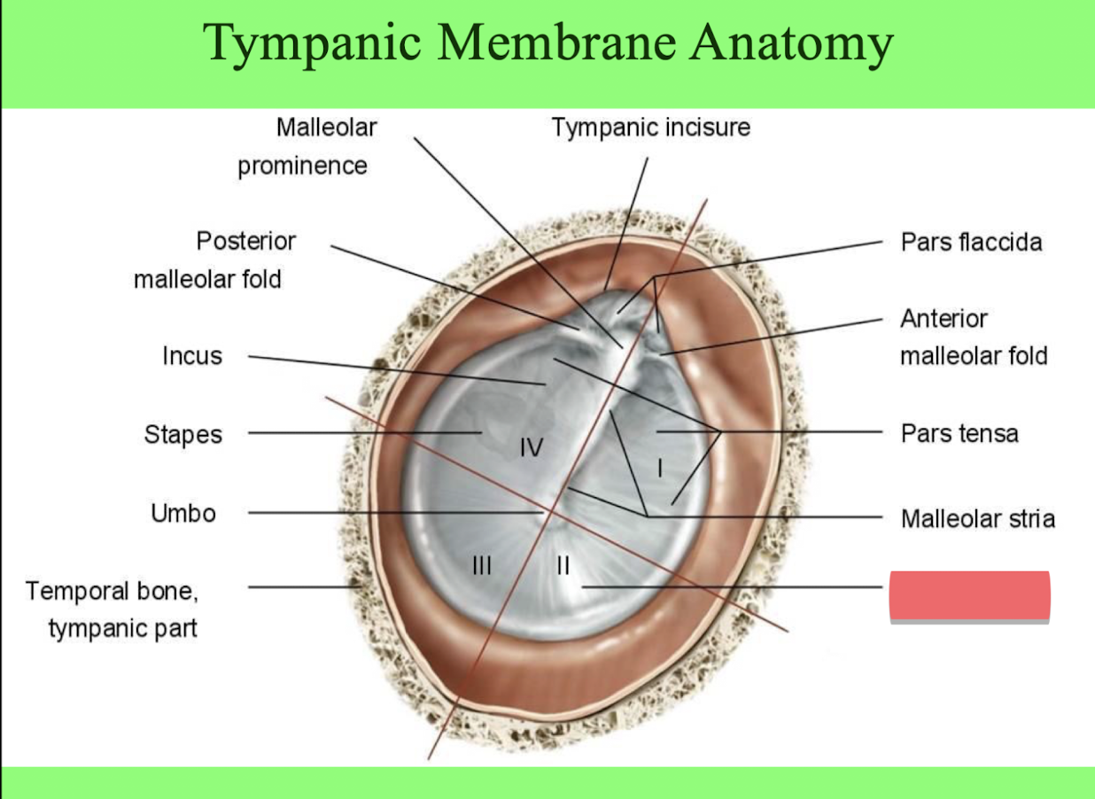
cone of light
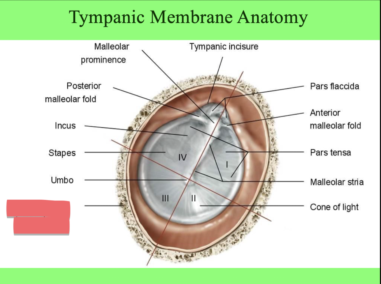
temporal bone
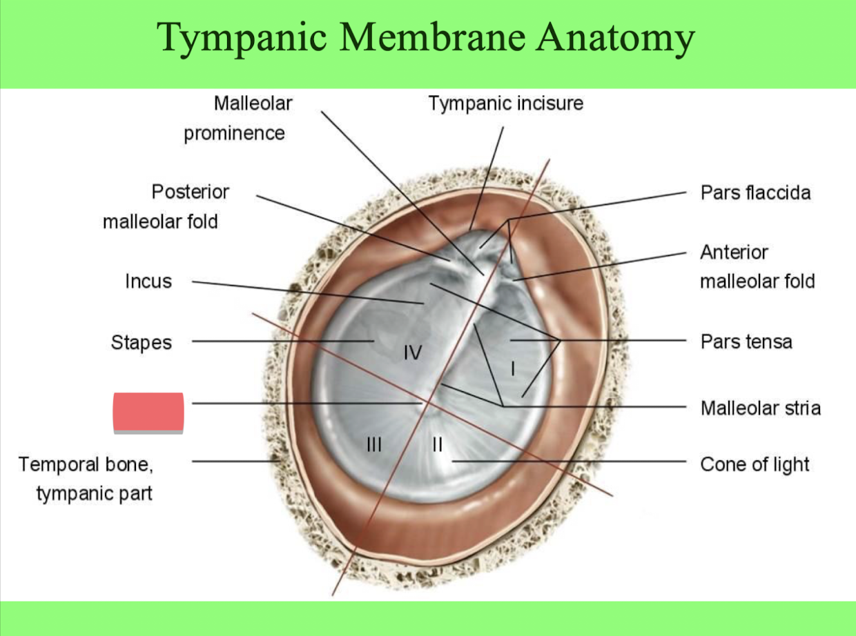
umbo
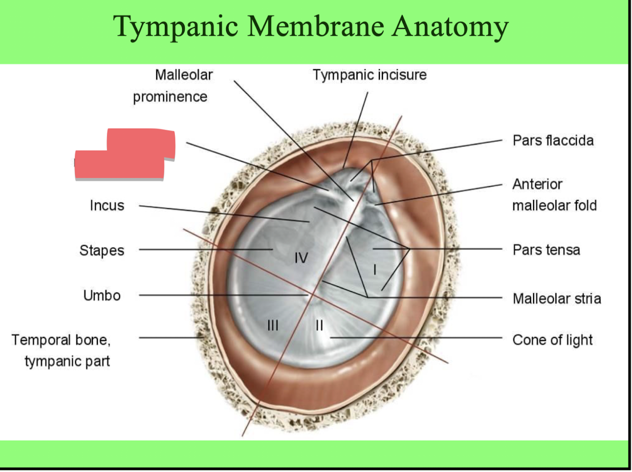
posterior malleolar fold
What is the point of the cone of light?
The reflection of the light of the otoscope off of the tympanic membrane signals a healthy ear drum
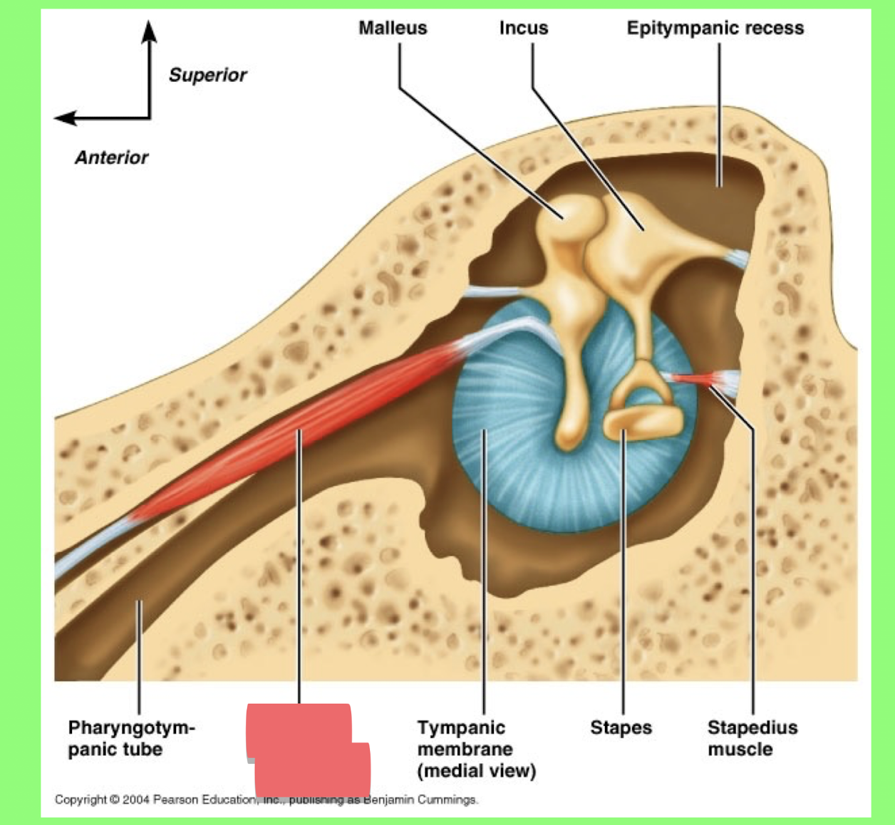
Tensor tympani muscle
Stapedius muscle
The middle ear cavity is filled with
air
The medial wall of the middle ear cavity contains the
oval window, round window and promontory
The posterior wall of the middle ear cavity contains the
pyramidal eminence containing the stapedius muscle
What fits into the oval window?
the footplate of the stapes
What is the purpose of the round window?
To displace fluid pushed inward by the stapes, and separate the air and fluid
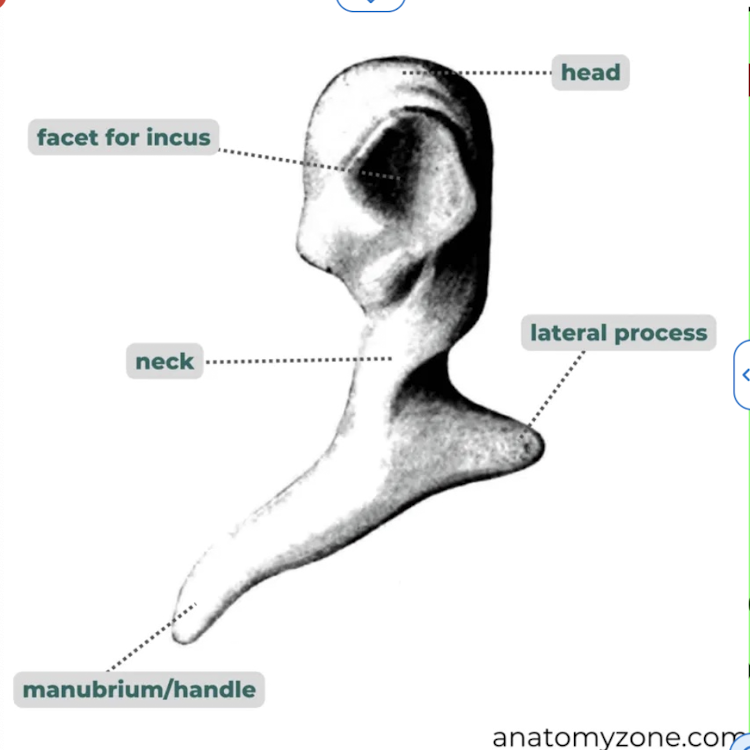
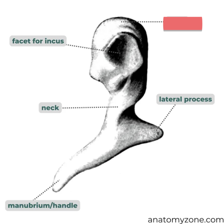
This whole bone
malleus
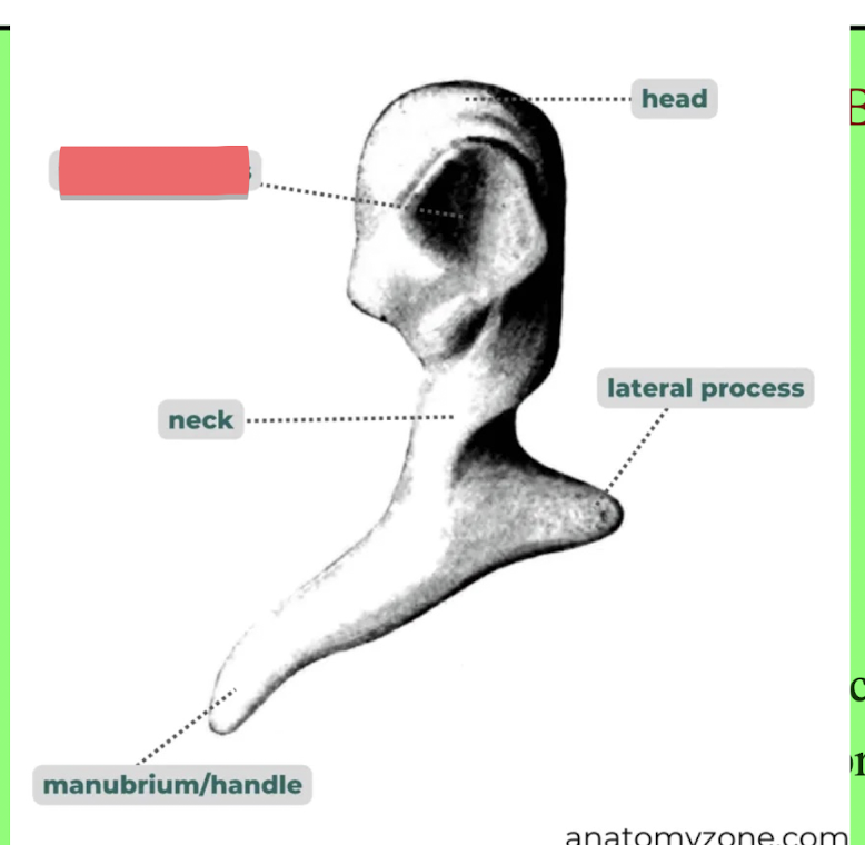
facet for incus
head of malleus
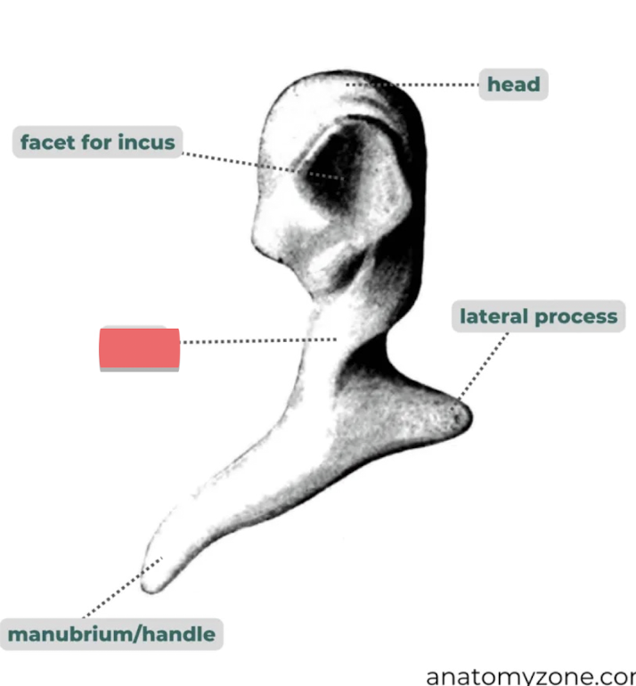
neck of malleus
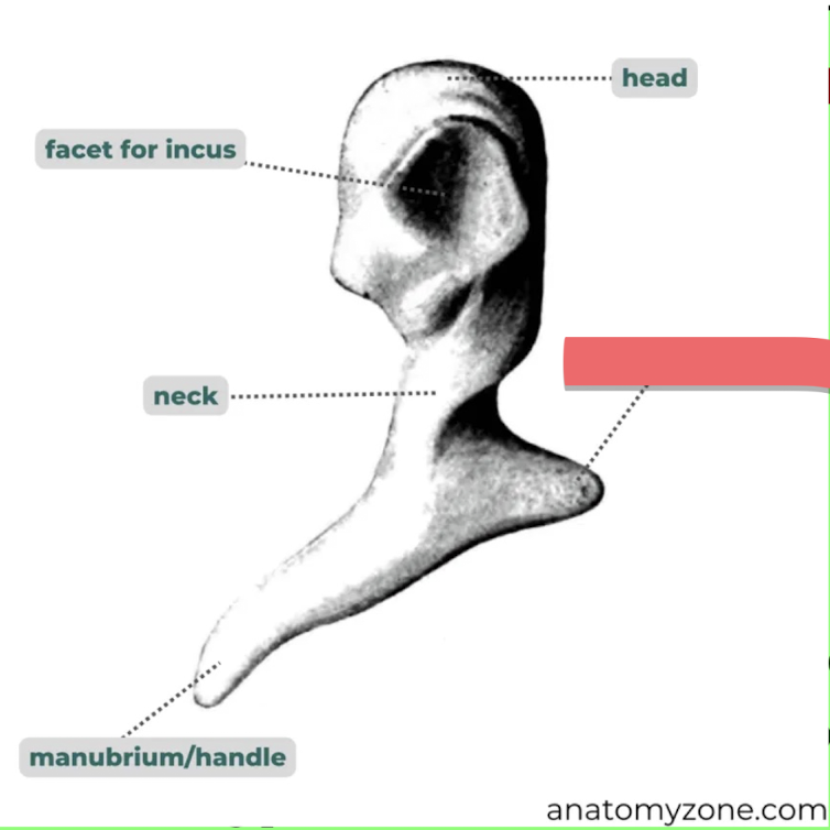
lateral process

facet for incus
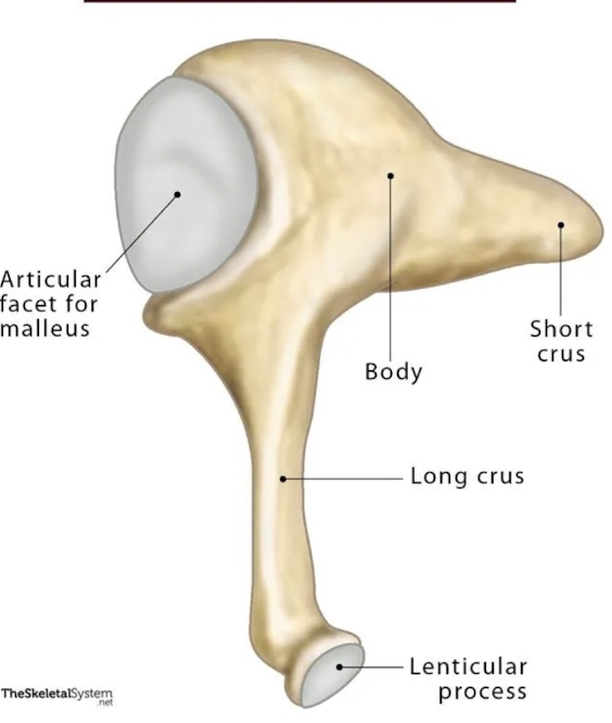
whole bone
incus
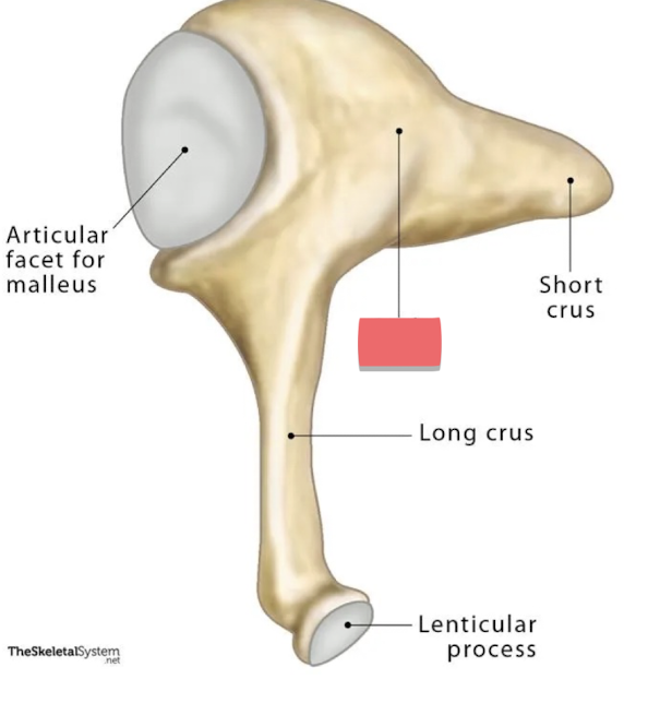
corpus (body)
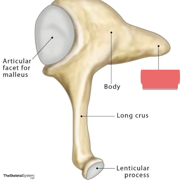
short process
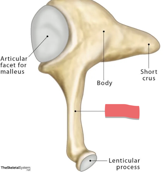
long process
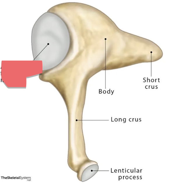
articular facet
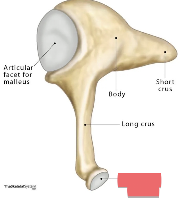
lenticular process

this whole bone
stapes
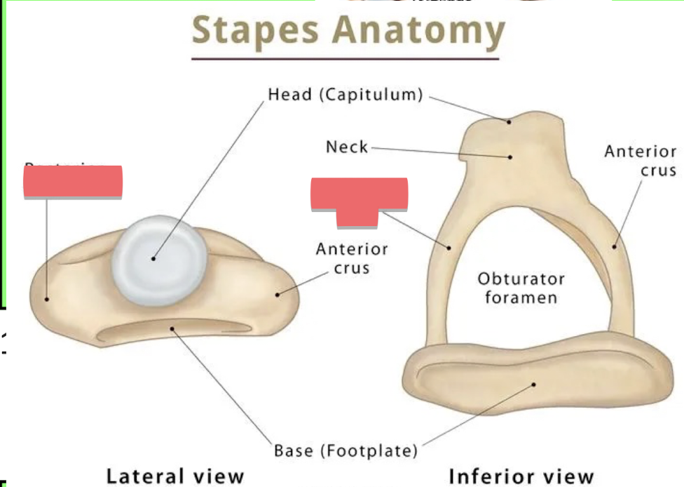
posterior crus
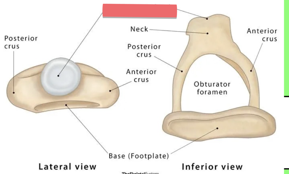
head of stapes
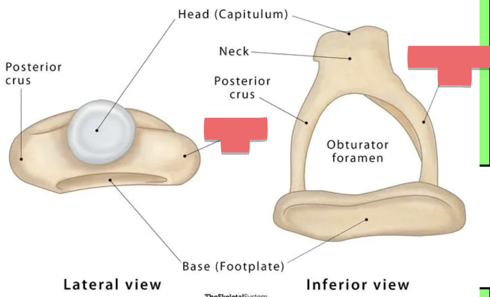
anterior crus
Give the insertion, function, and innervation of the stapedius muscle
Insertion: neck/ head of stapes
Function: primary muscle involved with the acoustic reflex
Innervantion: Facial nerve VII
Give the insertion, function, and innervation of the Tensor tympani muscle
Insertion: Upper manubrium/ anterior process of malleus
Function: involved in triggering the acoustic reflex
Innervation: trigeminal nerve CN V
What is the acoustic reflex
the reflex triggered by loud sounds to dampen transmission to avoid damages
where is the eustachian tube
establish connection between the middle ear and the nasopharynx
What is the function of the eustachian tube?
opens to match the pressure outside with the pressure in the middle ear and remove harmful fluid
What muscles open the eustachian tube
levator veli palatini and the tensor veli palatini
Why are ear infections easier for kids
Children’s eustachian tube are more horizontal and can get infections from fluid in the middle ear easier
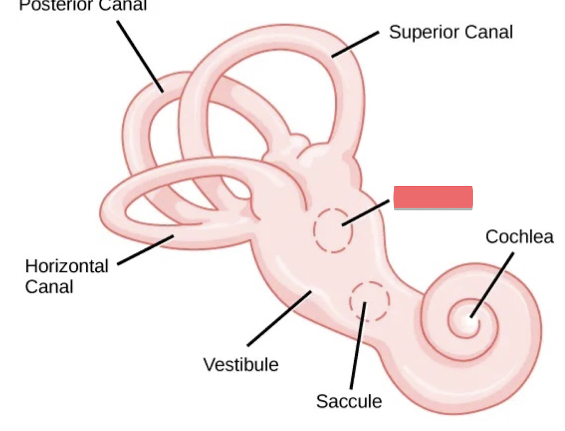
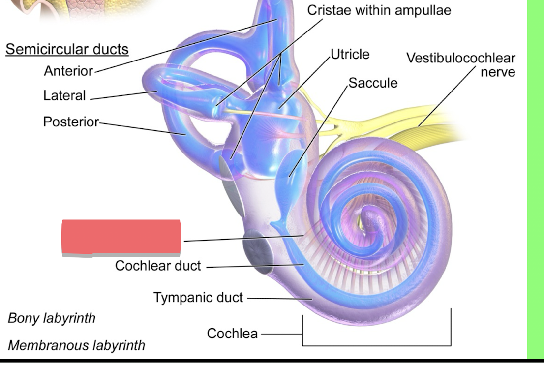
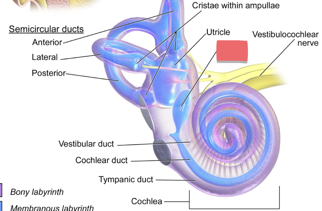
utricle
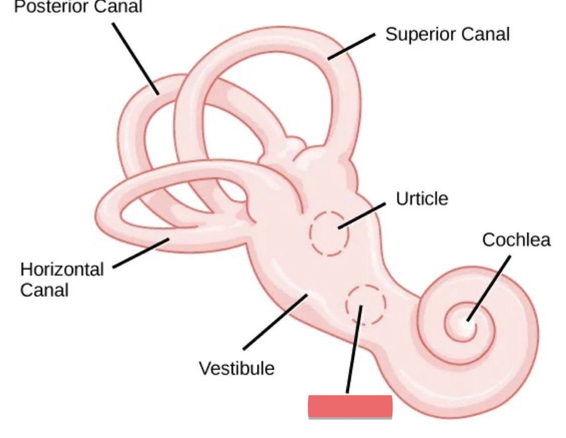
saccule
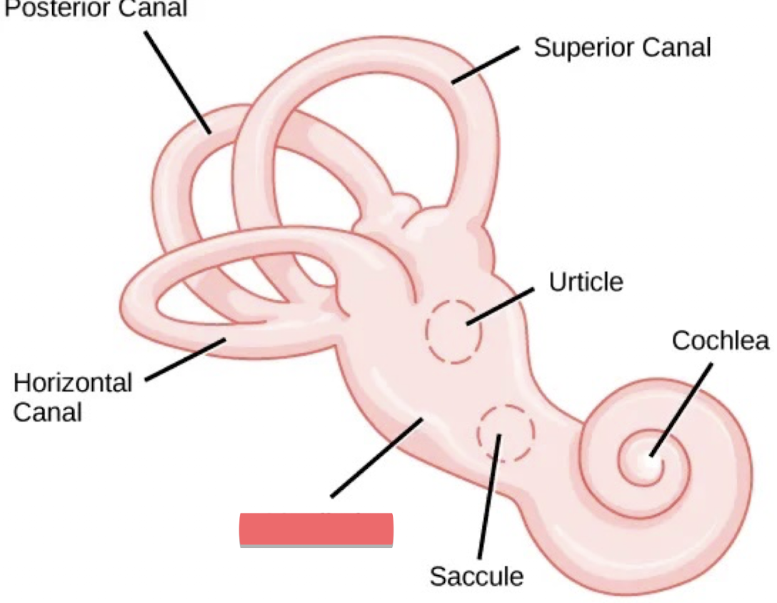
Vestibule
What two main parts make up the Inner ear
The bony labyrinth
The membranous labyrinth
What 3 parts make up the bony labyrinth
Semicircular canals
Vestibule
Cochlea
What 3 parts make up the membranous labyrinth
the membranous semicircular canals
utricle and saccule
cochlear duct
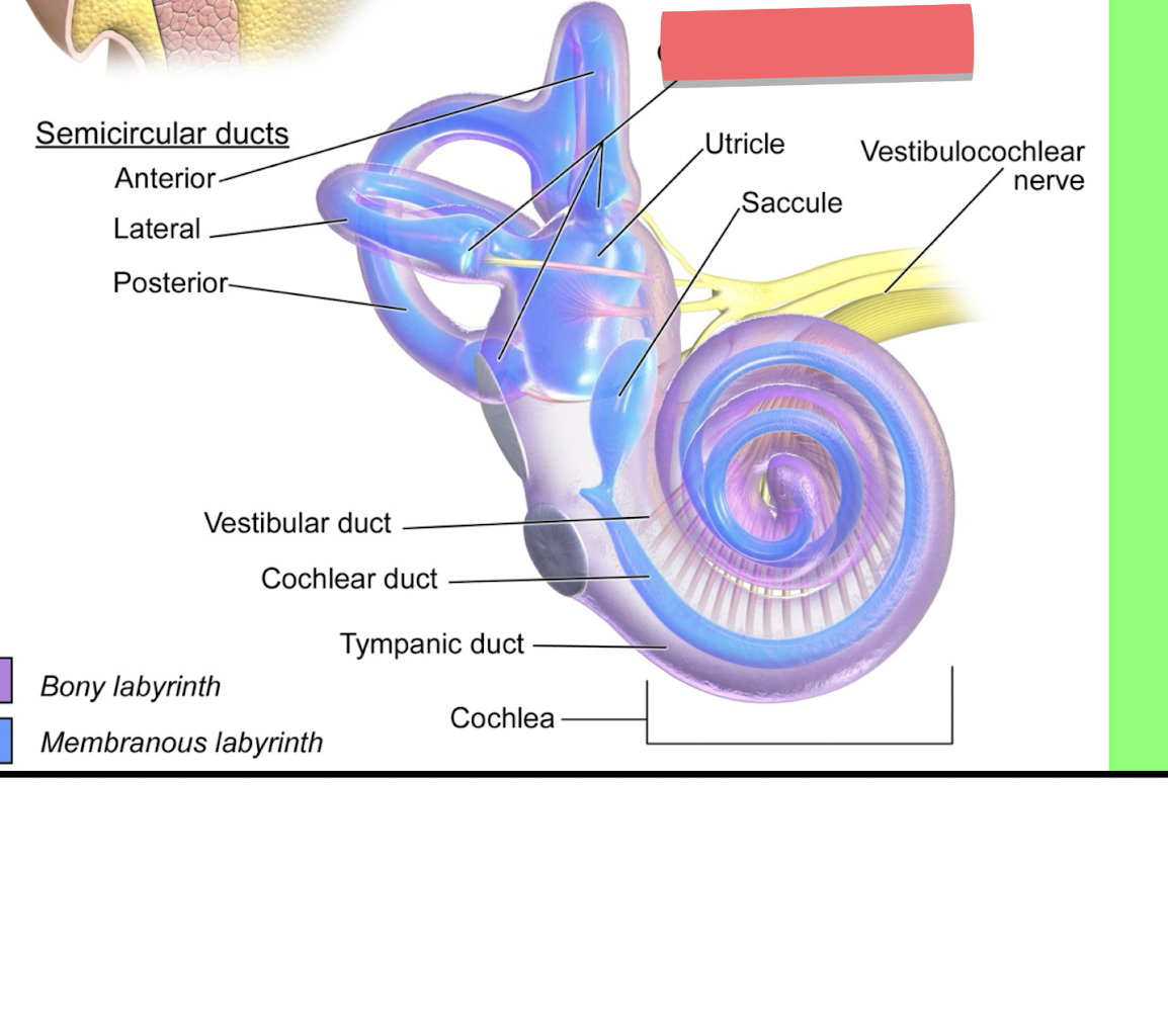
Chrisae within ampillae
vestibular duct
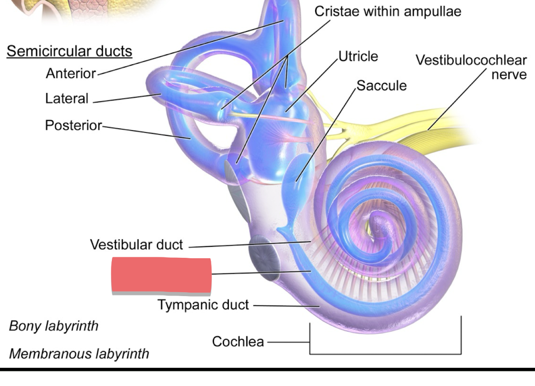
cochlear duct
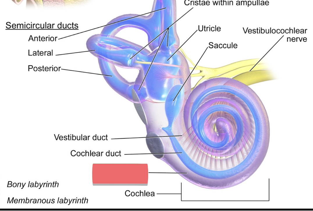
tympanic duct
saccule
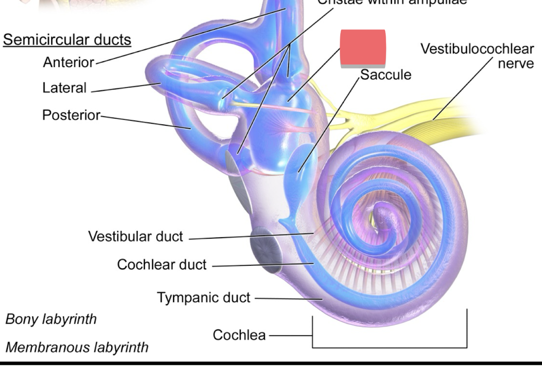
utricle
what do the ampulla contain
sensory cells of the semicircular canal that tell movement
perilymph
bony labyrinth (makes up most of the cochlea)
endolymph
membranous labyrinth
the cochlea is ________?
tonotopic
the cochlea’s tonotopic nature means that
sounds (tones) are mapped on the cochlea by a frequency on each part
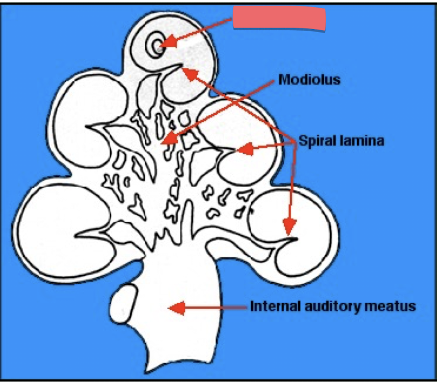
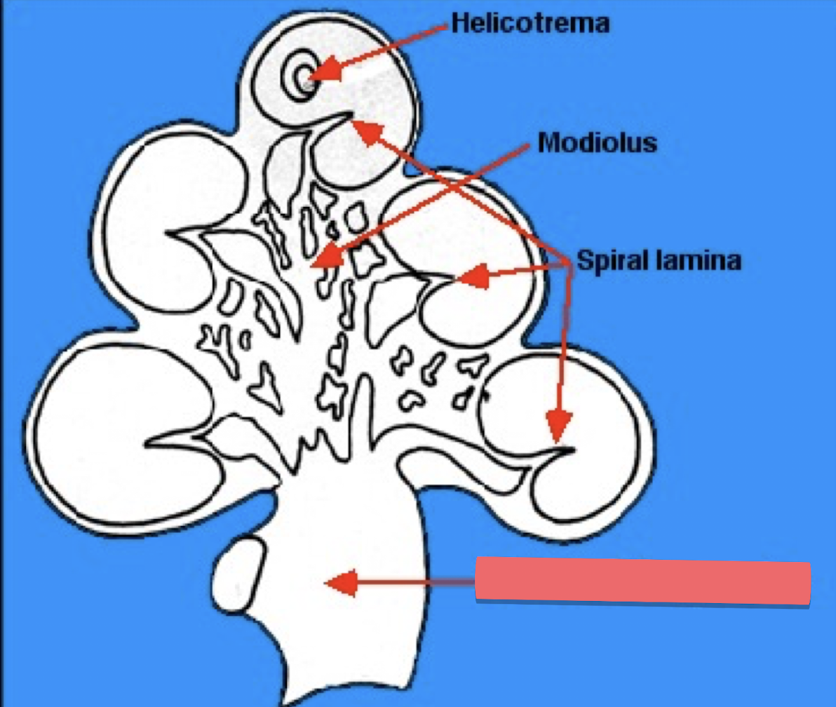
top cinnamon roll portion of the cochlea
helicotrema
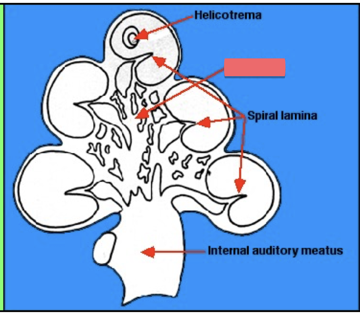
center pillar in cochlea
modiolus
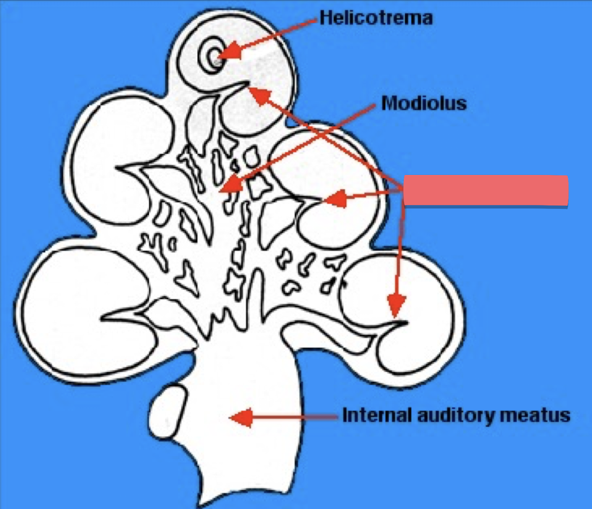
each layer of tube of cinnamon roll
spiral lamina
internal auditory meatus
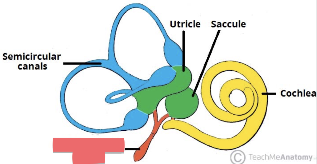
endolymphatic duct
The function of the endolymphatic duct is to
connect the saccule and utricle

Everything in diagram except for the cochlea is part of the ________
vestibular system (equilibrium)
What two parts make up the vestibular system? (and their functions)
Kinetic part
the perception of the rotation and acceleration of the head
Static part
the perception of the position in the vertical plane (sense of gravity and where you are in space)
What are the crista ampillaris
sensory cells of the semicircular canal
Where does the perception of sound in the body transfer from mechanical into electrochemical energy?
hair cells in cochlea
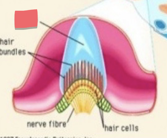
The top, gelatinous portion inside the crista ampullaris where bundles of hairs are
cupola
The jumping of the eyes from side to side is called____ and is characteristic of…
a nystagmus ; vertigo
The damage of vestibular system is manageable with
sight information from eyes
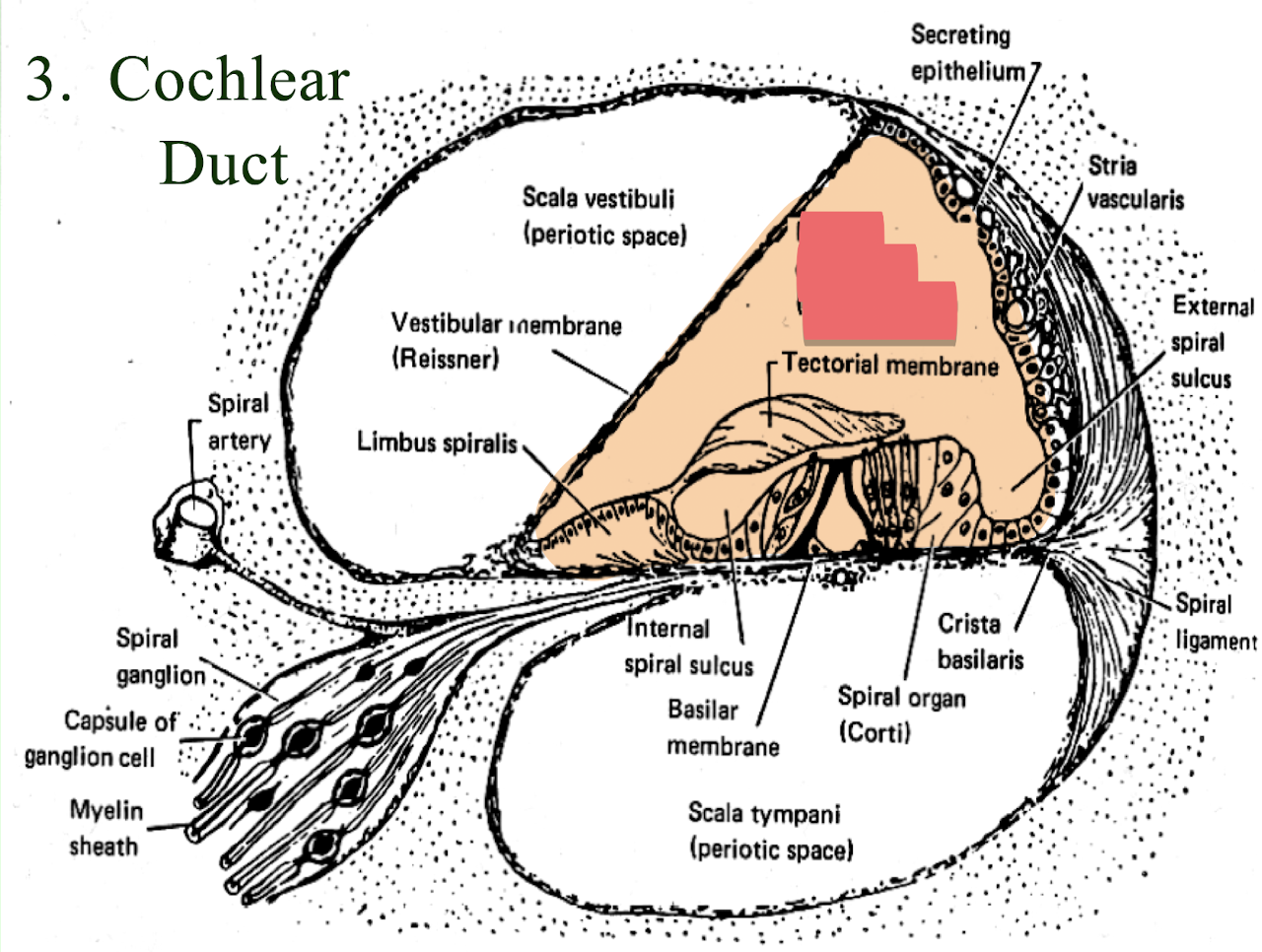
List the membrane, sensory cells, and membrane cover of the following bone:
Cochlea
Cochlear duct
Spiral organ of Corti
tectorial membrane
List the membrane, sensory cells, and membrane cover of the following bone: Semicircular Canals
Membranous semicircular canals
crista ampullaris
cupola
List the membrane, sensory cells, and membrane cover of the following bone:
utricle saccule
macula
otolithic membrane
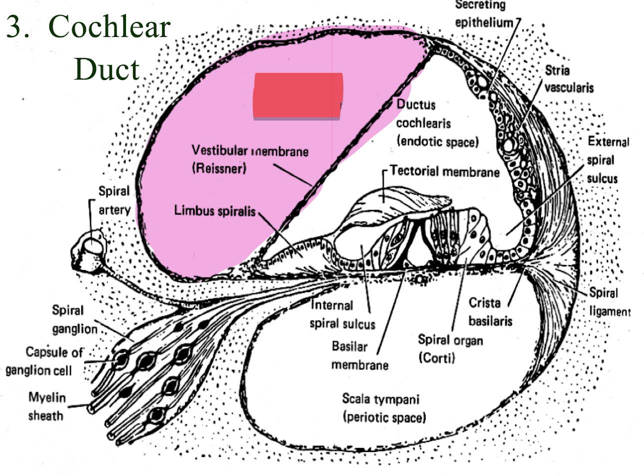
upper triangle of perilymph
scala vestibuli
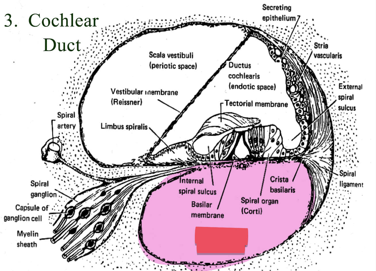
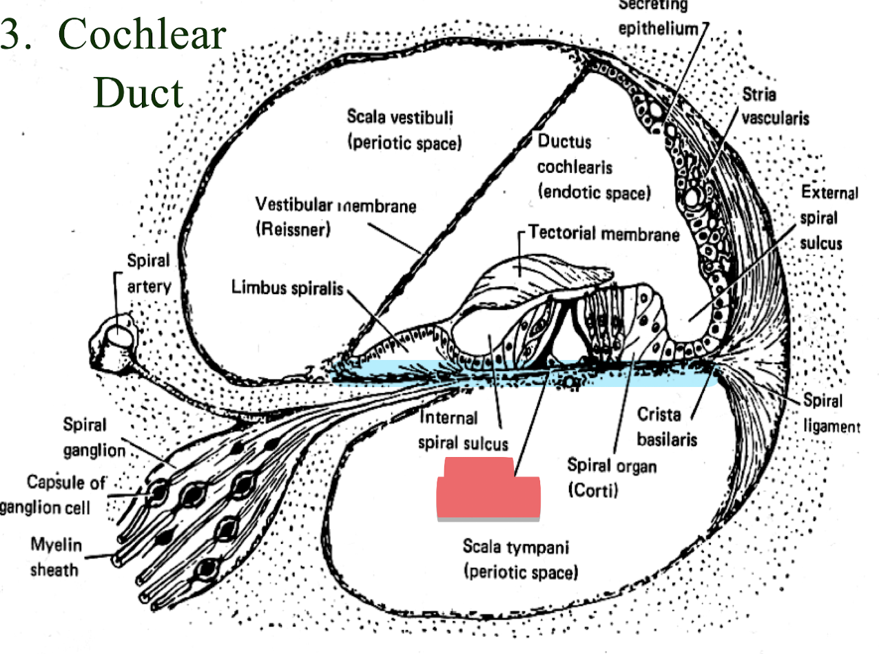
bottom triangular space under the basilar membrane
scala tympani
middle space made of endolymph
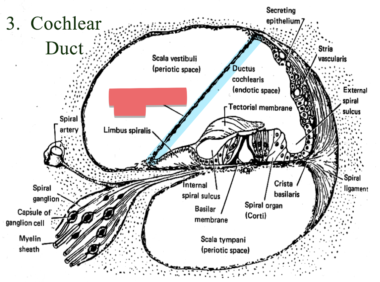
cochlear duct
top membrane separating membrane and bone
Reissner’s membrane
bottom membrane holding organ of corti
Basilar membrane
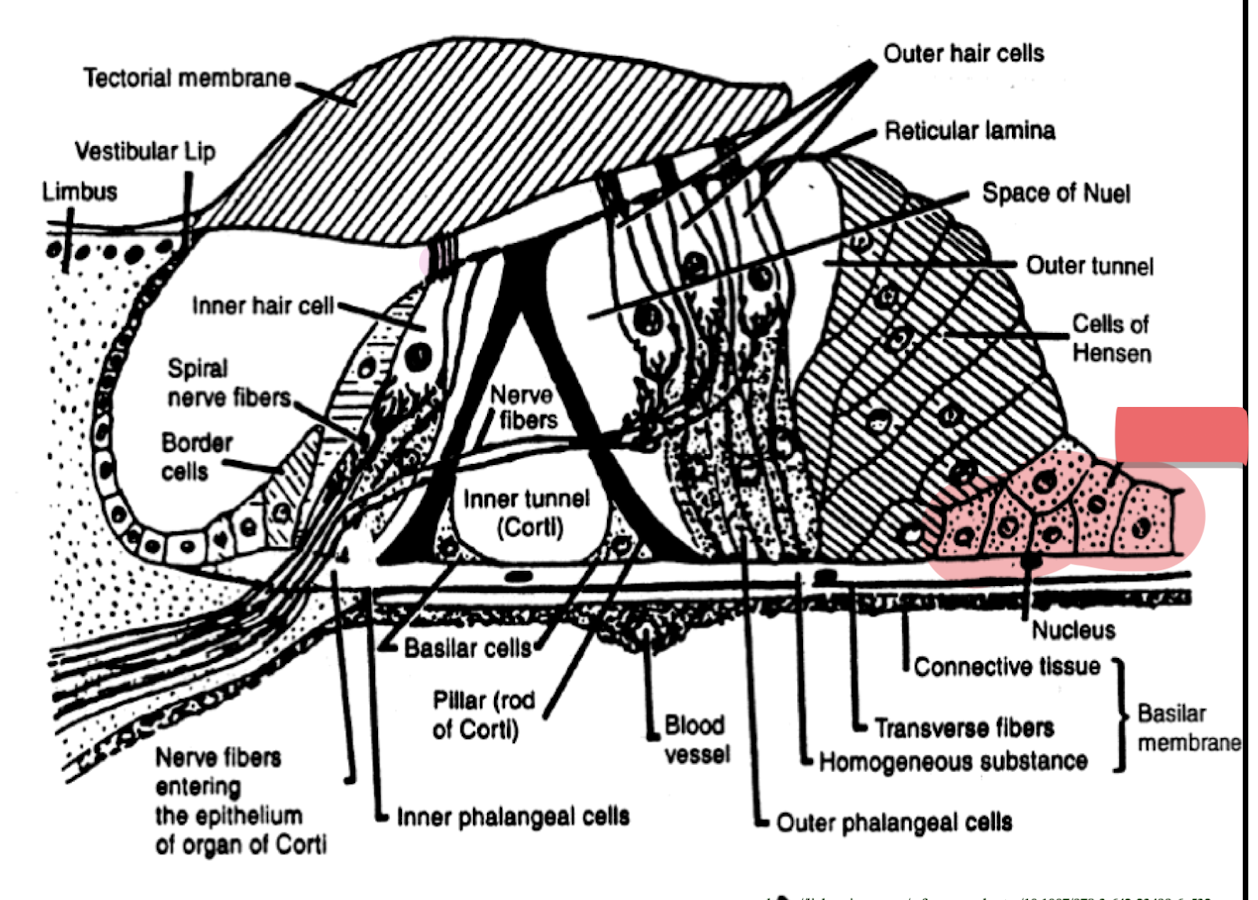
Cells of Claudius
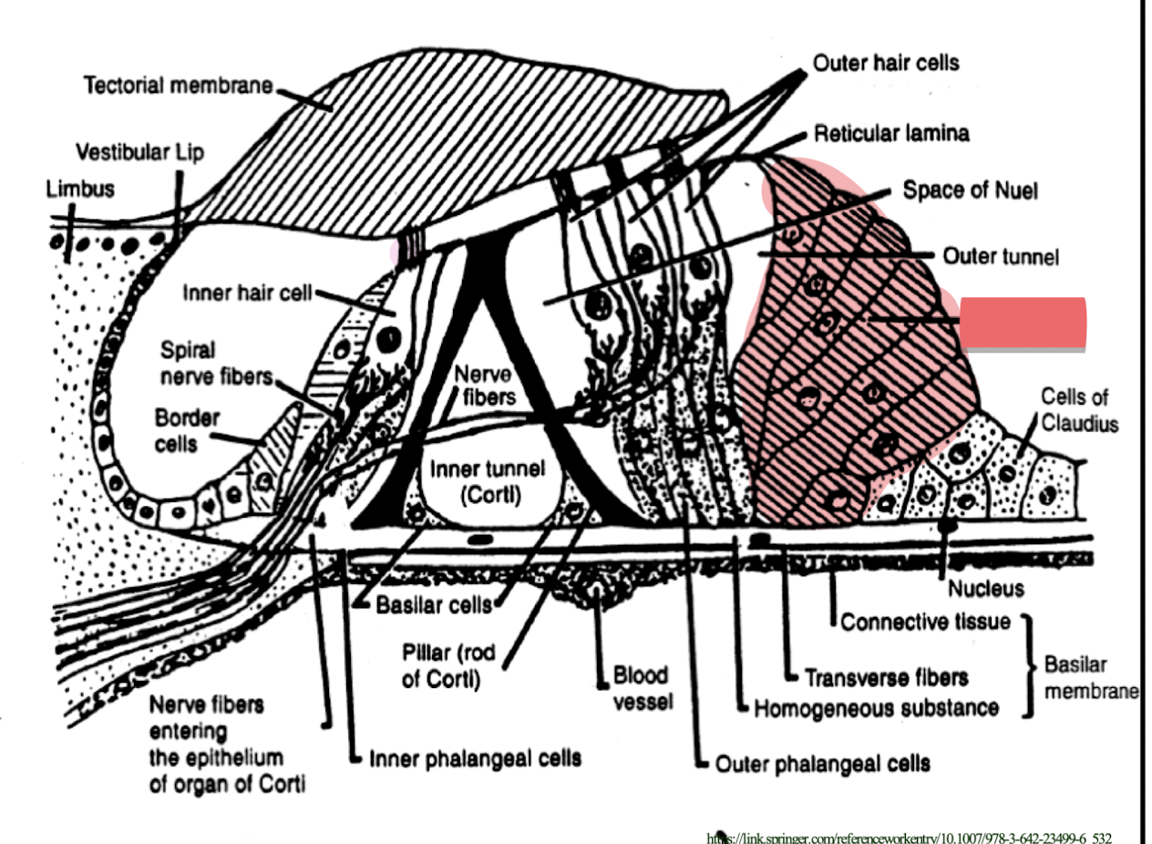
Cells of Hansen
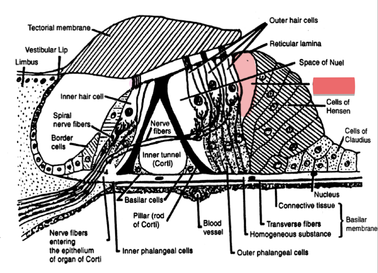
Outer tunnel
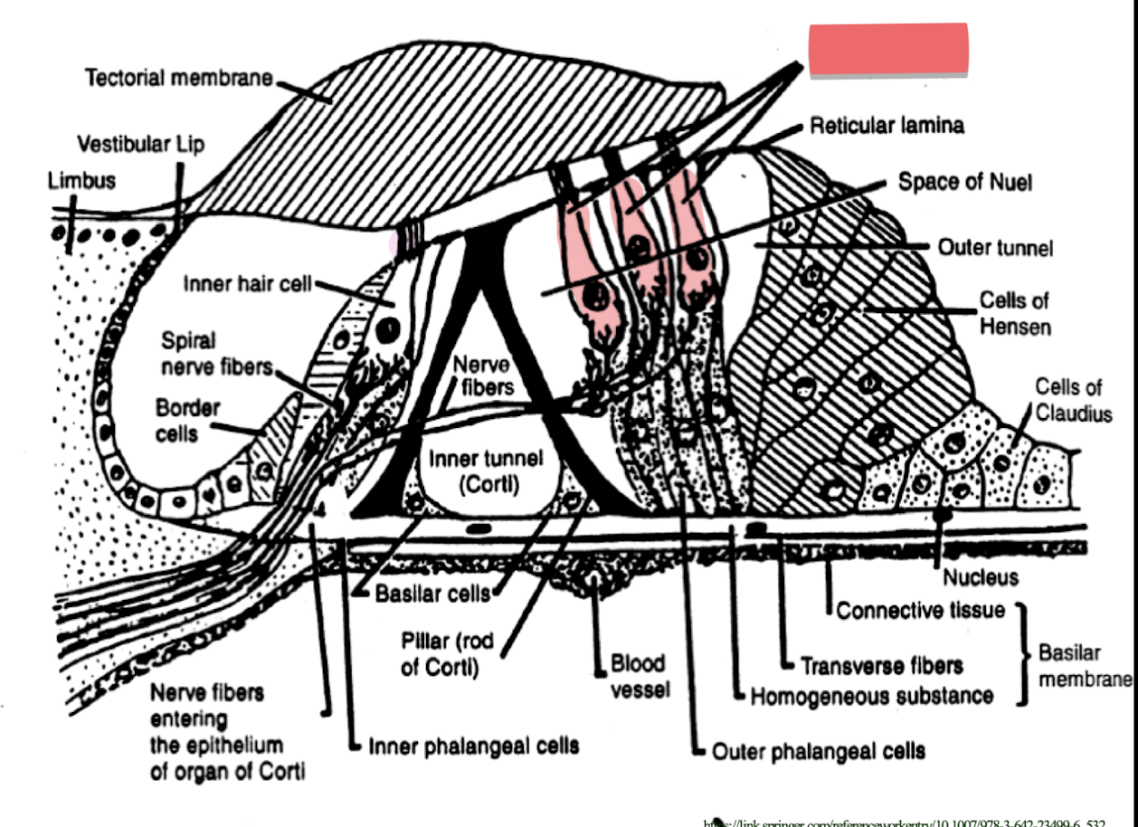
outer hair cells
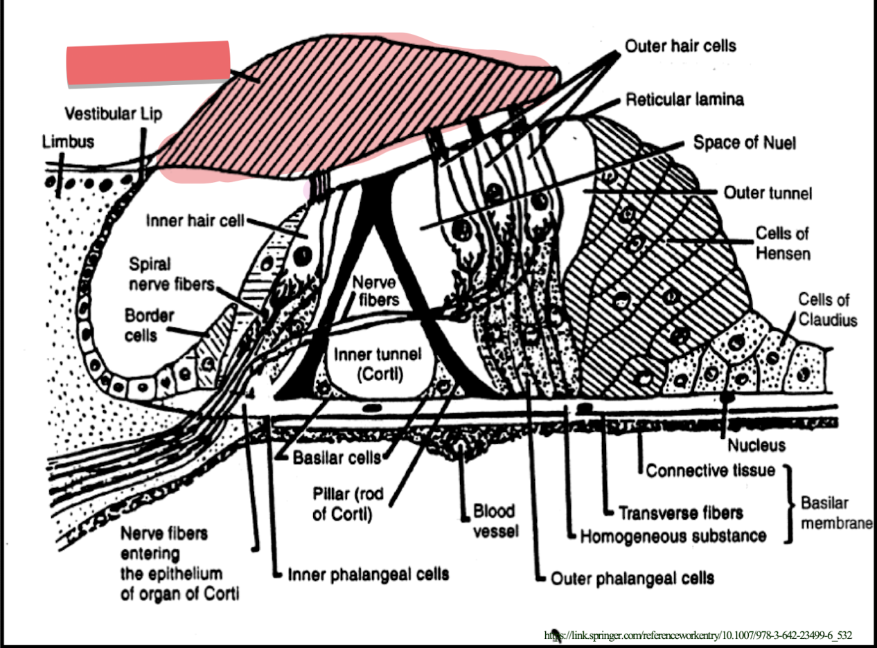
tectorial membrane
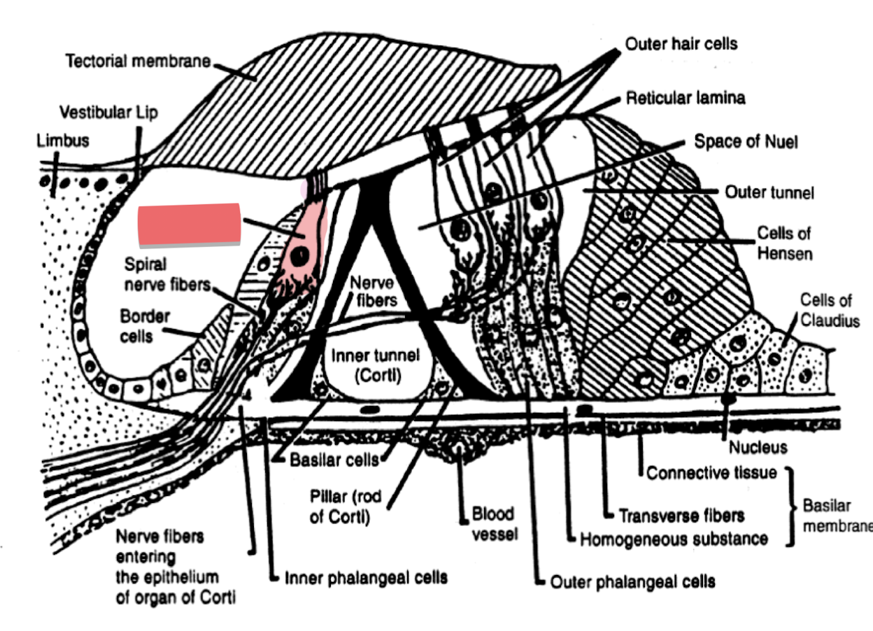
inner hair cell
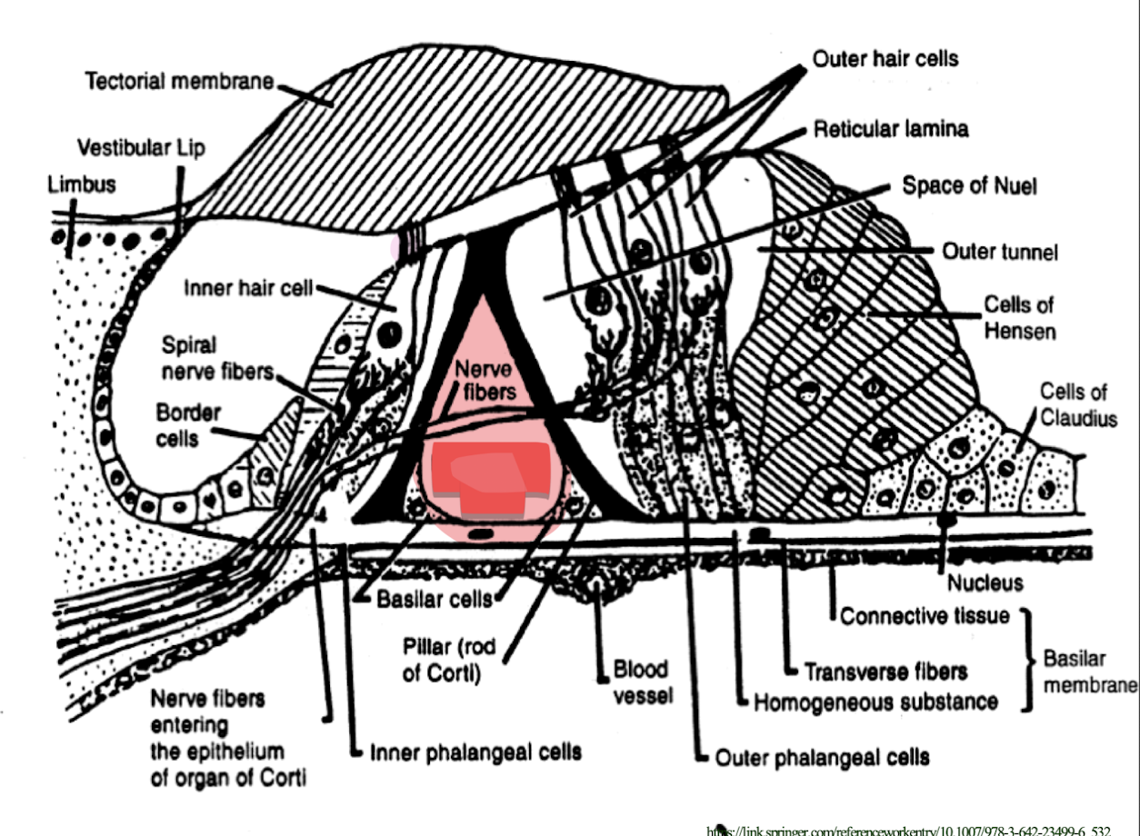
inner tunnel (tunnel of Corti)
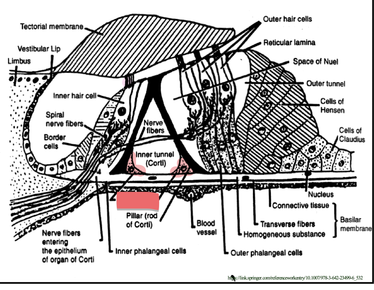
basilar cells
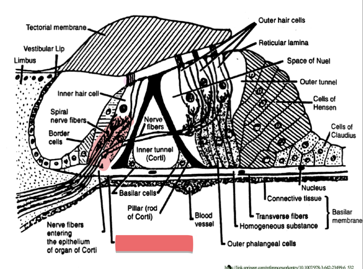
inner phalangeal cells
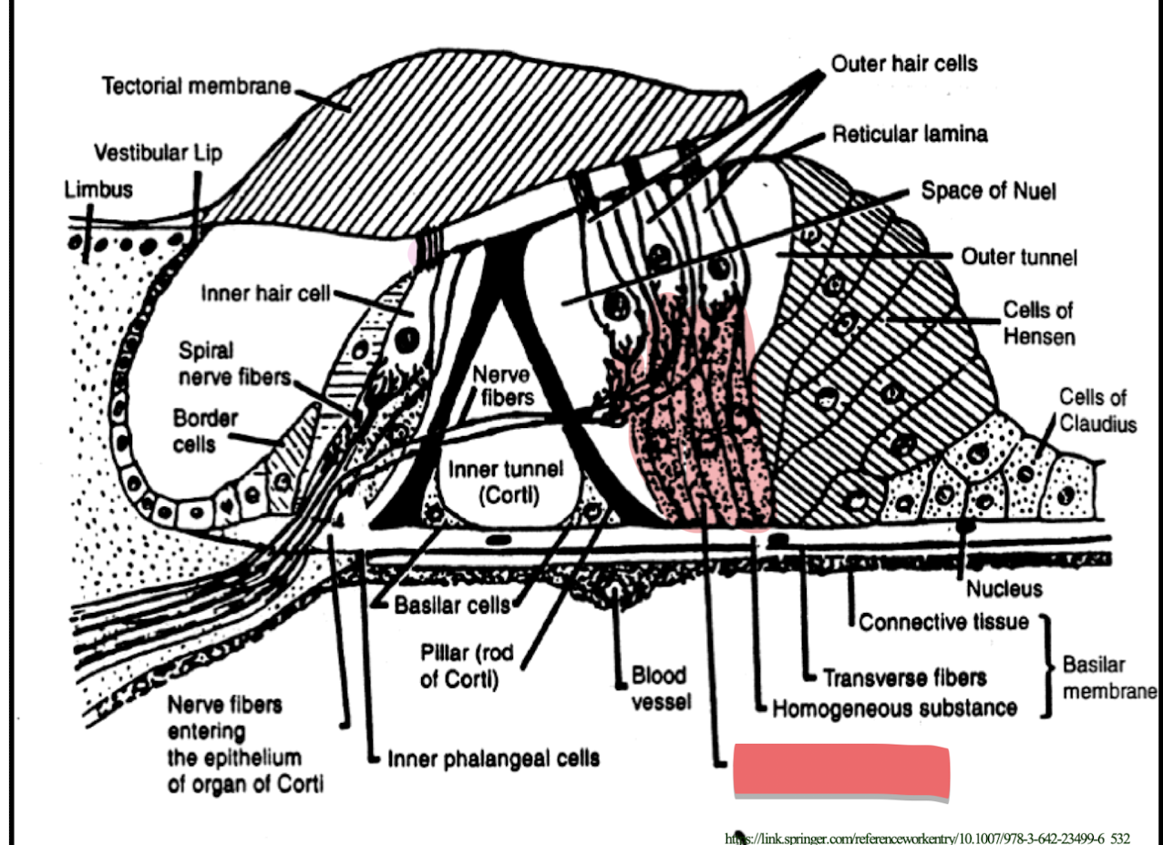
outer phalangeal cells

basilar membrane
What are the two main cell divisions of the Spiral organ of Corti?
The supporting cells
The receptor cells
What makes up the supporting cells of the Spiral organ of Corti?
a. inner phalangeal cells
b. rods of corti
c. outer phalangeal cells (cells of Deiters)