Lateral Geniculate Nucleus & Functional Testing - Visual Neurophysiology and Perception Spring 2026
1/166
There's no tags or description
Looks like no tags are added yet.
Name | Mastery | Learn | Test | Matching | Spaced | Call with Kai |
|---|
No analytics yet
Send a link to your students to track their progress
167 Terms
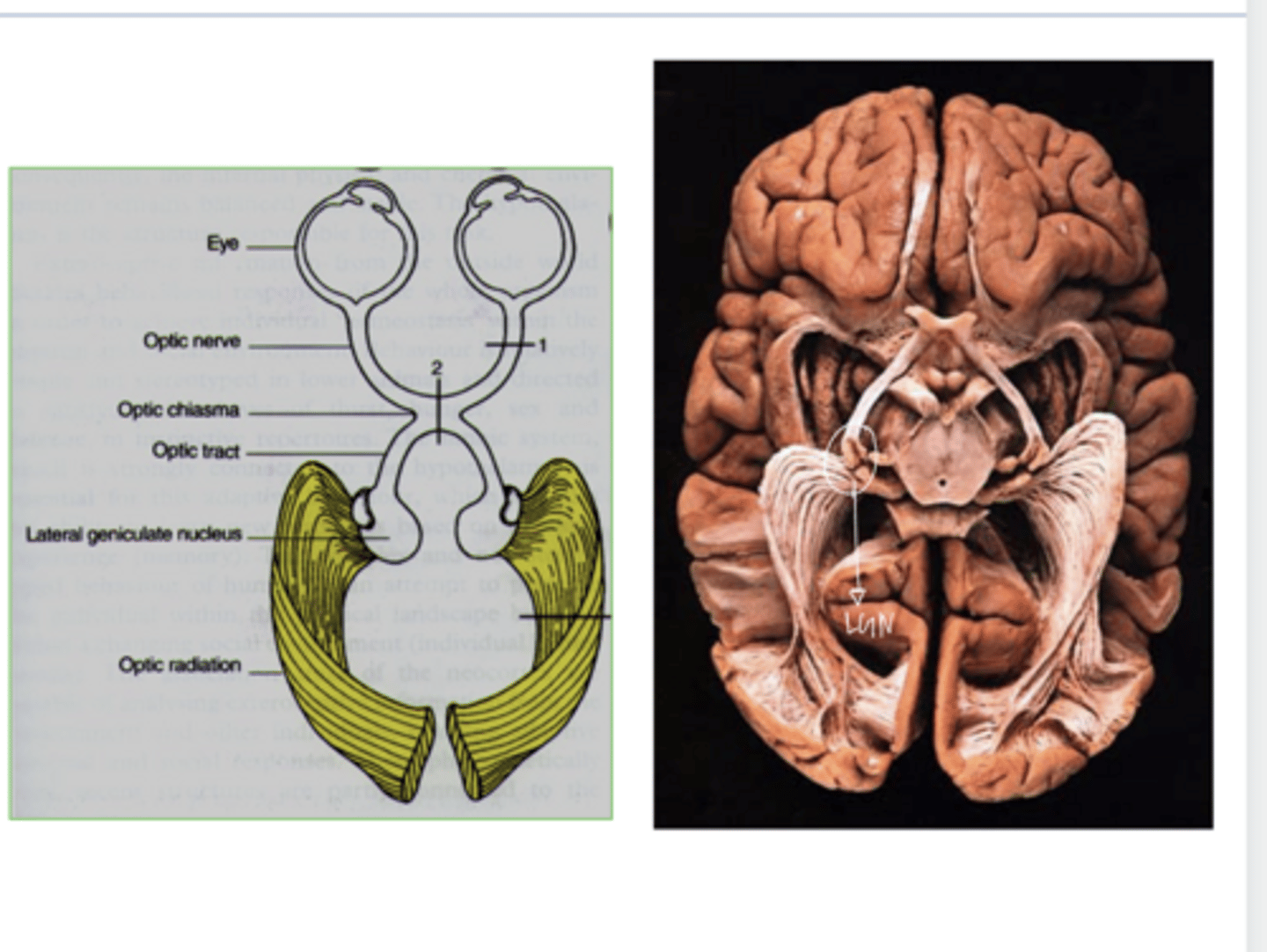
along their axons and out of the optic nerve
The action potentials generated in RPCs are carried where?
to the dorsal lateral geniculus (LGN) of the thalamus
In primates, 90% of the RGCs project to where?
glutamate
RGCs release ______ onto target neurons
LGN in Brain (Pic)
LGN in Brain (Pic)
true
True or False:
In general, the response of the LGN neuron is similar to that of the RGC that provides input to it

Yes
Do LGN neurons have center-surround spatial antagonism?

LGN neurons have stronger center-surround antagonism
Is center-surround antagonism stronger in RGCs or LGN nuerons?

minimal response
Diffuse light to LGN neurons results in what d/t strong center-surround antagonism?
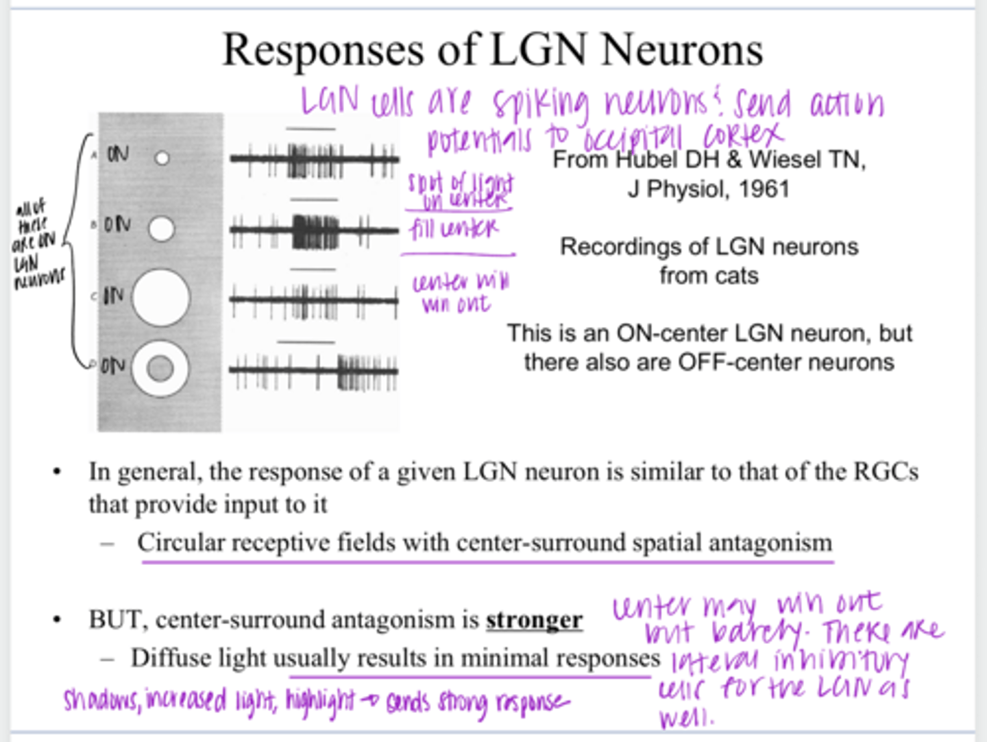
When shadows, increased light, or highlights are present
When will the LGN neurons send stronger responses to the visual cortex?

No -- there is not dramatic processing of the visual signal here but it is not only a relay station either
Is the LGN a "relay station"?
Yes -- increased lateral inhibition (center-surround antagonism) at this structure
Is there modification to the visual signal at the LGN?
gate
The LGN can be thought of as a "____-"
Controlling the flow of info to higher brain centers so that only the most relevant or interesting info passes through
Why can the LGN be thought of as a gate?
inhibitory
In addition to excitatory input from the retina, LGN also receives _____ input
-From the visual cortex
-Mesencephalic reticular formation for saccadic suppression
-Inter-neurons within the LGN -- Magno neurons can inhibit Parvo
Where does the LGN receive feedback inhibition from?
true
True or False:
Are different qualitative attributes of the visual image separated out within the retina into and carried onto the brain (LGN) via different pathways?
Separate RGC classes transmit different sensory messages to the brain
What is the basic organizational principle that is often applied to the visual pathway?

True
True or False:
The cells of the LGN have a specific anatomical orientation

6
How many layers are in the LGN in cross-section?

each layer received input from either the ipsilateral or contralateral eye
What is the importance of each of the 6 LGN layers?

true
True or False:
The cells in each of the different layers of the LGN have a different physiological property
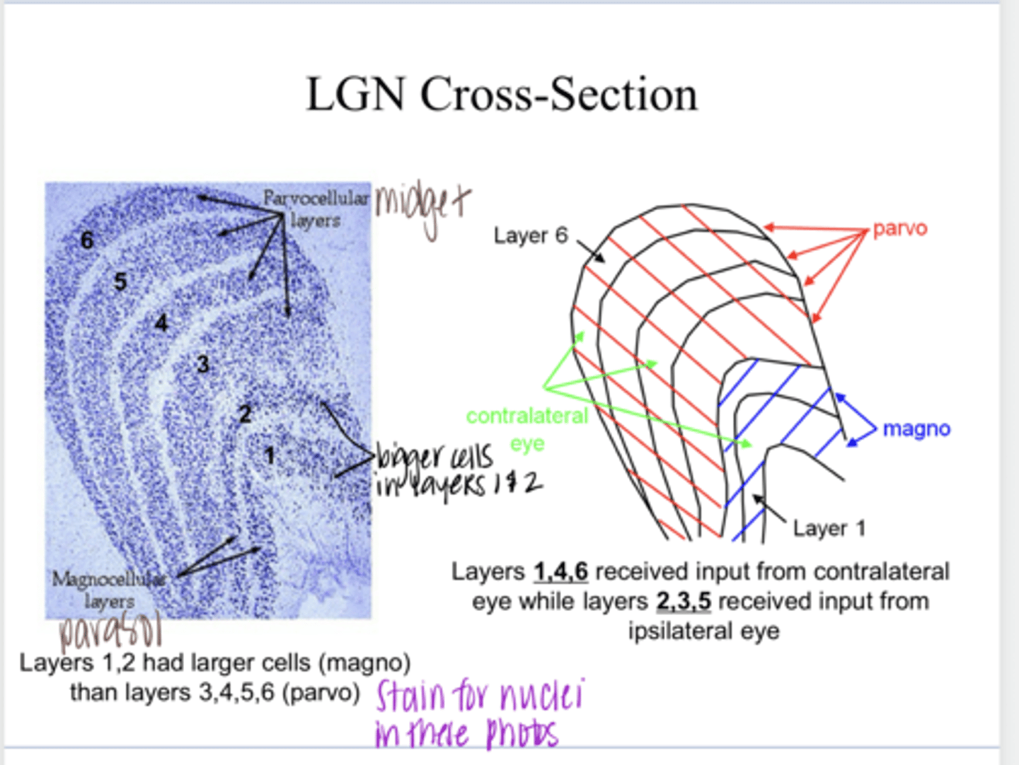
Magno Layers
What is the name of the 2 bottom-most layers of the LGN?

larger cells
What are the characteristics of the Magno layers compared to Parvo?

3, 4, 5, 6
What are the "parvo" layers of the LGN?

1, 4, 6
What layers of the LGN receive contralateral eye input?

2, 3, 5
What layers of the LGN receive ipsilateral eye input?

Parvo (90%); Magno (10%)
Magno vs Parvo Physiology of LGN Neurons
Which are more predominant?

None
Magno vs Parvo Physiology of LGN Neurons
Color Opponency of Magno Neurons present?

More transient
Magno vs Parvo Physiology of LGN Neurons
Response duration of Magno Neurons?

fast (high speed of transmission)
Magno vs Parvo Physiology of LGN Neurons
Response Timing of Magno Neurons?

Large receptive field w/ center-surround antagonism
Magno vs Parvo Physiology of LGN Neurons
Receptive field of Magno Neurons?
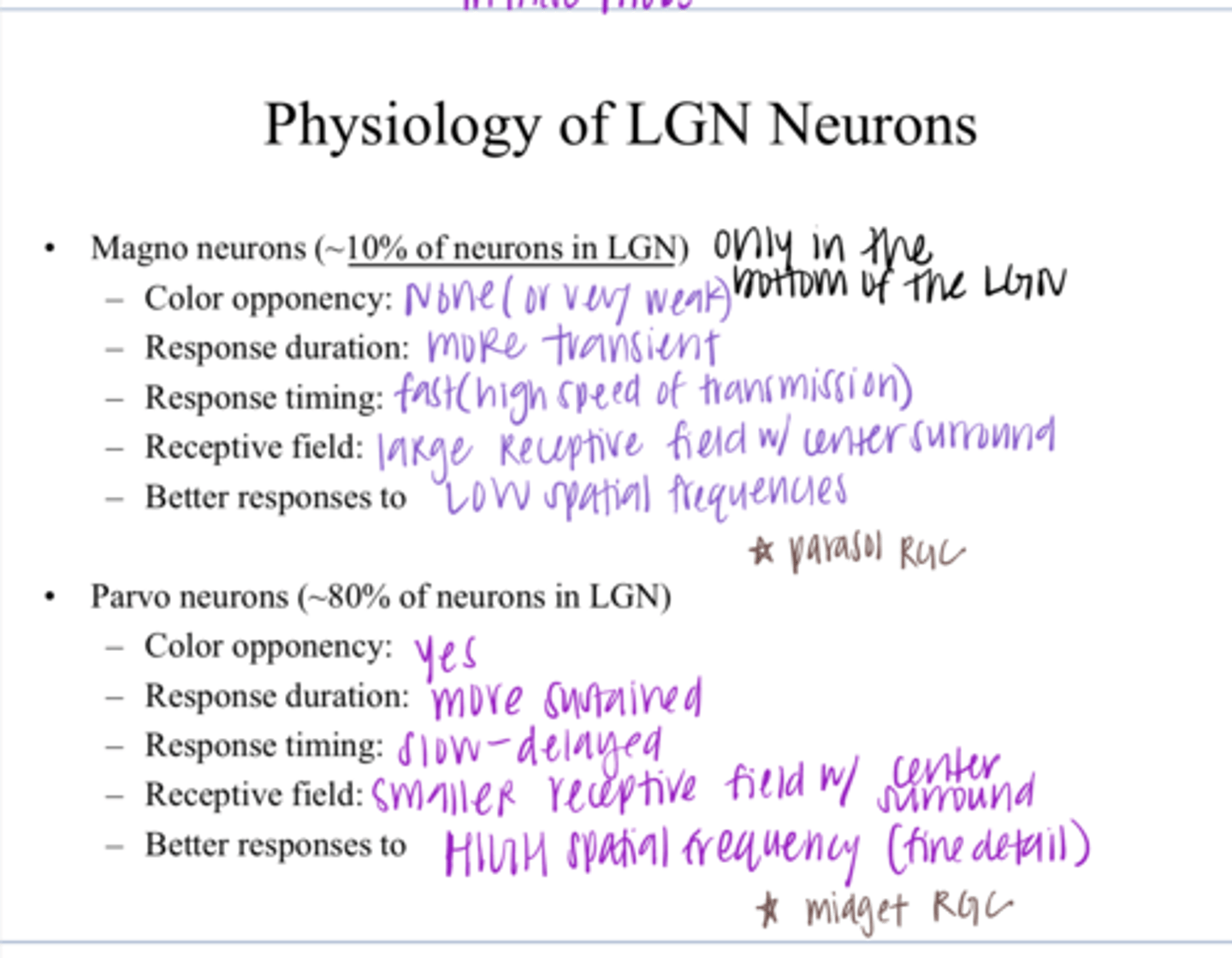
LOW
Magno vs Parvo Physiology of LGN Neurons
Magno: Better responses to what spatial frequencies?

Yes
Magno vs Parvo Physiology of LGN Neurons
Color Opponency of Parvo Neurons?

more sustained
Magno vs Parvo Physiology of LGN Neurons
Response Duration of Parvo Neurons?

slower -- more delayed
Magno vs Parvo Physiology of LGN Neurons
Response timing of Parvo Neurons?

Smaller receptive field w/ center-surround antagonism
Magno vs Parvo Physiology of LGN Neurons
Receptive Field of Parvo Neurons?

HIGH
Magno vs Parvo Physiology of LGN Neurons
Parvo: Better responses to what spatial frequencies?

true
True or False:
The differences in the responses of the two classes of LGN neurons should remind you of the differences between parasol and midget RGCs
Parvo (P pathway)
Midget RGCs project to what LGN neuron?
Magno (M pathway)
Parasol RGCs project to what LGN neuron?
where; what
The idea emerged in the 1980s that there where two major visual pathways -- a ____ system and a _____ system
-flicker
-movement
-alters us to the presence of a visual stimli
What is the Magno system good at detecting?
providing info regarding the spatial details of a stimulus
What is the Parvo system good at?
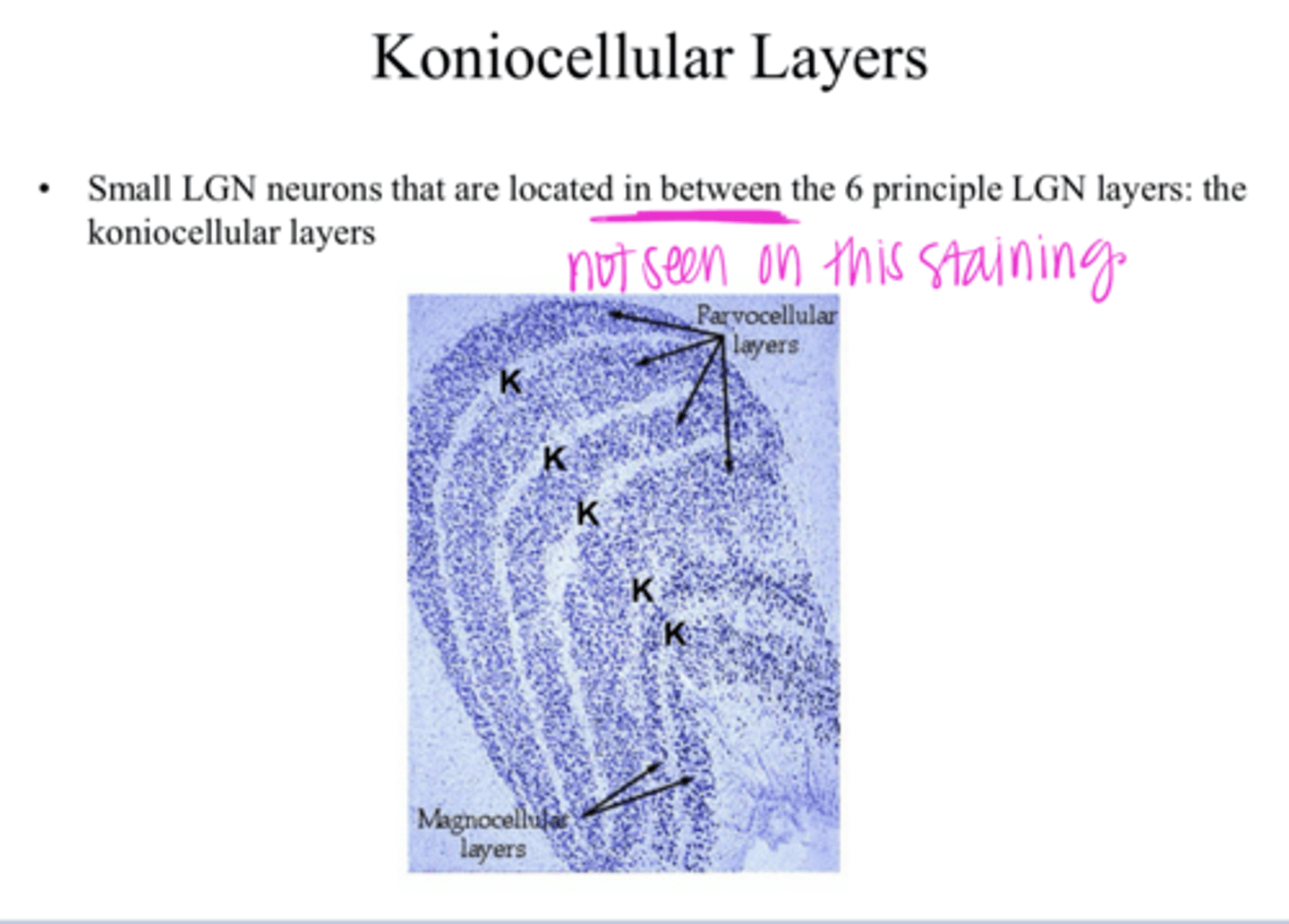
small bistratified RGCs
What is the retinal component that connects to the konicellular pathway of the LGN?

limited (Blue ON, Yellow OFF)
Does the konicellular pathway of the LGN show color opponency?
large receptive field
What is the size of the receptive field of the konicellular pathway of the LGN?
No
Does the konicellular pathway of the LGN have center-surround antagonism?
lower
What spatial frequency does the konicellular pathway of the LGN best respond to?
slow responses -- very slow speed of transmission
What is the speed of responses of the konicellular pathway of the LGN?
IN BETWEEN the 6 principle LGN layers
Where are the konicellular pathway neurons of the LGN located?
The idea is that different channels in the visual pathway are tuned into specific spatial frequencies that exist within a given image
Why is the visual system classified as a Fourier analyzer?
Yes
Does each cell of LGN have a peak sensitivity in terms of spatial frequency?
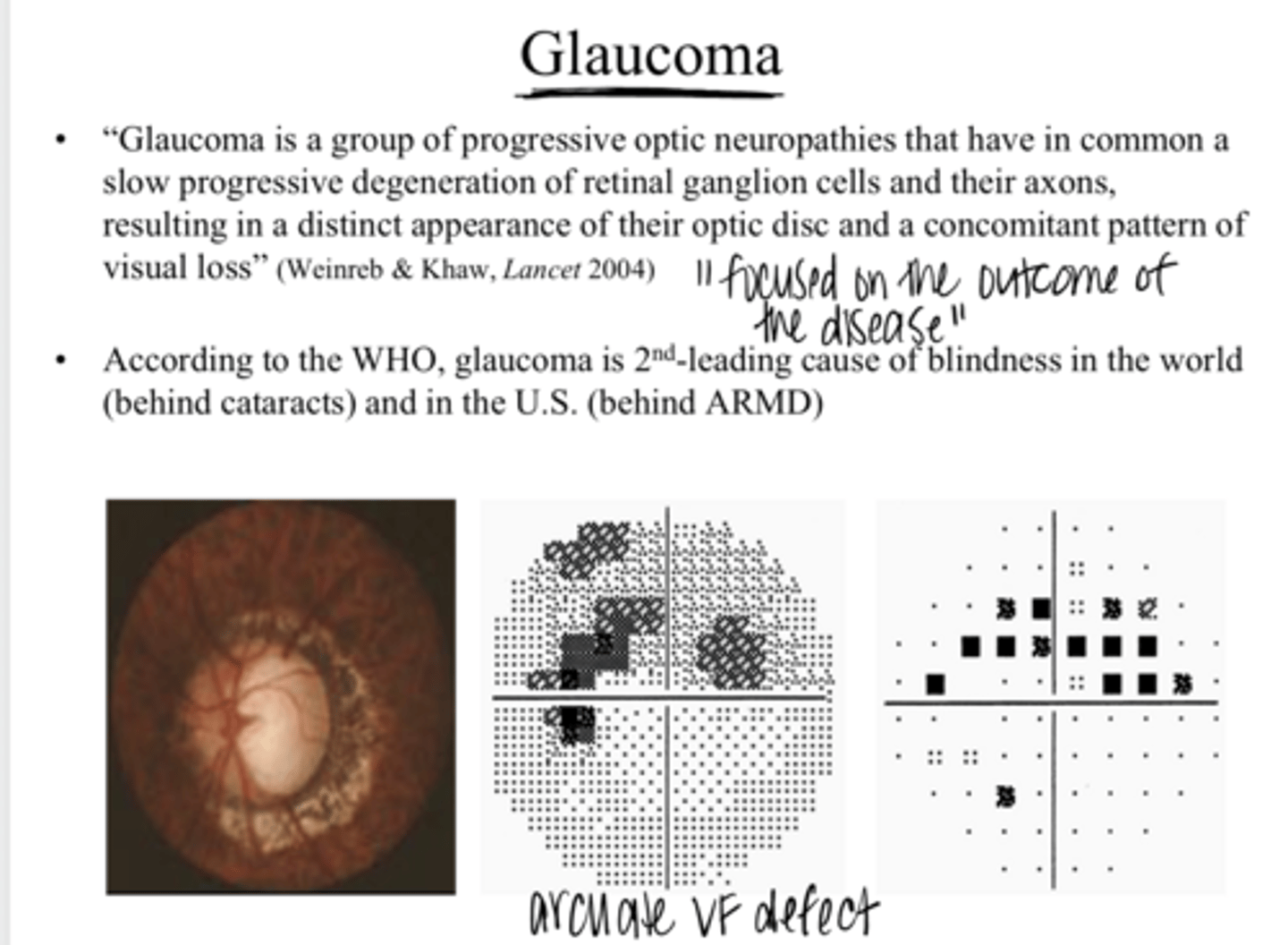
Glaucoma is a group of progressive optic neuropathies that have in common a slow progressive degeneration of RGCs and their axons; resulting in a distinct appearance of their optic disc and a concomitant pattern of visual loss
What is the definition given for glaucoma?

Intraocular pressure
What is the most important known risk factor for primary open angle glaucoma?

medical or surgical procedures designed to lower IOP
All current therapies for this disease involve what?
Yes
Do the results of wide-scale clinical trials support that IOP reduction is beneficial in decreasing POAG incidence/progression?

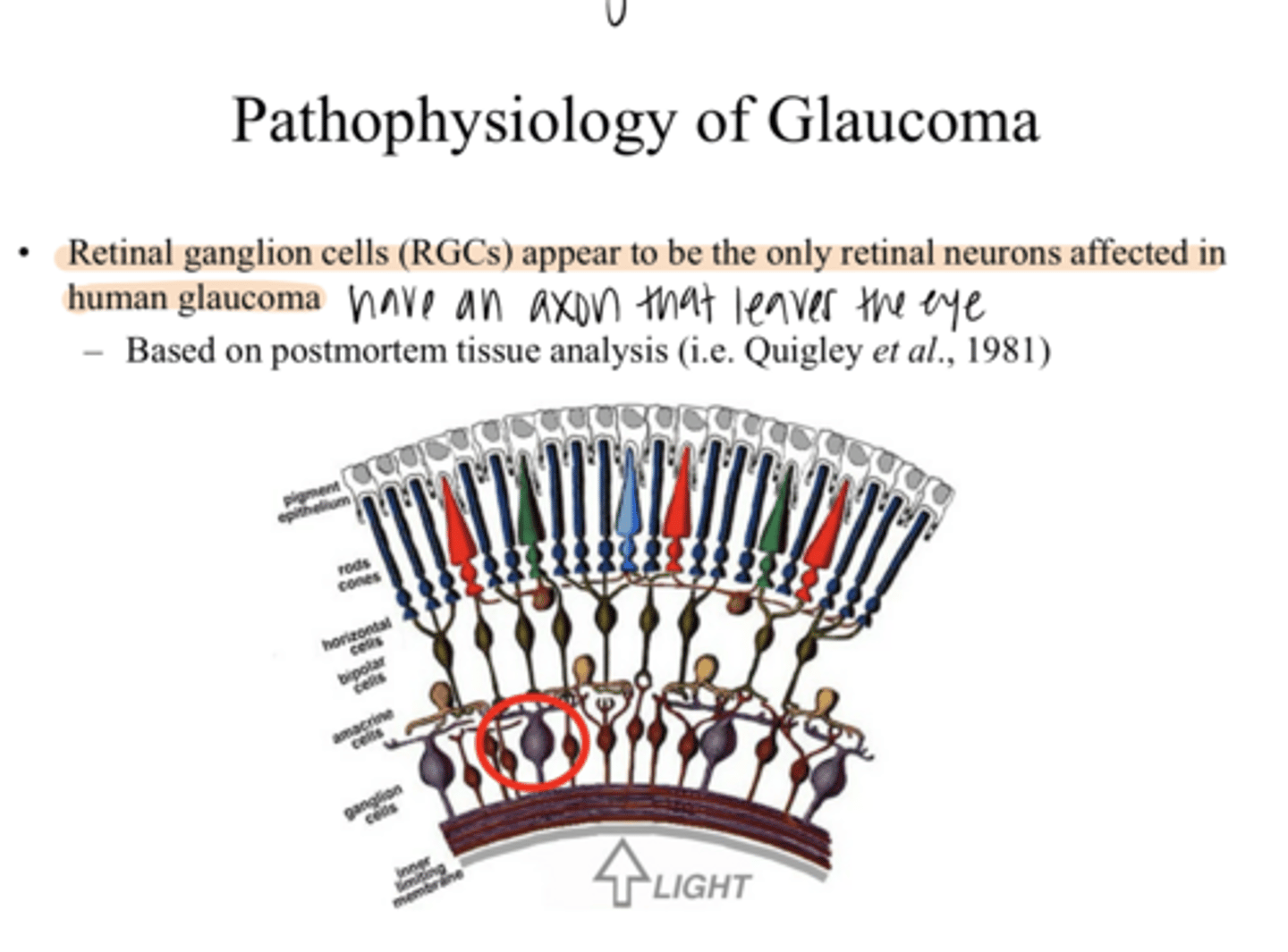
RGCs
_____ are the only cell that is affected in human glaucoma
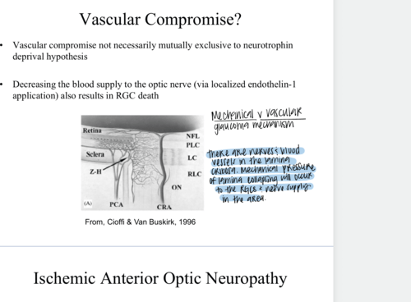
The initial damage to the RGC axon occurs at the lamina cribosa. This is AXONAL injury -- not CELL BODY injury.
Where are RGCs injured in glaucoma?

sheets of the lamina cribosa get compressed and pushed outward
Earliest changes in the optic nerve in POAG show what happening to the lamina cribosa?
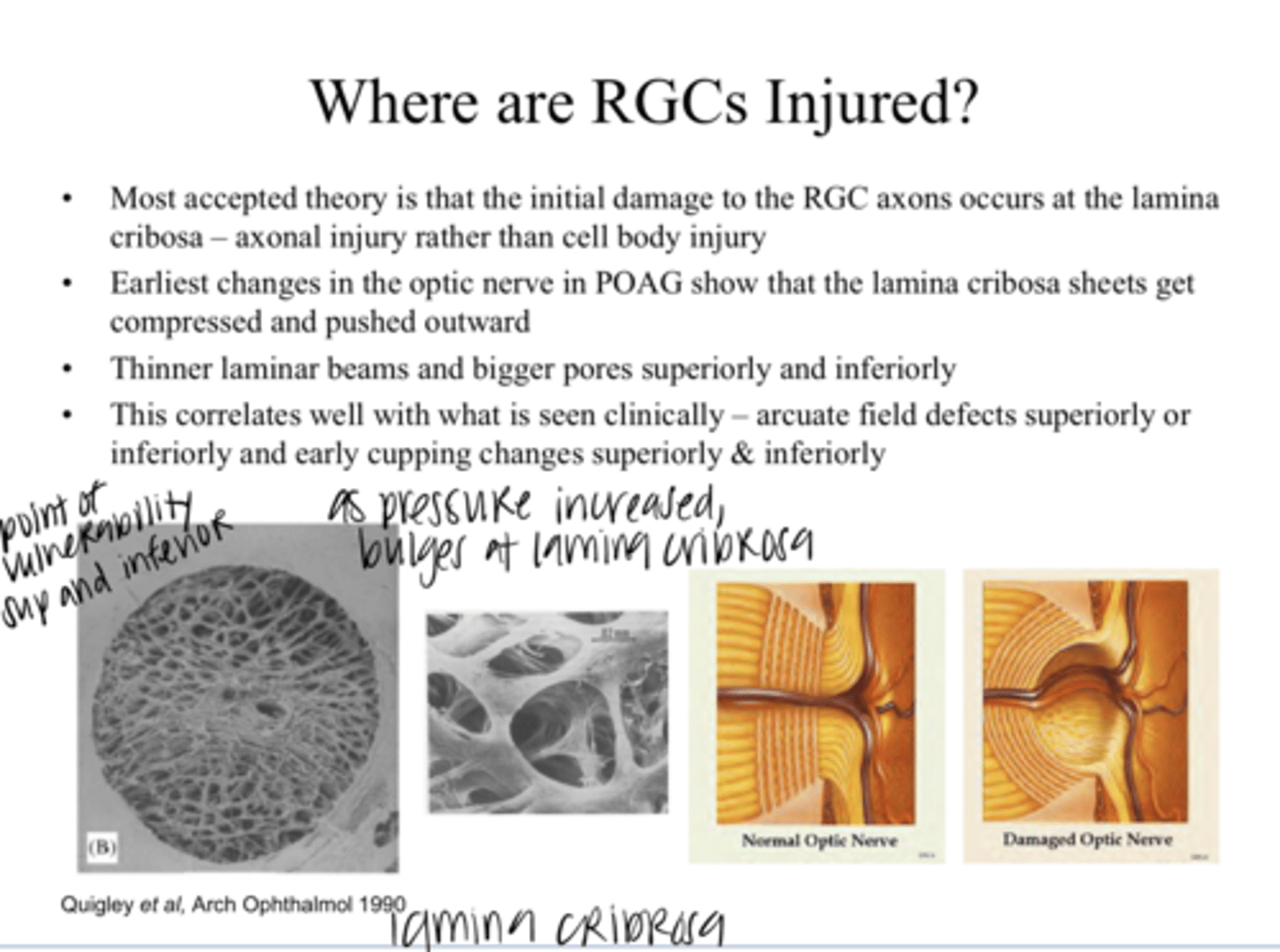
superior and inferior; arcuate defect superior and inferiorly and early cupping changes superior and inferior
Where are the thinner lamina cribosa beams & bigger pores? What is the clinical implication of this?

superior or inferiorly
In glaucoma, the earliest changes appear ____ or ____
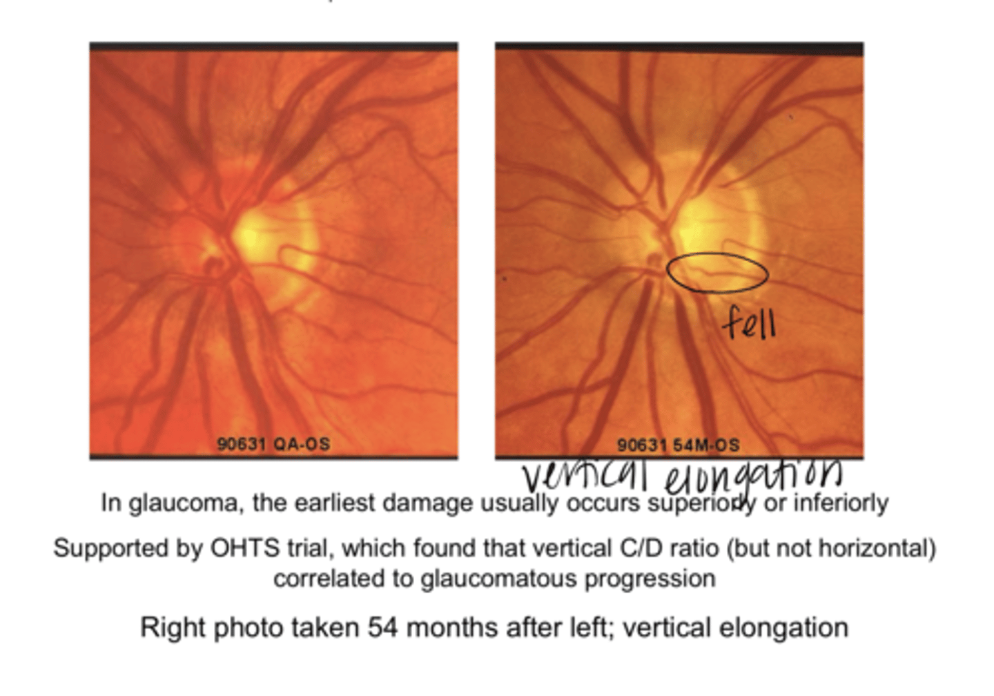
vertical
The (vertical/horizontal) C/D ratio correlates to glaucomatous progression

RGC axons are pinched at the lamina cribosa, disrupting axoplasmic flow. This flow constantly carries vital messengers (neurotrophins like BDNF) from the brain target back tot he RGC cell bodies of the retina. If the axonal transport is affected and BDNF does not reach the RGC cell bodies, the cell will initiate its own death.
What is the neurotrophic deprival hypothesis that explains why RGCs die in glaucoma?
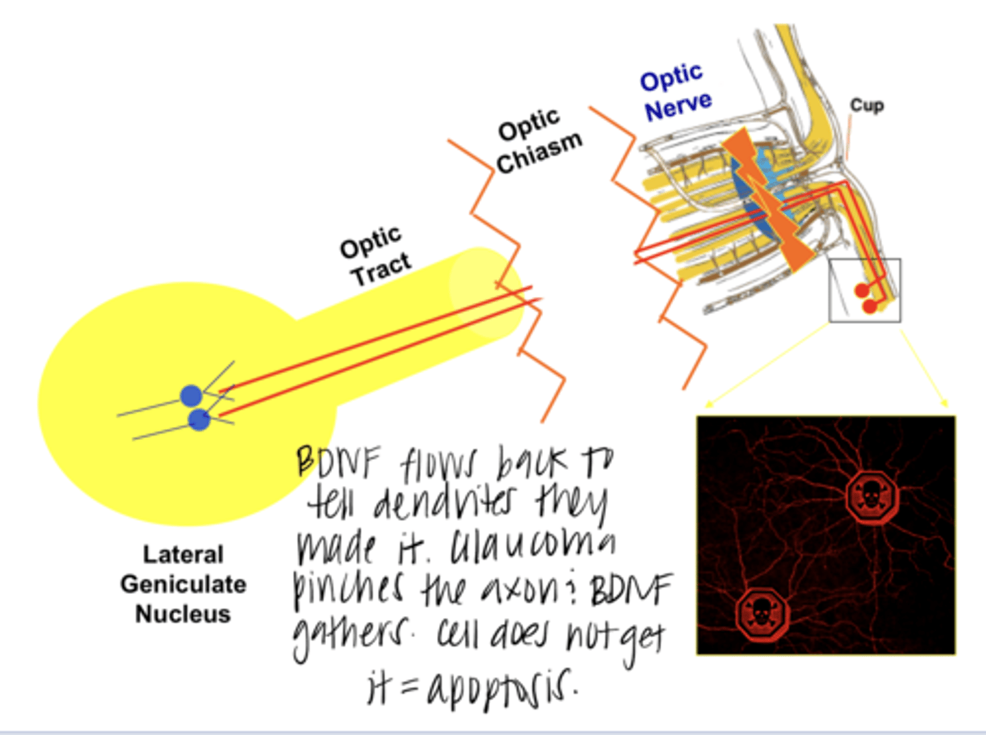
The RGC will initiate its own death
What happens if the BDNF neurotrophin is affected and does not reach RGC cell bodies?

Yes -- RGCs that do not reach the correct target eliminate themselves
Is BDNF important embryonically?

apoptosis
Neurotrophin deprivation will genetically program cell death called ______

-Each affected cell expresses enzymes to digest its own DNA and other cellular components
-The cell then fragments into small vesicles (no membrane rupture) and removed without inducing inflammation
How does a cell undergo apoptosis?
No
After apoptosis of a RGC, will inflammation result?
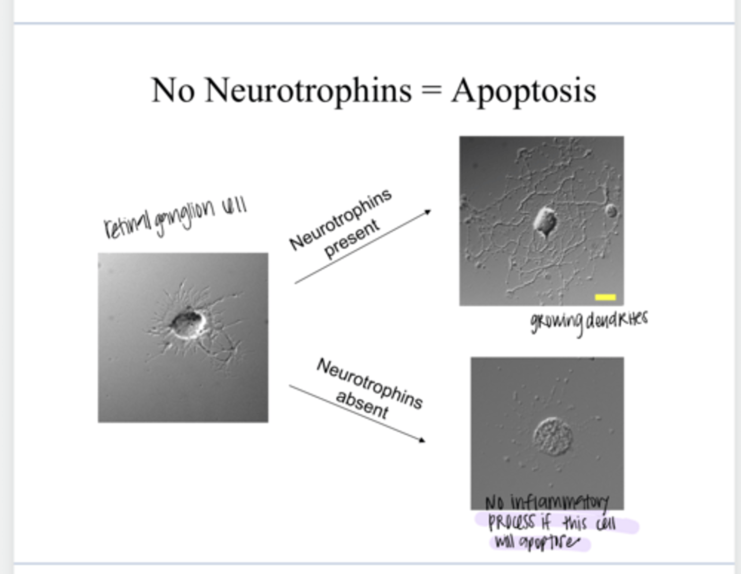
No Neurotrophins = Apoptosis
Neurotrophins Present = RGC Growth (Pic)
No Neurotrophins = Apoptosis
Neurotrophins Present = RGC Growth (Pic)

Yes -- meshes well with classical mechanical theory of damage
is neurotrophin deprivation an attractive theory for glaucoma?
by cutting the optic nerve (in vivo) or depriving cultured RGCs (in vitro)
How can you indice neurotrophin deprivation experimentally?
Apoptotic
________ RGCs have been detected in human glaucoma
No
Is vascular compromise mutually exclusive to neurotrophin deprival hypothesis?
Yes
Does decreasing the blood supply to the optic nerve also result in RGC death?

acute ischemia of the optic nerve
What is ischemic anterior optic neuropathy d/t?
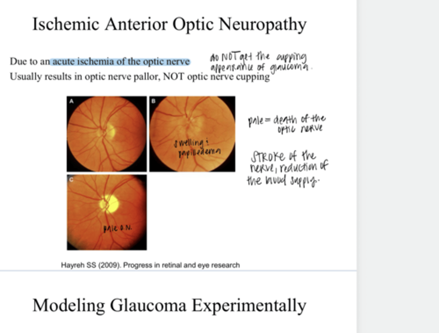
optic nerve pallor
What does ischemic anterior optic neuropathy result in?

No
Does ischemic anterior optic neuropathy result in optic nerve cupping?

Yes -- elevated IOP is a causative factor for glaucomatous damage
Does the experimental elevation of IOP in an animal induce RGC death?
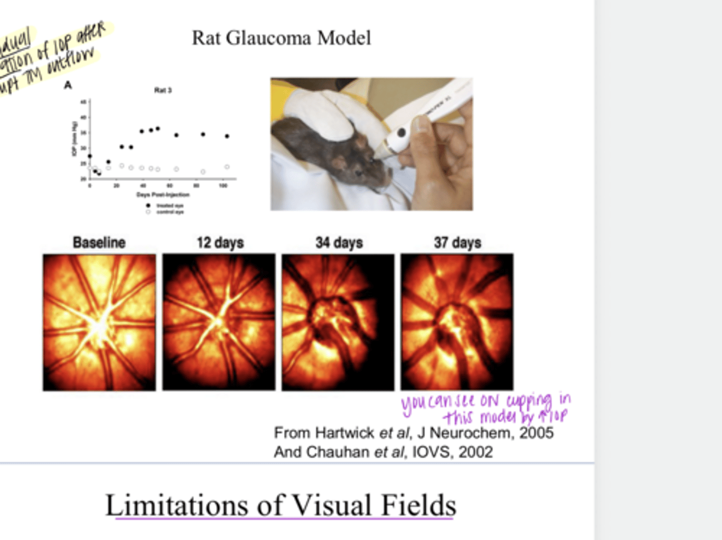
Yes
Does chronic and moderate elevation in IOP induce RGC death & optic nerve cupping?

Rat Glaucoma Model -- Disrupting TM outflow w/ Gradual Elevation of IOP (Pic)
Rat Glaucoma Model -- Disrupting TM outflow w/ Gradual Elevation of IOP (Pic)

50
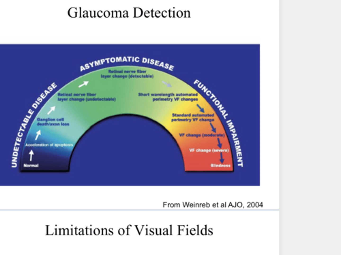
Research on human cadaver eyes from glaucoma patients suggested that VF defects were not detectable until about _____% of RGCs had died
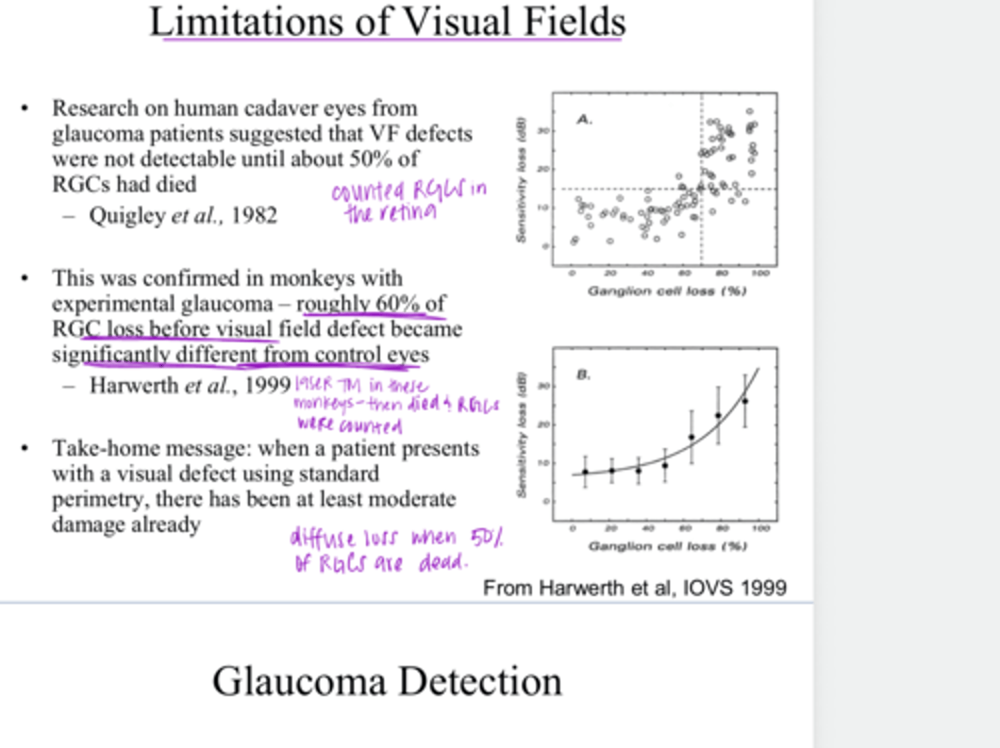
Yes -- loss seen when 50% of RGCs already gone
TAKE HOME MESSAGE: When a patient presents with a VF defect using standard perimetry, has there been a least moderate damage already?

Glaucoma Detection Model (Pic)
Glaucoma Detection Model (Pic)
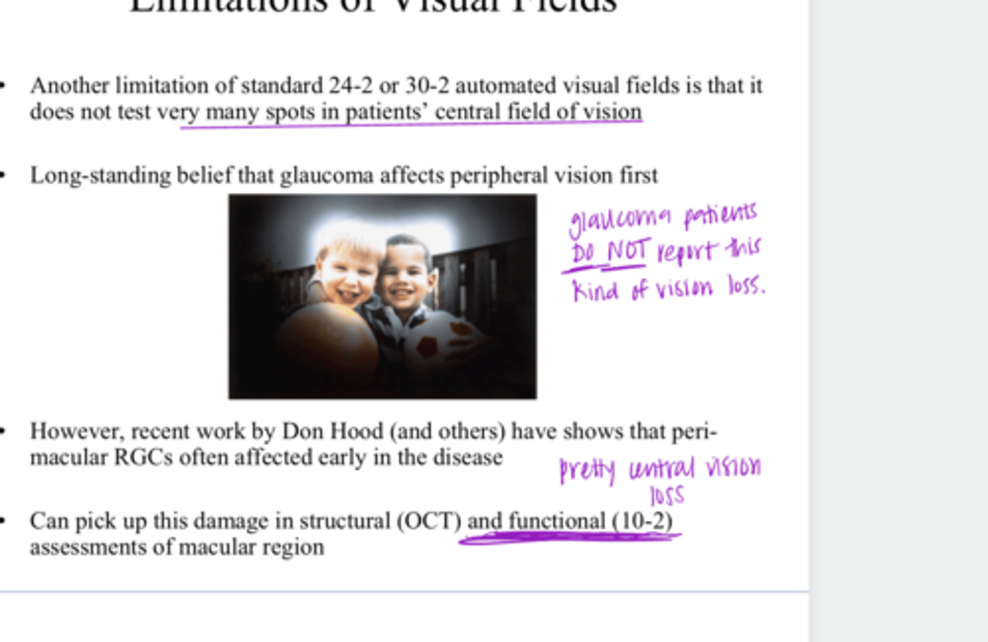
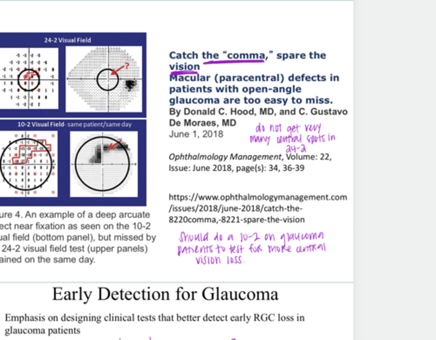
does not test very many spots in a patient's central field of vision
What is a limitation of standard 24-2 or 30-2 automated VFs?

peripheral
What is the long standard belief? Glaucoma affects _____ vision first

perimacular RGCs
Recent work by Don Hood shows that _____ are affected early in glaucoma

structural OCT and 10-2 functional assessment of the macular region
On what tests can you pick up the early changes in vision and perimacular RGC loss in glaucoma?

Yes -- do not necessarily show up on 24-2 VF
Are macular defects in patients with POAG easy to miss?
designing clinical tests that better detect early RGC loss in glaucoma patients
What is the emphasis on in early detection for glaucoma?

-OCT
-Scanning Laser Polarimetry
-HRT
-FAF
What are the structural tests that are involved in the early detection of glaucoma?
-VF (speed, reliability)
-SWAP
-Frequency doubling
What are the functional tests that are involved in the early detection of glaucoma?

-clinician grading of the ONH
-fundus photo
20-40 years ago, _____ were the main structural assessments of glaucoma

Yes
Can certain diseases impact one pathway more than another?
You might be able to detect the disease earlier with special clinical tests
Why is it important to know which pathway of vision a certain disease will impact?
S cone driven konio
_______ pathway shows reduction in diabetic retinopathy and glaucoma
stimulus/background
You can adjust the ______ parameters to selectively identify a specific cone class
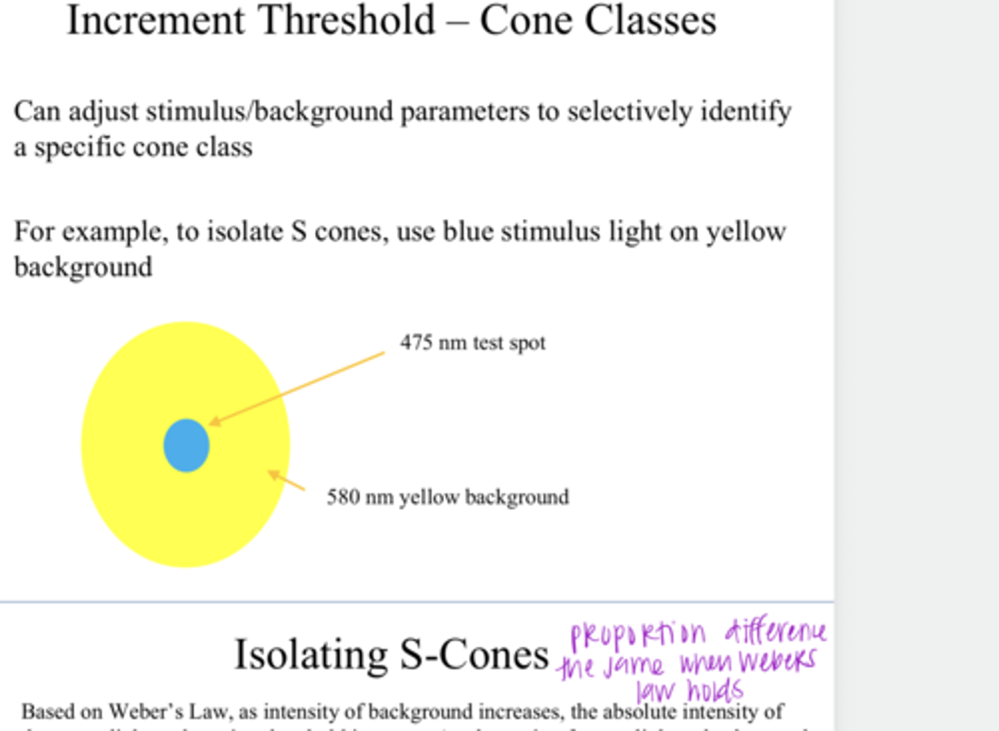
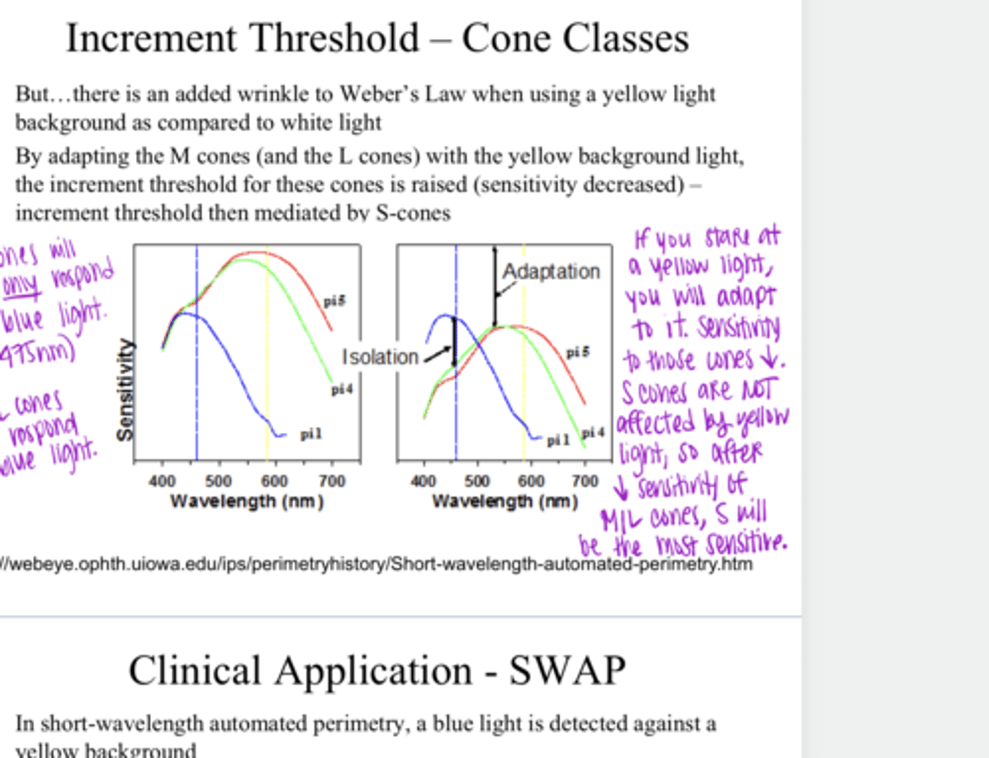
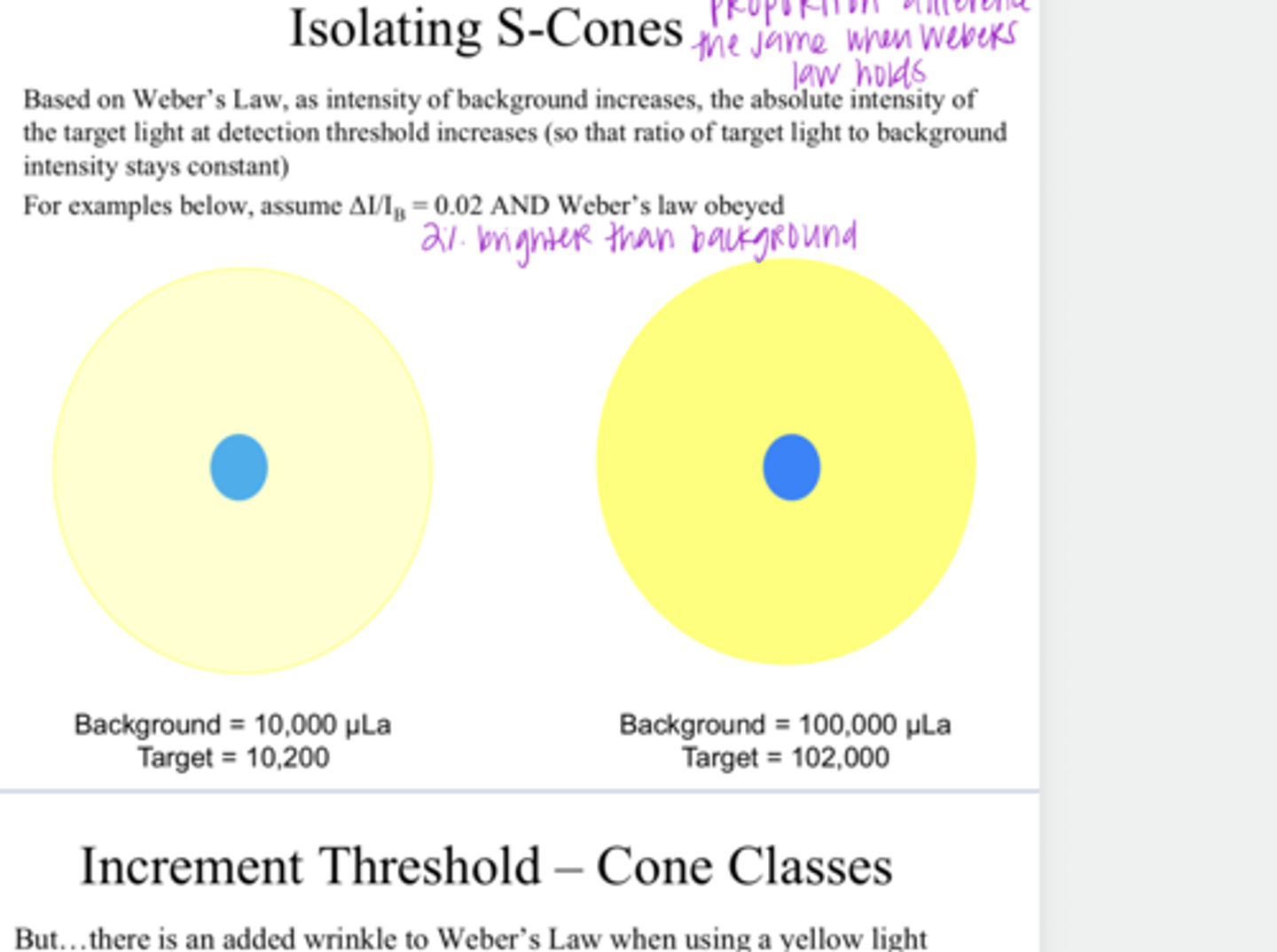
blue; yellow
To isolate S cones, use a ____ stimulus light on a ____ background

increases
Isolating S Cones
Based on Weber's Law, as intensity of a background increases, the absolute intensity of the target light at detection threshold (increases/decreases) -- so that the ratio of the target light to the background intensity would change
Yes
Do cones adapt to light?

M and L cones will adapt to the yellow light background, the threshold is raised (sensitivity decreased), and increment threshold will then be mediated by S cones
What is the wrinkle to Weber's Law when using a yellow light background compared to a white light?