Final Anatomy Lab Exam
1/37
There's no tags or description
Looks like no tags are added yet.
Name | Mastery | Learn | Test | Matching | Spaced | Call with Kai |
|---|
No analytics yet
Send a link to your students to track their progress
38 Terms
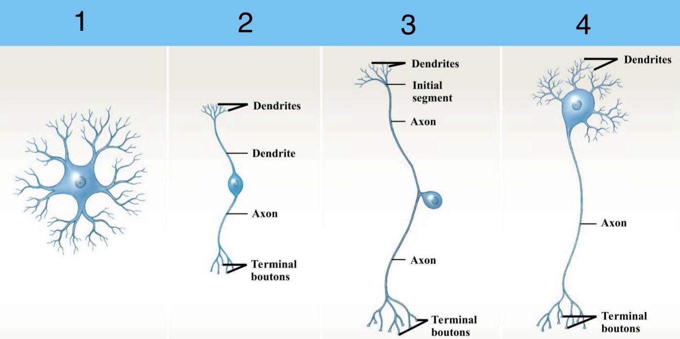
Identify the structural classifications of Neurons
Anaxonic Neuron
Bipolar Neuron
Unipolar Neuron
Multipolar Neuron
What makes up the CNS & PNS?
CNS: Brain, Spinal Cord
PNS: Peripheral nerves
What is the area between 2 myelinated internodes called?
Node of Ranvier

Identify the parts of the Neuron
Dendrites
Cell Body
Axon
Terminal Boutons
Mitochondrion
Nucleus
Nucleolus
Nissl Bodies
Dendritic Spines
Axon Hillock
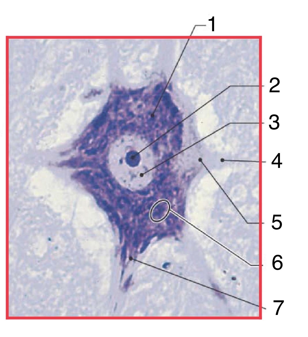
Identify the parts of the Neuron (histology)
Nerve Cell Body
Nucleolus
Nucleus
Initial segment of Axon
Axon Hillock
Chromatophilic Substance
Neurofilament
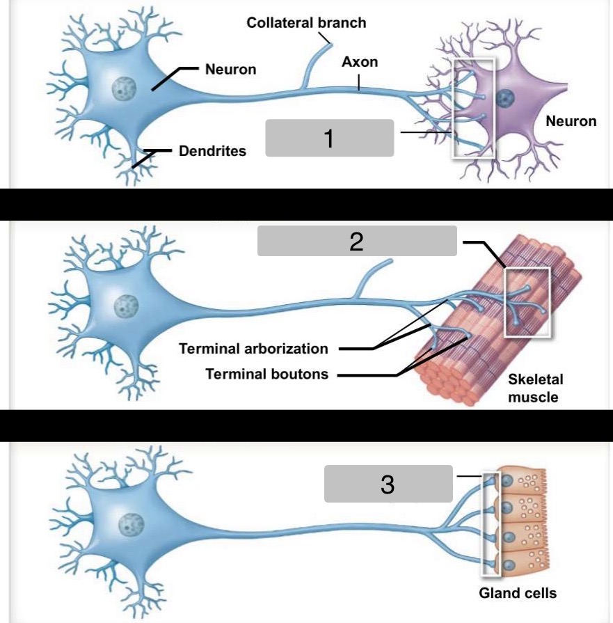
Identify the types of Synapses
Synapses with another Neuron
Neuromuscular Synapses
Neuroglandular Synapses
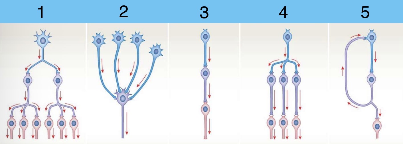
Identify the Neuronal Organization Circuits (the way neurons communicate)
Divergence
Convergence
Serial Processing
Parallel Processing
Reverberation
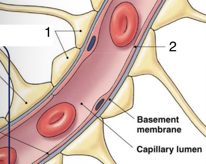
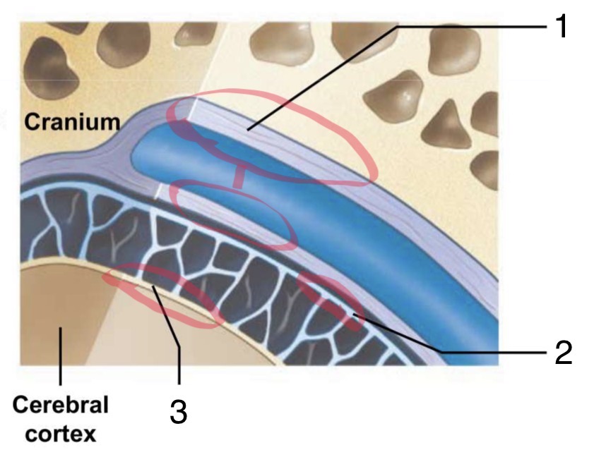
Label parts of the Blood-Brain Barrier
Astrocyte Foot Processes
Tight Junction
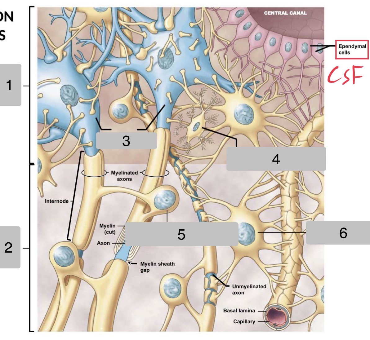
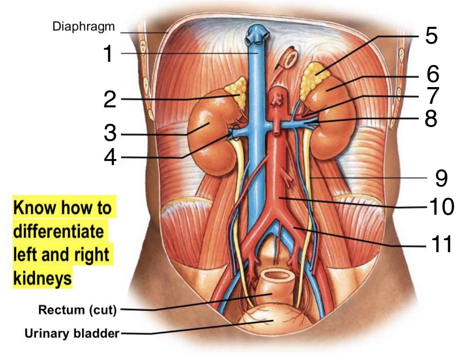
Label the parts of the CNS
Gray Matter
White Matter
Neurons
Microglial Cell
Oligodendrocyte
Astrocyte
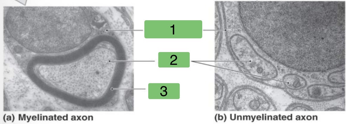
Identify the parts of the different Axons
Neurilemma
Axons
Myelin
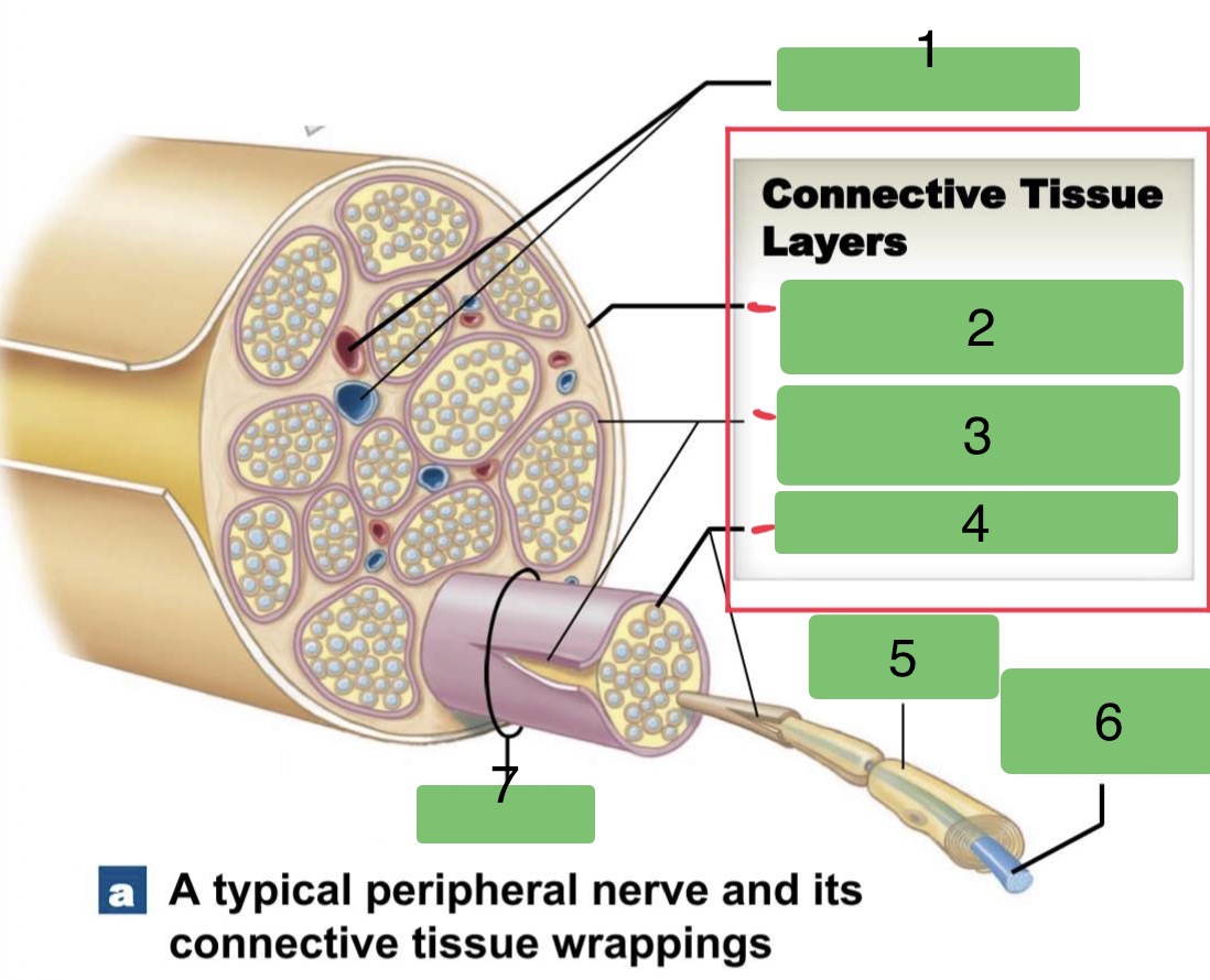
Label the parts of a Peripheral Nerve
Blood Vessels
Epineurium (covering peripheral nerve)
Perineurium (around one fascicle)
Endoneurium
Schwann Cell
Myelinated Axon
Fascicle
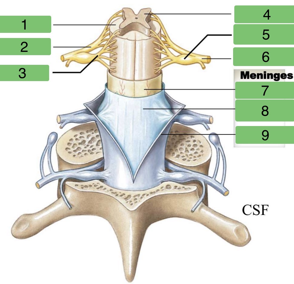
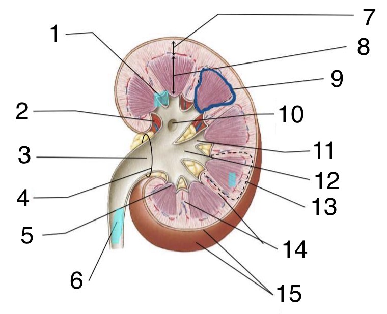
Identify the regions of the Spinal Cord
White Matter
Ventral Root
Dorsal Root
Gray Matter
Dorsal Root Ganglion
Spinal Nerve
Pia Mater
Arachnoid Mater
Dura Mater
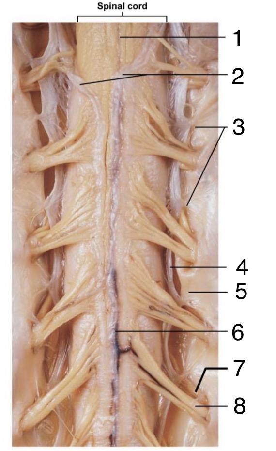
Label the parts of the anterior Spinal Cord
Anterior Median Fissure
Pia Mater
Denticulate Ligaments
Arachnoid Mater
Dura Mater
Spinal Blood Vessel
Dorsal Root
Ventral Root
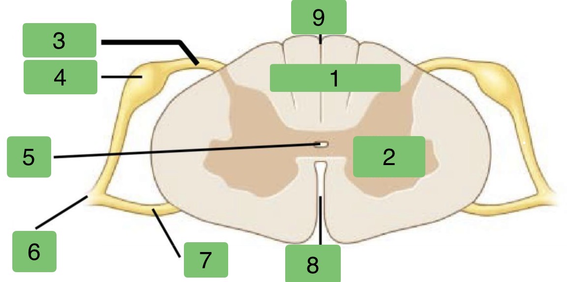
Label the parts of the Transverse Plane Spinal Cord
White Matter
Gray Matter
Dorsal Root
Dorsal Root Ganglion
Central Canal
Spinal Nerve
Ventral Root
Anterior Median Fissure
Posterior Median Sulcus
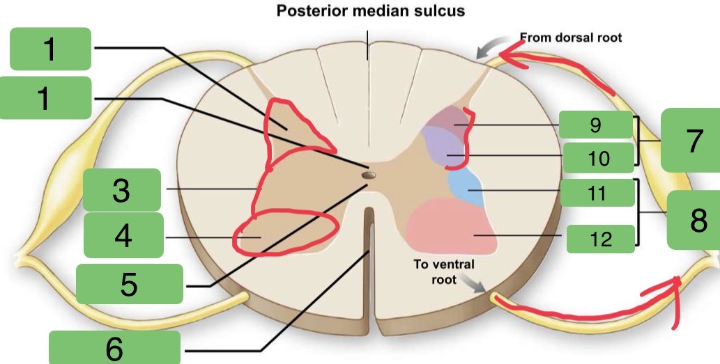
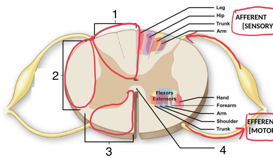
Identify the sections of the Spinal Cord
Posterior Gray Horn
Posterior Gray Commissure
Lateral Gray Horn
Anterior Gray Horn
Anterior Gray Commissure
Anterior Median Fissure
Sensory Nuclei
Motor Nuclei
Somatic
Visceral
Visceral
Somatic
Identify the sections of the Spinal Cord
Posterior White Column
Lateral White Column
Anterior White Column
Anterior White Commissure
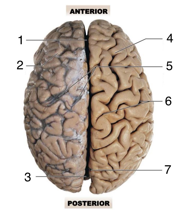
Label the superior anatomy of the Brain
Longitudinal Fissure
Left Ceribral Hemisphere
Cerebellum
Right Cerebral Hemisphere
Cerebral Blood Vessels (covered by arachnoid mater)
Central Sulcus
Parieto-Occipital Sulcus
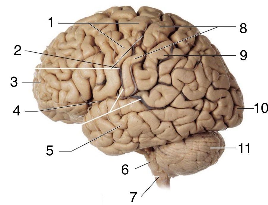
Label the lateral anatomy of the Brain
Precentral Gyrus
Central Sulcus
Frontal Lobe (left cerebral hemisphere)
Lateral Sulcus
Temporal lobe
Pons
Medulla Oblongata
Postcentral Gyrus
Parietal Lobe
Occipital Lobe
Cerebellum
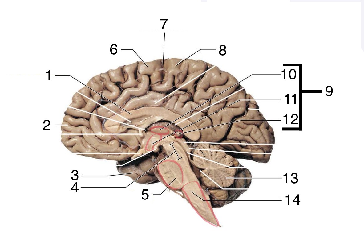
Label the lateral anatomy of the Brain (including brain stem)
Corpus Callosum
Frontal Lobe
Temporal Lobe
Mesencephalon
Pons
Precentral Gyrus
Central Sulcus
Postcentral Gyrus
Diencephalon
Thalamus
Hypothalamus
Epithalamus
Cerebellum
Medulla Oblongata
Label the Cranial Meninges
Dura Mater
Arachnoid Mater
Pia Mater
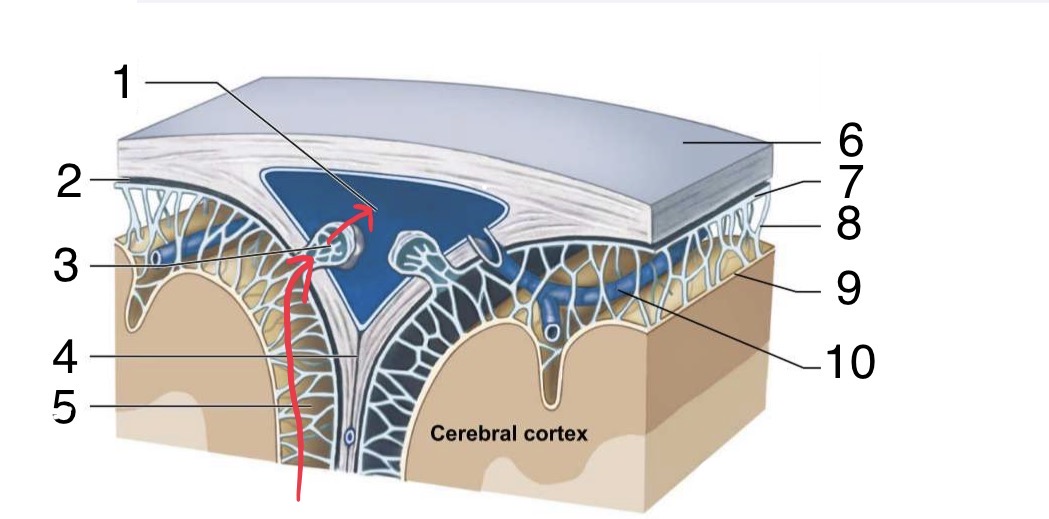
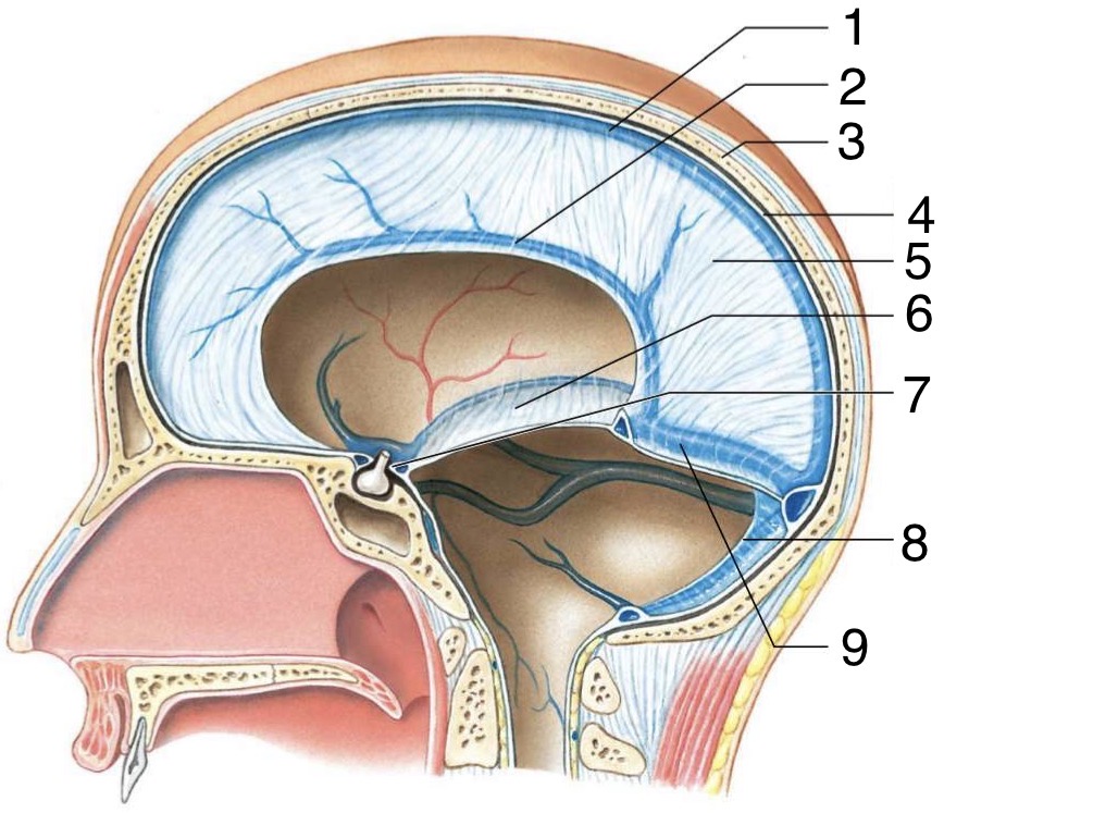
Identify the parts of the superior Cranium
Superior Sagittal Sinus
Subdural Space
Arachnoid Granulation
Flax Cerebri
Subarachnoid Space
Dura Mater
Arachnoid Mater
Arachnoid Trabeculae
Pia Mater
Cerebral Vein
Identify the parts of the sagittal Cranium
Superior Sagittal Sinus
Inferior Sagittal Sinus
Cranium
Dura Mater
Falx Cerebri
Tentorium Cerebelli
Diaphragma Sellae
Falx Cerebelli
Transverse Sinus
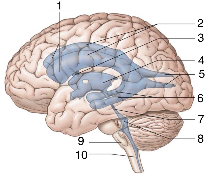
Label the Ventricles and other parts of the Brain
Anterior Horns of Lateral Ventricles
Lateral Ventricles
Interventricular Foramen
Third Ventricle
Posterior Horns of Lateral Ventricles
Inferior Horns of the Lateral Ventricles
Aqueduct of Midbrain
Fourth Ventricle
Medulla Oblongata
Central Canal
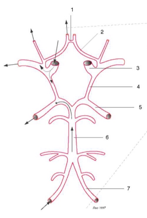
Label the Arteries of the Circle of Willis
Anterior Communicating
Anterior Cerebral
Internal Carotid
Posterior Communicating
Posterior Cerebral
Basilar
Vertebral

Label the 12 Cranial Nerves
Olfactory Nerve
Optic Nerve
Oculomotor Nerve
Trochlear Nerve
Trigeminal Nerve
Abducens Nerve
Facial Nerve
Vestibulocochlear Nerve
Glossopharyngeal Nerve
Vagus Nerve
Accessory Nerve
Hypoglossal Nerve

Label the 2 important structures of the Spinal Cord
Conus Medullaris
Filum Terminale

Label the sections of the Brain
Frontal Lobe
Precentral Gyrus
Central Sulcus
Postcentral Gyrus
Parietal Lobe
Parieto-Occipital Sulcus
Lateral Sulcus
Occipital Lobe
Temporal Lobe
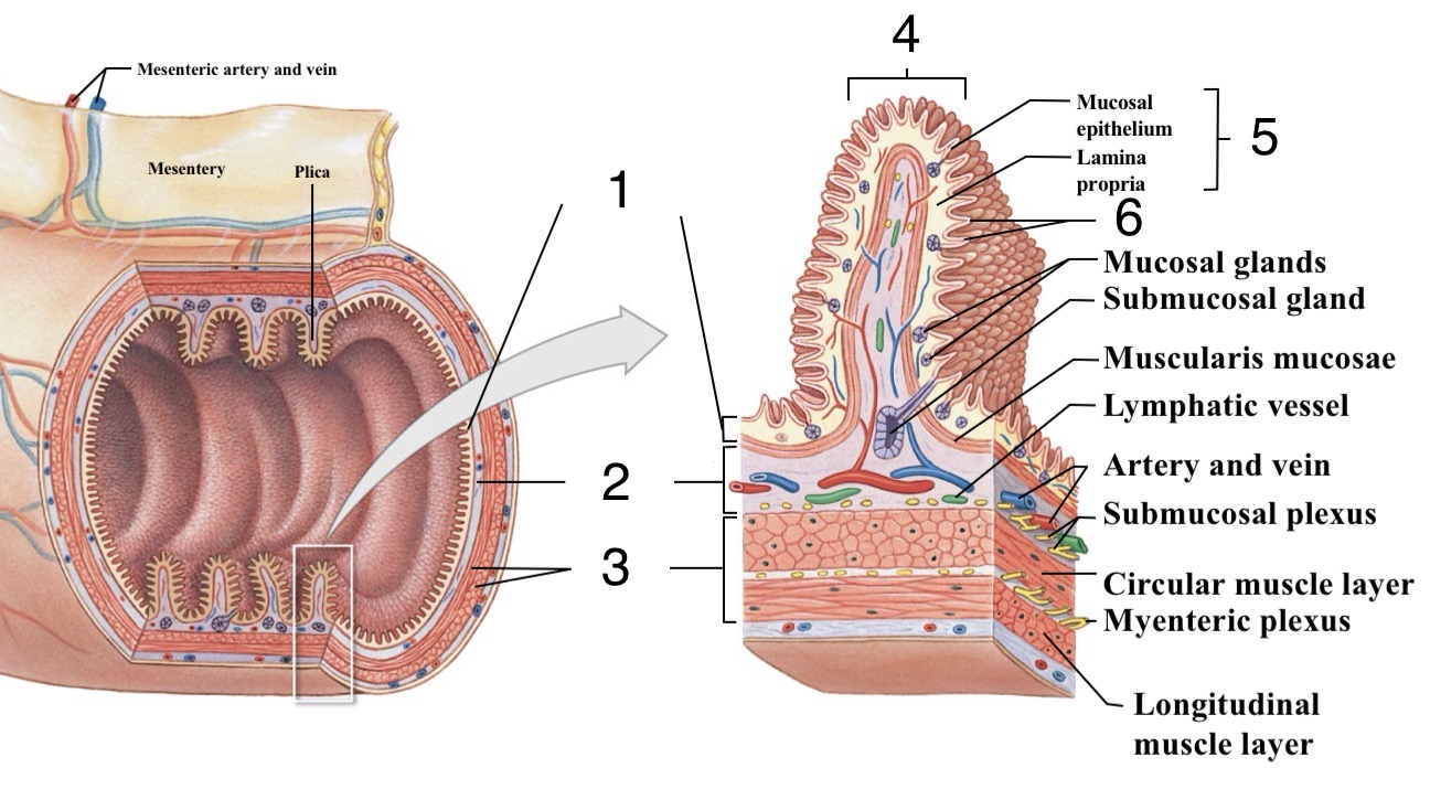
Identify the structures of the Digestive Tract
Mucosa
Submucosa
Muscularis Externa
Plica
Mucosa
Villi
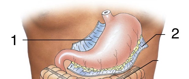
Label the stabilizers of the Stomach
Lesser Omentum
Greater Omentum
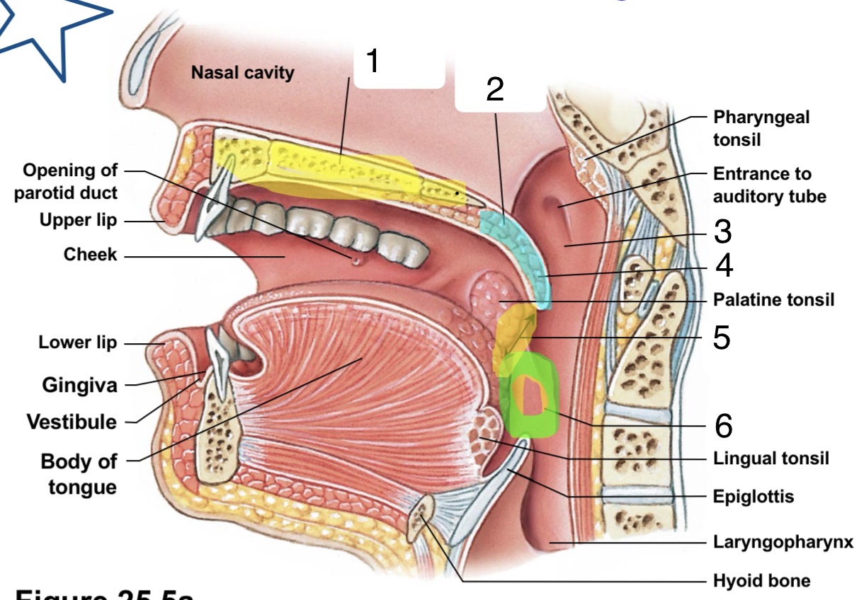
Identify the structures of the Oral Cavity
Hard Palate
Soft Palate
Nasopharynx
Uvula
Fauces
Oropharynx
Identify the structures of the Oral Cavity
Fauces
Palatoglossal Arch
Palatopharyngeal Arch
Hard Palate
Soft Palate
Uvula
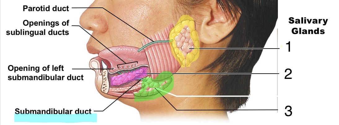
Identify the Salivary Glands of the Mouth
Parotid Salivary Gland
Sublingual Salivary Gland
Submandibular Salivary Gland

Identify the parts of the Liver
Right Lobe
Quadrate
Left Lobe
Caudate
Spleen
Pancreas
Pancreatic Duct
Gallbladder
Right Haptic Duct
Cystic Duct
Common Haptic Duct
Common Bile Duct
Left Haptic Duct

Identify the parts of the Stomach
Esophagus
Longitudinal Layer
Circular Layer
Oblique Layer
Lesser Curvature
Pyloric Sphincter
Duodenum
Pyloric Canal
Pyloric Antrum
Cardia
Fundus
Serosa
Body
Lumen
Rugae of Mucosa
Greater Curvature
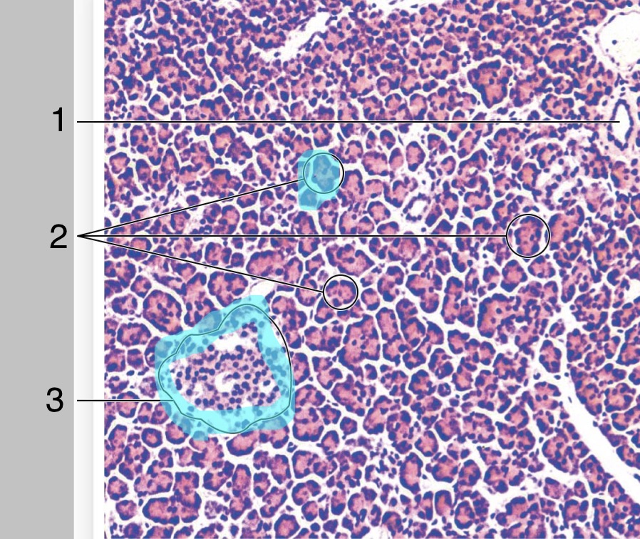
Identify the regions of the Pancreas (histology)
Duct
Pancreatic Acini
Pancreatic Islet

Label the structures of the Gallbladder & Pancreas
Minor Duodenal Papilla
Major Duodenal Papilla
Hepatopancreatic Sphincter
Main Pancreatic Duct
Hepatopancreatic Ampulla
Accessory Pancreatic Duct
Duodenojejunal Flexure