Anatomy weeks 1-4
1/173
There's no tags or description
Looks like no tags are added yet.
Name | Mastery | Learn | Test | Matching | Spaced | Call with Kai |
|---|
No analytics yet
Send a link to your students to track their progress
174 Terms
Regions of the vertebral column
Cervical, thoracic, lumbar, sacrum, coccyx
Functions of the vertebral column
framework
Support/ weight transmission
Protection
Muscle attachment
General bone formation
Number of vertebrae in each region
Cervical:
Thoracic:12
Lumbar: 5
Sacrum: 5 (fused)
Coccyx 4 (fused)
body
Major part of a bone (where ossification commences)
Protuberance (bumps)
Attachment sites for muscles or ligaments
Articular surface
For articulation (joint) with another bone
Depressions/ holes
For a passage of other structures
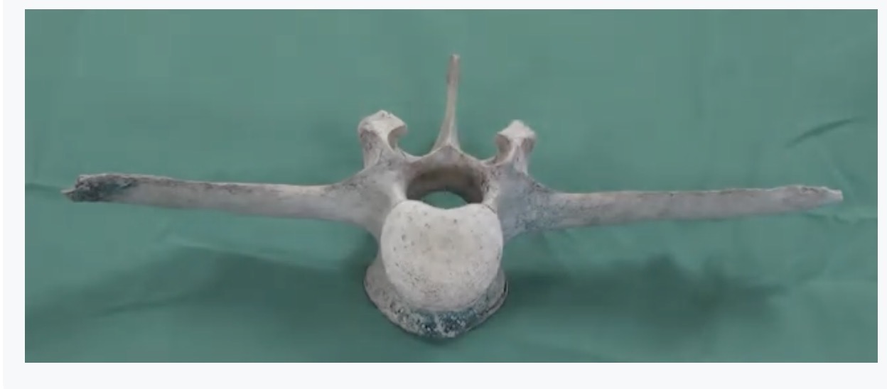
Name the landmarks
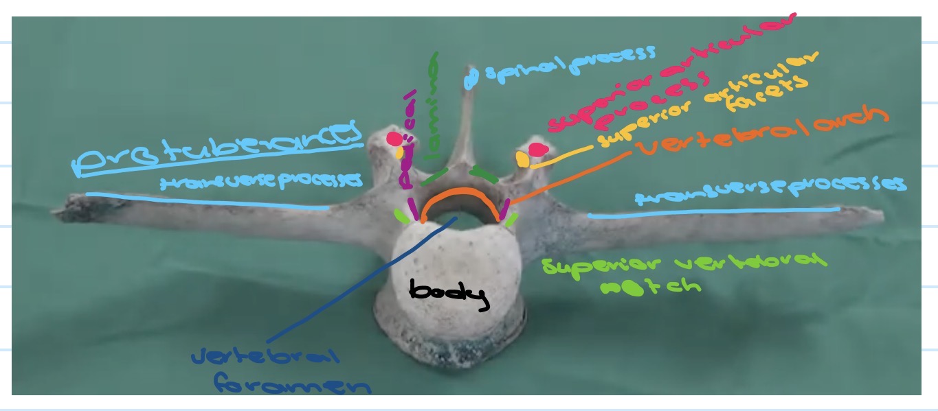
What is the function of a body in the vertebrae?
The major weight bearing part of the vertebra. It is also an articular surface
What is the function of the vertebral arch in the vertebral column?
The arch consists of a pedicle and lamina on each side. They make up the boundary of an anatomical space that is called the vertebral foramen (Passage for the spinal cord)
What is the function of the spinous process and transverse process in the vertebral column?
The attachment of muscles and ligaments
What is the function of superior and inferior articular processes?
Bony projections on vertebrae that form facet joints, enabling, guiding and limiting spinal movement while providing stability. Superior processes project upwards to connect with inferior processes projecting downwards from the vertebrae above. The orientation of the processes determines the range of motion in different spinal regions
What is the function of the inferior and superior notch of the vertebrae?
Inferior notch if the vertebra above and superior notch of vertebra below make up the intervertebral foramen
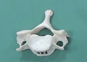
What region of the vertebrae is this:
Cervical
Regional classification of cervical vertebrae
Body: it is a small body. C1 doesn’t have a body
Spinous process: bifid (larger attachment area)
Transverse process: short with a hole in it (transverse foramen - passage for vertebral artery)
Articular facets: oriented in oblique plane (allows movement in all three planes)
What region of the vertebrae is this?
Thoracic
Regional classification of thoracic vertebrae
Body: it is a bit bigger than the cervical region to hold more body weight
Spinous process: long pointy and angled downwards (limits extension)
Transverse process: projected posteriorly. Coastal facets for rib articulation
Articular facets: in a frontal plane (limited in flexion and extension but rotation is possible)
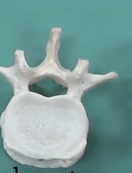
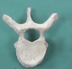
What part of the vertebrae is this?
Lumbar
Regional classification of lumbar vertebrae
Body: large kidney shaped body - to support the weight of the body above it
Spinous process: large and hatched shaped (large surface area for attachment)
Transverse process: in more of a frontal plane. They are long and skinny (provide attachment for larger muscles)
Articular facets: they are sagittal (allow flexion and extension)
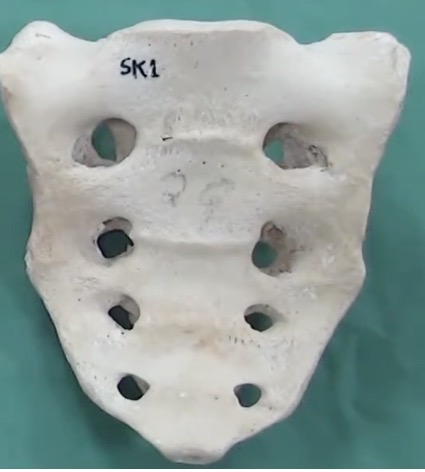
What part of the vertebrae is this?
Sacrum
Regional classification of sacrum vertebrae
Body: they are fused together. It decreases in size as you go down (weight is transmitted out to hip bones)
Spinous process: fused forming a crest in the midline on the back of the sacrum (muscle attachment)
Transverse process: fused and enlarged (create lateral mass for sacroiliac joint)
Articular facets: fused together so just see bumps (vertebrae don’t need to move relative to each other)
What region are transverse foramen found?
Cervical
What is the function of the transverse foramen?
A passageway for the vertebral artery, vertebral vein and sympathetic nerve plexus
What region are costal facets found?
Thoracic region
What is the function of the costal facets?
Attach the ribs to the vertebral column
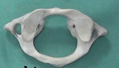
What is this?
Atlas (C1)
Differences of the atlas to typical vertebrae and the functional significance
No body: joints above and below don’t have an intervertebral disc therefore mean it is far more mobile
Superior articular facets are quite large: large mass to hold
Absence of a spinous process: little muscle attachment
Elongated transverse process: increased mechanical advantage to muscles that attach to it
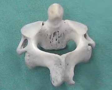
What is this?
The axis
Differences of the axis to typical vertebrae and the functional significance
Large superior articular facets: they are the weight bearing joints
Dens (adhen toy process): critical for the articulation between the two vertebrae (Atlanta axial joint). The dens allows rotation
Curvatures in vertebral column
Cervical: secondary, concave posteriorly, allow upright posture for head
Thoracic: primary, concave anteriorly, more room for thoracic organs
Lumbar: secondary, concave posteriorly, allow upright posture of the trunk
Sacral: primary, concave anteriorly, more room for pelvic organs
What is the purpose of joints in our body?
They enable us to change the shape of our skeleton.
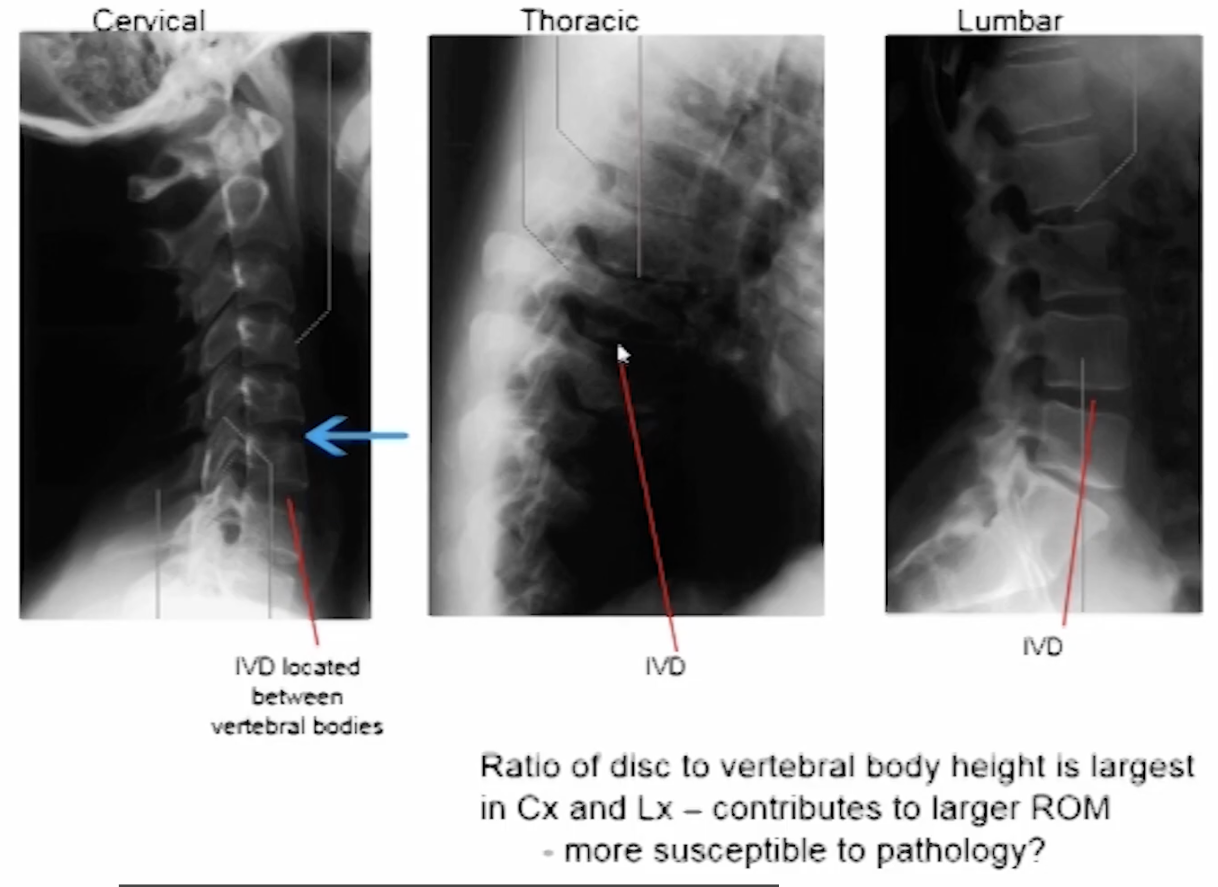
What is the purpose of the intervertebral discs?
Helps to bind each of the vertebra to one another so that there is a greater amount of overall mobility but still maintaining stability.
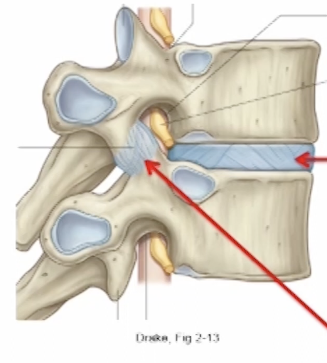
Anterior intervertebral joints
Located between superior and inferior articular surfaces of adjacent vertebral bodies
Posterior intervertebral joint
(Facet or zygapophyseal) located between superior and inferior articular facets of adjacent vertebrae
Can you name these?
Features of the anterior intervertebral joints
Secondary cartilaginous in design
Between the articular surfaces is a fibrocartilagenous disc (intervertebral disc or IVD)
Hyaline cartilage lines articular surfaces
Designed for shock absorption
Allow only some mobility in all planes
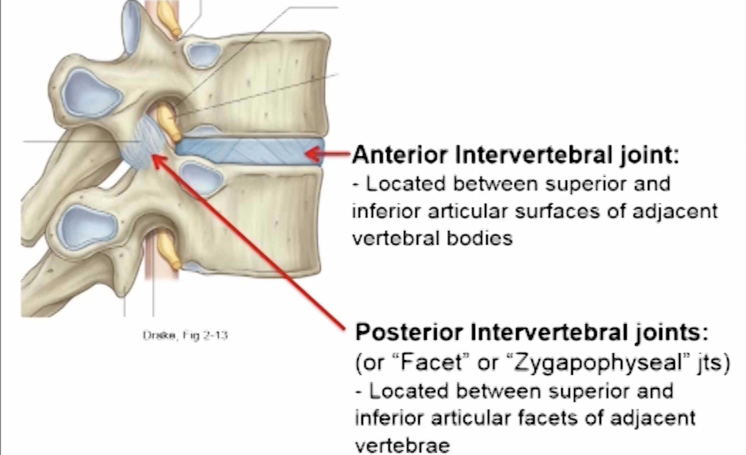
What are the two parts of the IVD (intervertebral disc)?
Annulus fibrosis: outside layer for strength
Nucleus pulposis: inner layer for shock absorption
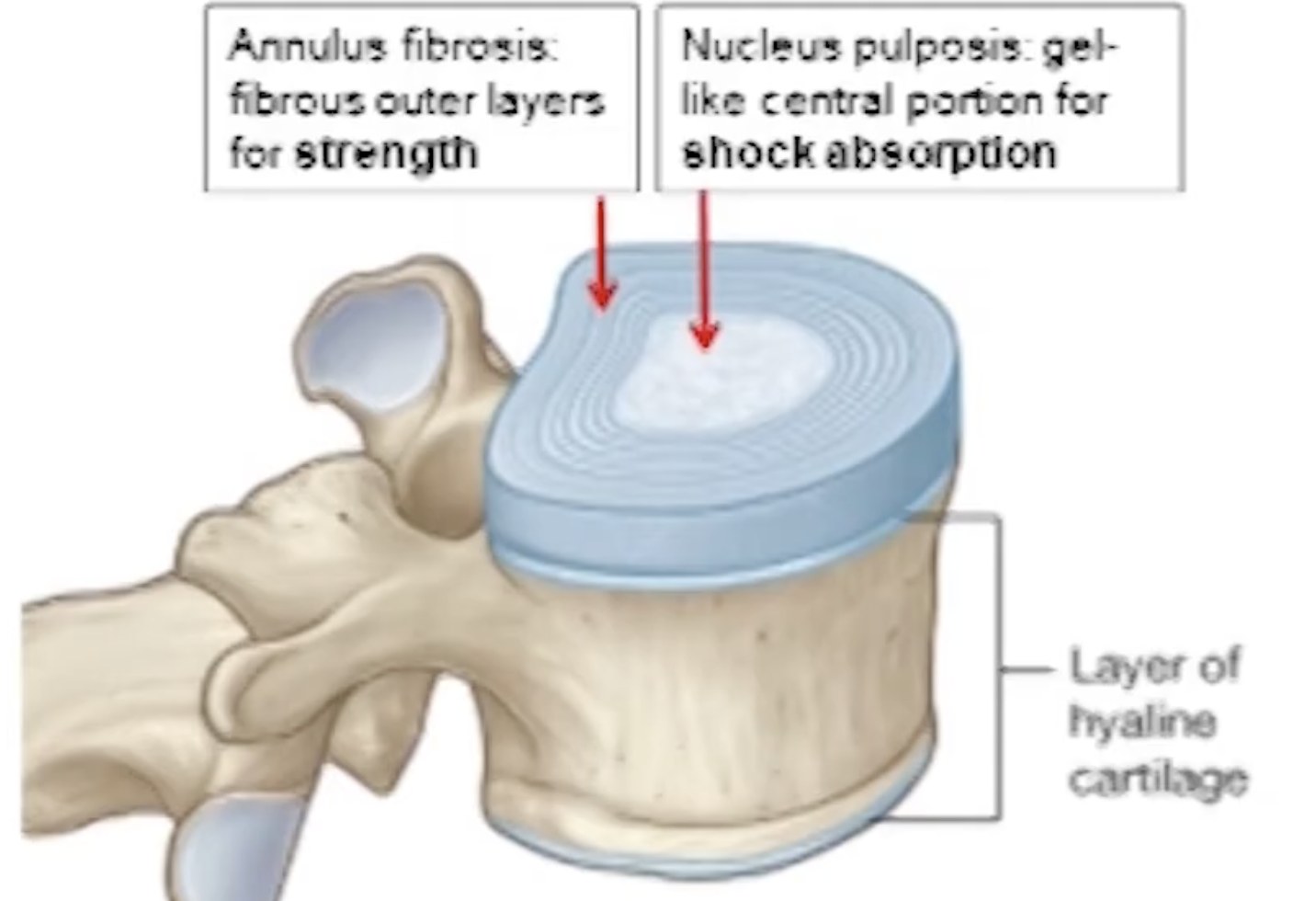
What is IVD herniation?
Means that part of the annulus fibrosis has been overstretched and damaged and the nucleus pulposus is able to push out. It often happens with flexion combined with rotation movements. Most common in cervical and lumbar regions because they have the greatest range of movement
When looking at an X-ray where would you see the IVD disc?
It is the black space between vertebral bodies
What does the lumbosacral joint consist of?
1 x anterior intervertebral joint - IVD between L5 and S1
2 x posterior intervertebral joints - between facets of L5 and S1
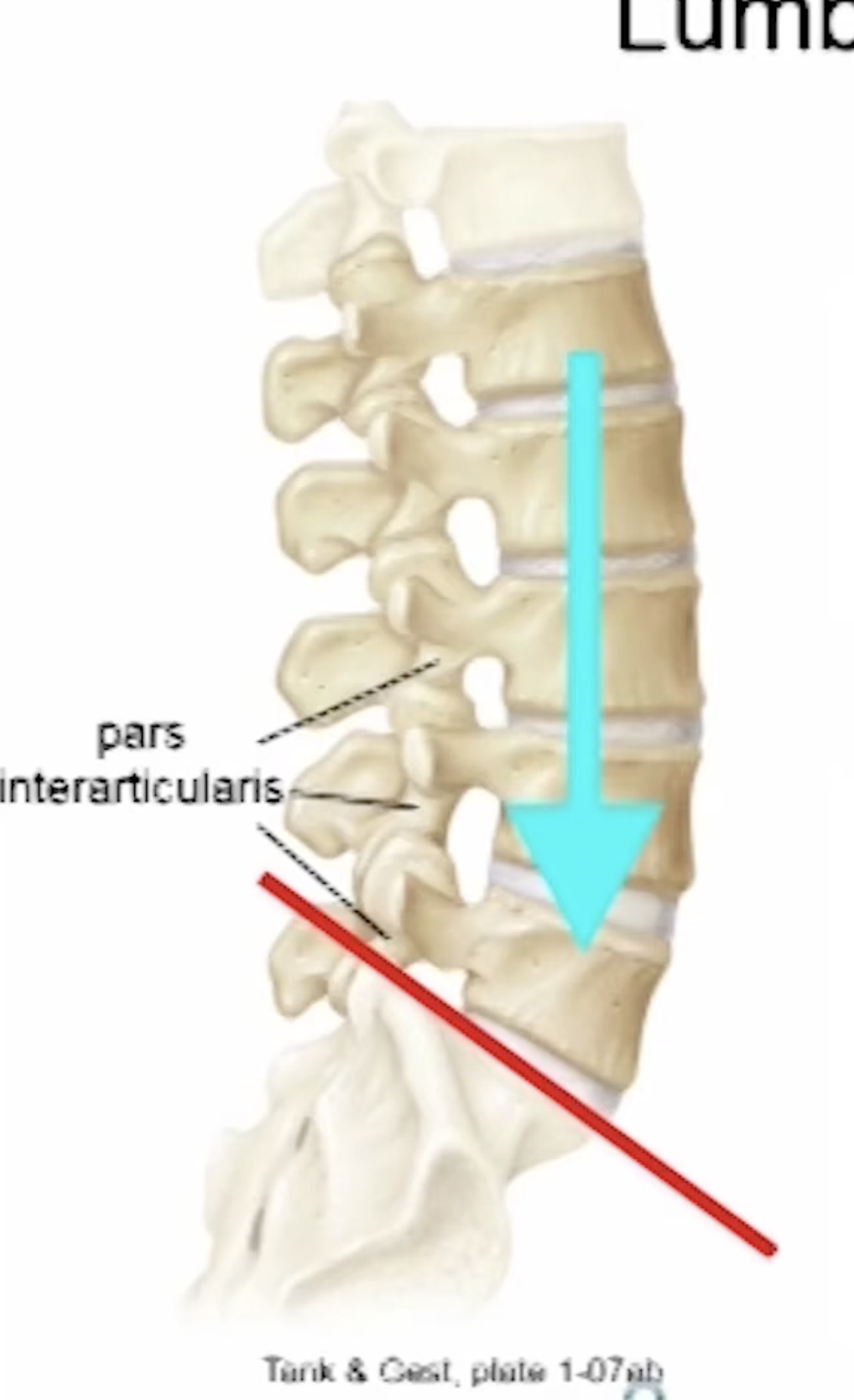
Why is the lumbosacral joint susceptible susceptible to injury’s and pathologies?
Due to the angle (lumbosacral angle) combined with the fact that the joint is supporting the weight of the whole body above it. The angle results in an anterior shearing force placed on the lumbosacral joint
What part of the vertebrae is stressed due to the anterior shearing force?
Pars interarticularis. It is located between the superior and inferior articular process on both sides of each vertebrae. Due to the anterior shearing force it’s most commonly the L5 pars interarticularis that is stressed
What is the condition called when one pars interarticularis fractures as a result of the stress?
Spondylolysis
What is the condition called if both pars interarticularis fractures on the one L5 vertebra?
Spondylolysthesis - forward slip of one vertebrae on another due to bilateral pars interarticularis fracture
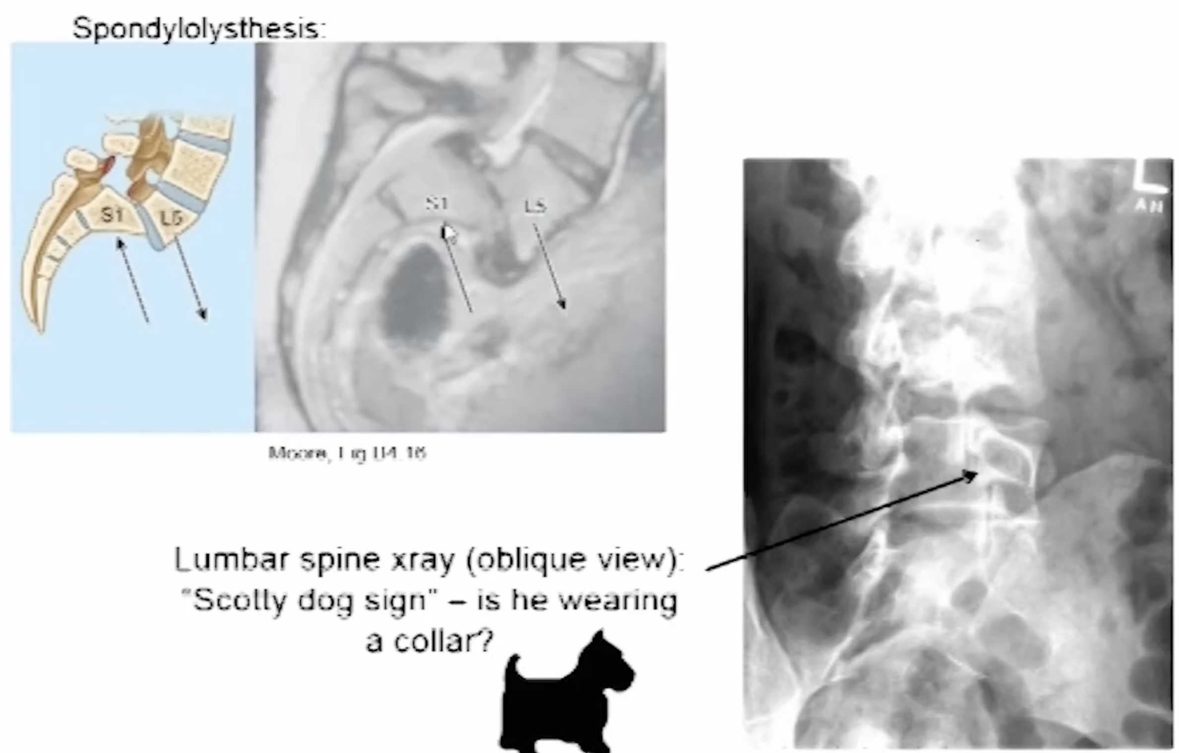
How do you diagnose a defect of the pars interarticularis?
Looking at an oblique view of the lumbar spine in an X-ray. You will see in outline of a Scotty dog. If there were a defect such as a fracture there would be a black line across the neck. It is called the Scotty dog sign if he is wearing a collar
Features of a posterior intervertebral joint
synovial joint (joint cavity filled with synovial fluid making them highly mobile)
Plane/gliding joints (limits the direction of movement determined by orientation of facets)
Overall good mobility in limited direction
What are the orientations of the facets in each region of the vertebrae and what movements do they allow?
Cervical:
facets slope down anterior to posterior (oblique plane)
Movements allowed = flexion, extension, rotation
Thoracic:
facets orientated (almost) in frontal plane
Movements allowed = rotation
Lumbar:
facets orientated (almost) in sagittal plane
Movements allowed = flexion, extension
What are the superior cervical that are in the cervical region?
Atlanto - occipital (A-O) joint
Atlanta - axial (A-A) joint
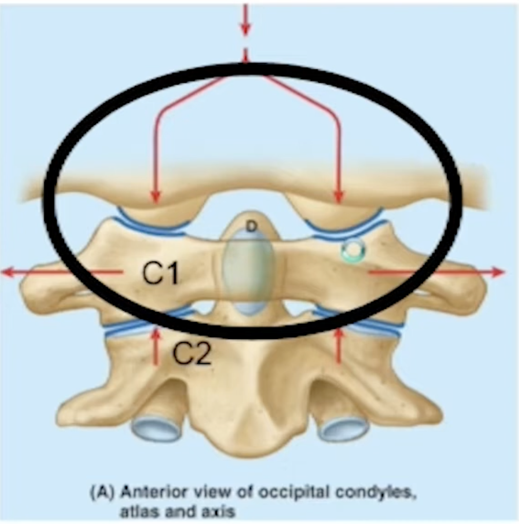
Features of the A-A (atalto-occipital) joint
Between the skull and C1
Synovial
Involves two enlarged codyloid joints (for weight bearing)
There are two superior facets of C1 and the occipital condyles on the inferior aspect of the skill and they sit on top of the facets of C1
Major movement is flexion and extension. The condyles can easily rock forwards and backwards
Features of the A-A (atlato-axial) joint
Between C1and C2
It is three different articulation which are all synovial
2 enlarged facet (plane) joints = lateral A-A joints
1 pivot joint between dens and anterior arch of the atlas = median A-A joint
Major movement is rotation (C1 can pivot around the dens)
What maintains the integrity of the joints?
The ligaments
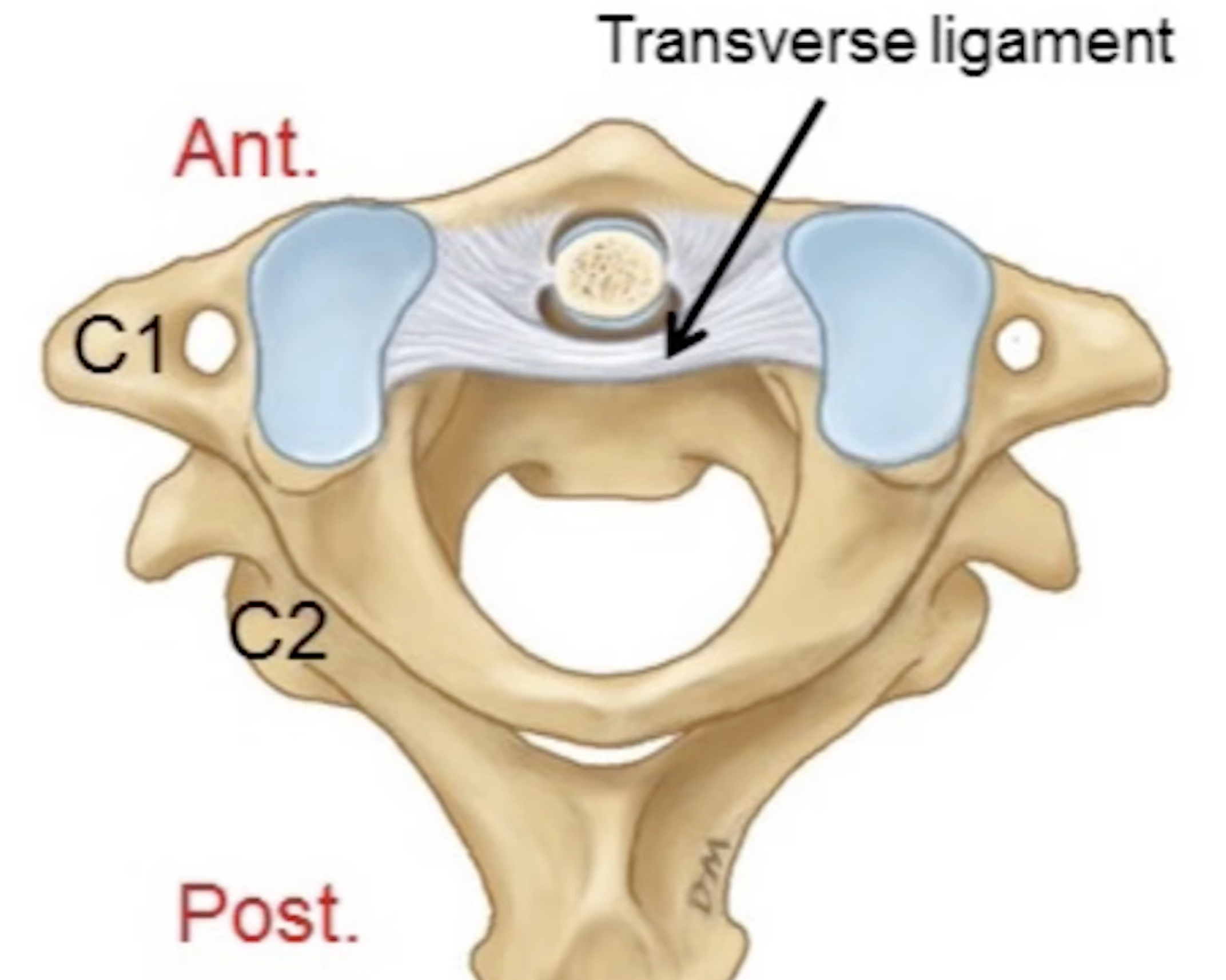
What is the transverse ligament?
Lies behind the dens, coming across from the inner aspects of the anterior arch of C1. Its job is to hold the dens in place. It ensures that the two vertebrae (C1 and C2) move as one
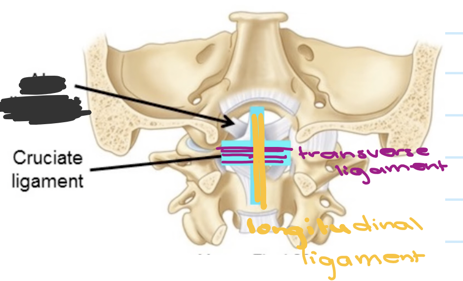
What is the cruciate ligament?
(Resembles a cross) it consists of the transverse ligament along with a longtidudinal ligament
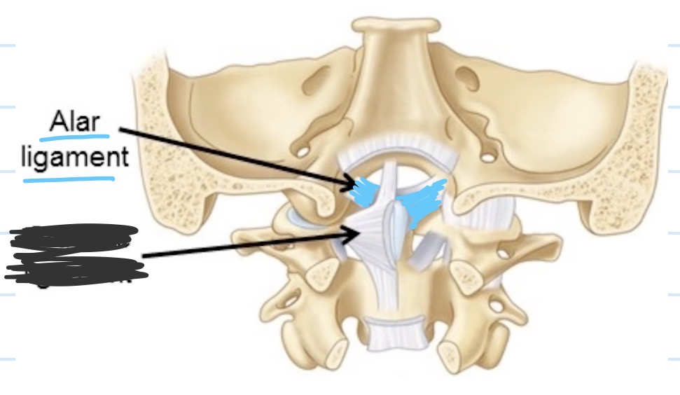
What is the alar ligament?
It has two parts to it. Each part attaches from the tip of the dens up to the edge of the foramen magnum of the skull
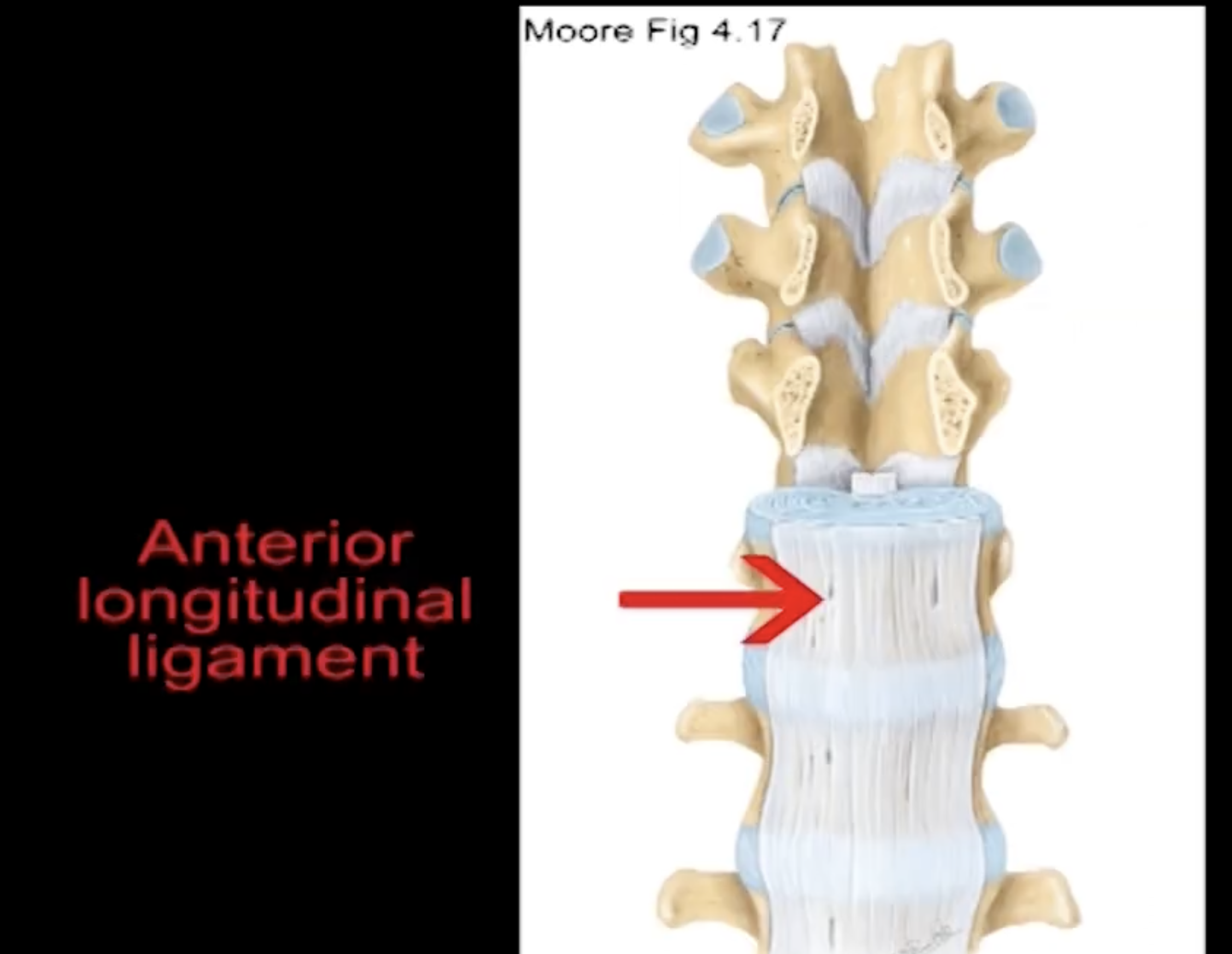
The function of the anterior longitudinal ligament and the relative to the anterior IV joints
It lies anterior to the vertebral bodies and in a longitudinal orientation. It extends longitudinal all the way from the pelvic surface of the sacrum up to the occipital bone anterior to the foramen magnum. Along the way it is attaching to every vertebral body and intervertebral disc that it passes. It is broad and cover the entire aspect of the vertebral bodies. It helps to support the intervertebral discs in the anterolateral aspects
Position: anterior
Function: limits extension
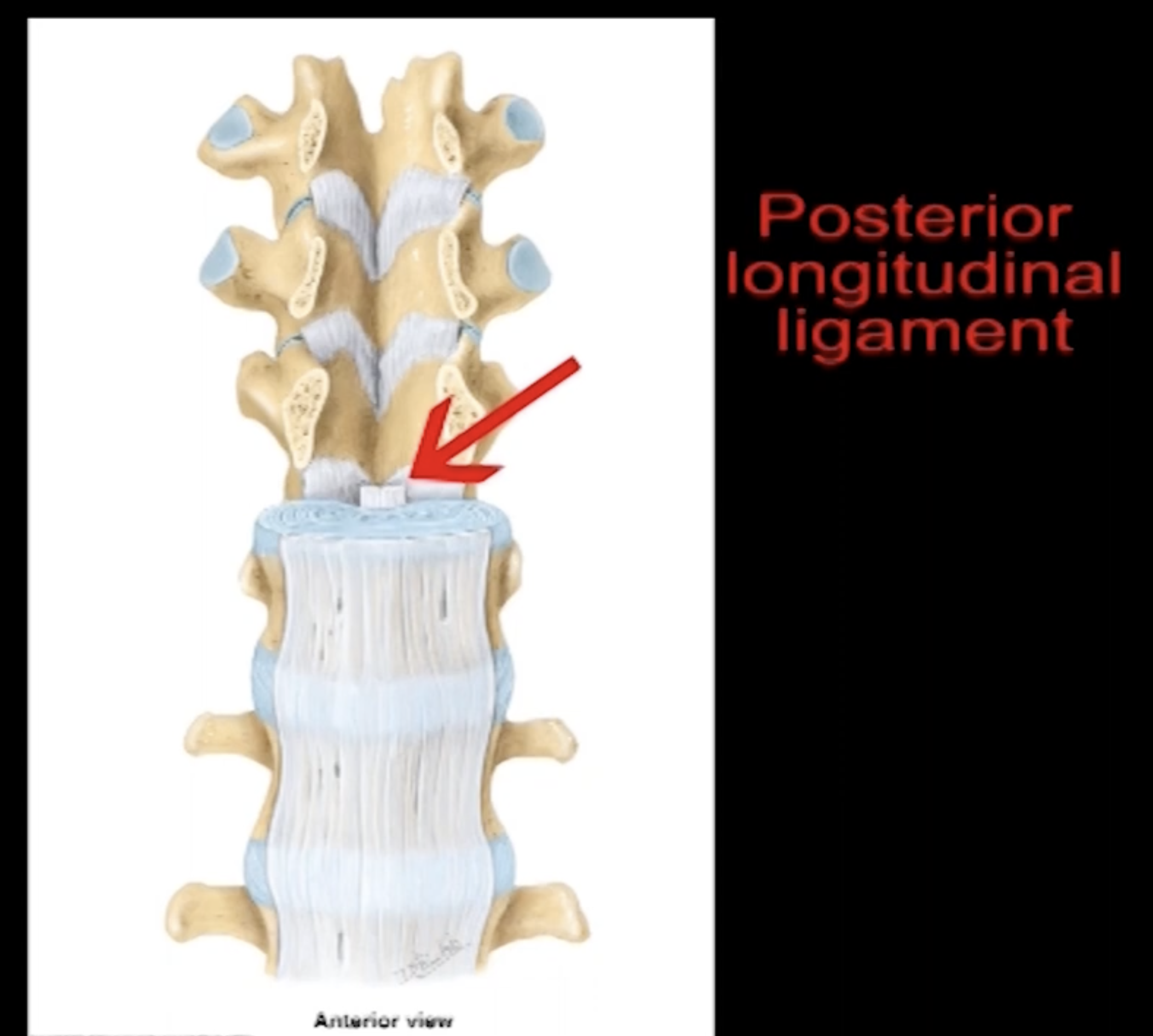
The function of the posterior longitudinal ligament and the relative to the anterior IV joints
It extends longitudinally from the sacrum to the occiput where it becomes the tectoral membrane. Lying posterior to the vertebral bodies means that its inside the vertebral canal. The ligament is narrow as it is restricted by the dimensions of the vertebral canal
Position: posterior
Function: limit flexion
The function of the supraspinous ligament and the relative to the anterior IV joints
It lies on top of the spinous processes. It is like a cord that runs from C7 spinous processes down to the sacrum. After C7 this ligament forms a larger ligament called the ligamentum nuchae
Position: posterior
Function: limits flexion
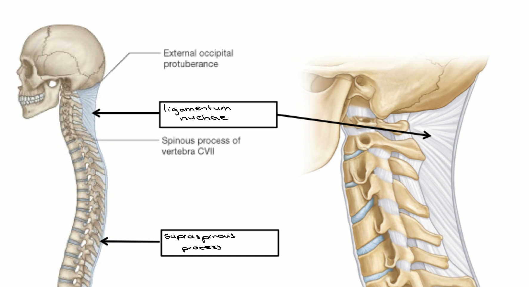
The function of ligamentum nuchae and the relative to the anterior IV joints
Extends as a median band from the external occipital protuberance and the posterior border of foramen magnum down to the spinous processes of the cervical vertebra up until C7. It forms surface area for muscles to attach to that would otherwise attach to the spinous processes of vertebrae at other levels
Position: posterior
Function: limits cervical flexion

The function of the interspinous ligament and the relative to the anterior IV joints
Position: between spinous processes
Function: limits flexion
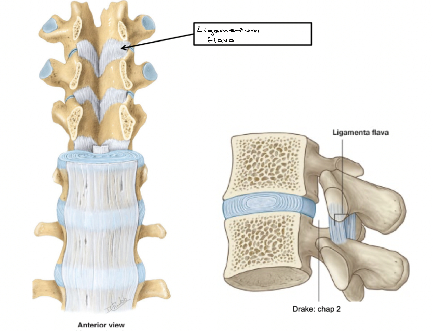
The function of the ligamentum flava ligament and the relative to the anterior IV joints
It lies on the inner aspect of the inner aspects of the lamina of adjacent vertebra. It is located within the vertebral canal
Position: posterior
Function: limits flexion
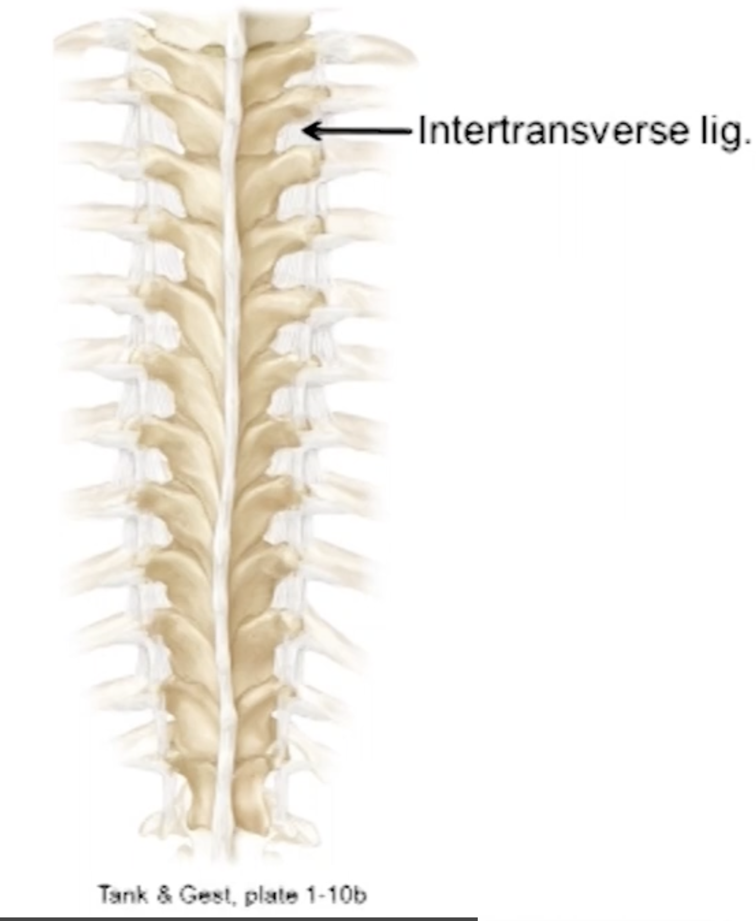
The function of the intertransverse ligament and the relative to the anterior IV joints
Most substantial in the thoracic and lumbar regions
Position: lateral to the intervertebral between the transverse processes
Function: limits lateral flexion
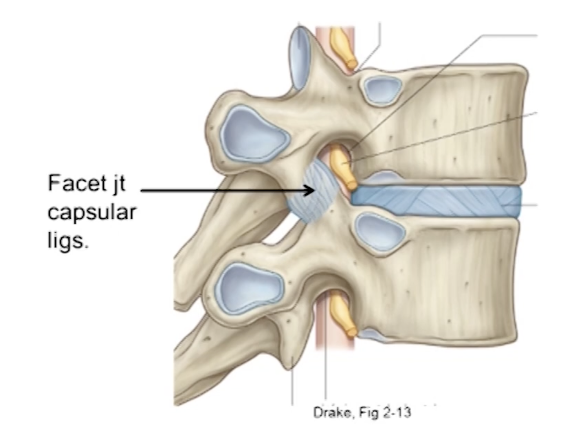
The function of the facet joint capsular ligament and the relative to the anterior IV joints
Help to reinforce the joint capsule of any synovial joint
Position: posterior to intervertebral joints surrounding the facet joints
Function: limit excessive flexion and rotation
Vertebral column movements and muscle location in the sagittal plane
Movements: flexion and extension
Muscle positions: (orientation vertical)
Flexors: anterior
Extensors: posterior
Vertebral column movements and muscle location in the coronal plane
Movements: lateral flexion (l/r)
Muscle position: lateral (orientation vertical)
Vertebral column movements and muscle location in the horizontal plane
Movements: rotation (l/r)
Muscle orientation: horizontal/oblique
What are unilateral actions?
Produce movements in the transverse and/or coronal plane. sagittal plane actions (associated with bilateral contractions) will still occur
What are bilateral actions?
Produce movements in the sagittal plane. Synergistic actions cancel out movements in the other planes
What are ipsilateral movements?
Are to the same side of the body as the body lies e.g. a unilateral contraction of the left erector spinae produces ipsilateral lateral flexion (to the left)
What are contralateral movements?
Are to the opposite side of the body to where the muscle lies e.g. unilateral contraction of the left external oblique produce contralateral rotation (to the right)
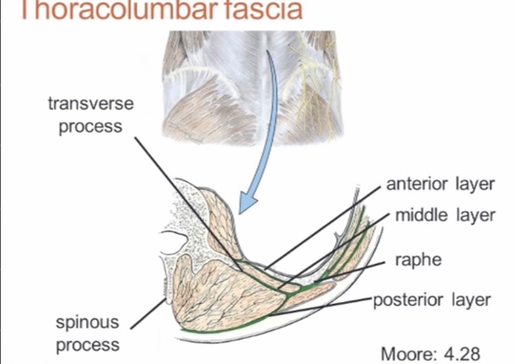
What is the thoraculmber fascia?
A broad sheet of white dense connective tissue that lies posteriorly. Provides attachment for muscles in the upper limb.
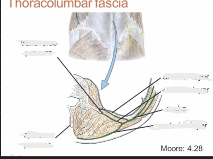
Label the diagram of the thoraculmar fascia?
Origin and insertion of the spinalis
Origin: spinous process
Insertion: spinous process
Origina and insertion of the longissimus
Origin: posterior sacrum
Insertion: laterally situated transverse process
Origin and insertion of iliac costalis
Origin: posterior part of iliac crest
Insertion: ribs
Unilateral actions
Produce movements in the transverse and/or coronal plane. Sagittal plane actions (associated with bilateral contractions) still occur
Bilateral actions
Produce movements in the sagittal plane actions
Ipsilateral movements
Are to the same side of the body as the body lies e.g. a unilateral contraction of left erector spinae produces ipsilateral lateral flexion (to the left)
Contralateral movements
Are to the opposite of the body to where the muscle lies e.g. unilateral contraction of the left external oblique produce contralateral rotation (to the right)
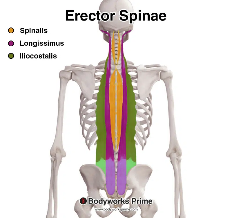
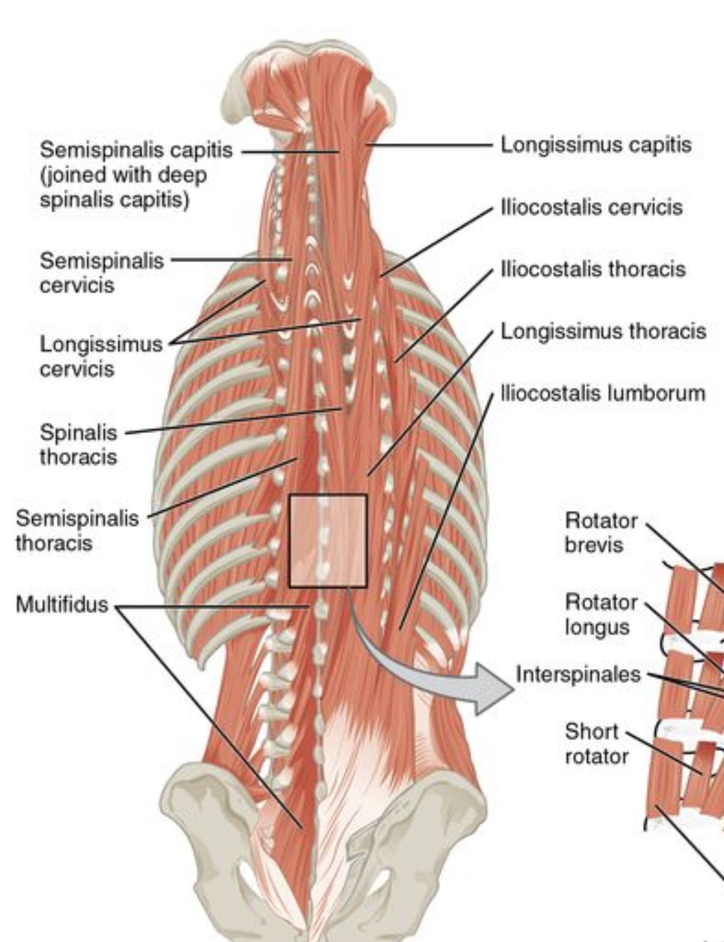
Muscles in the erector spinae group
iliocostalis
Longissumus
Spinalis

Name the muscles in the erector spinae and label on diagram
What region of the vertebral column are the components of the erector spinae groups found?
What is epimere?
Dorsi, muscles of the back, extension typically
What is hypomere?
Flexors, anterior muscles
Embryological origin and innervation of the erector spinae group
Epimere, dorsal rami on segmental basis (cervical dorsal rami for cervicis components)
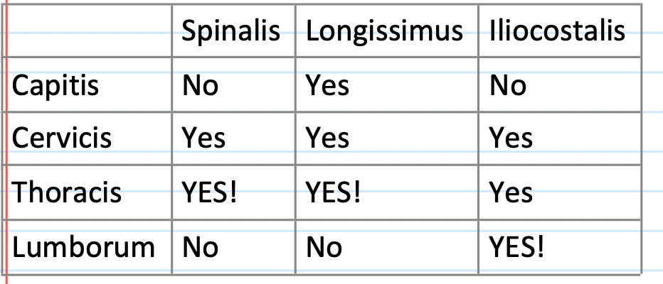
Inferior attachments of the iliocostalis regions
Capitis: n/a
Cervicis: angles of upper 6 ribs
Thoracis: angle of lower 6 ribs
Lumborum: posterior sacrum and iliac crest, lumbar SPs and supraspinous ligament (most important)
Inferior attachments of longissimus regions
Capitis: upper thoracic and cervical TP
Cervicis: Upper thoracic TP
Thoracis: Blends with iliocostalis lumborum and attaches to lumbar TPs (most important)
Lumborum: N/A
Inferior attachments of spinalis regions
Capitis: usually blends with semispinalis capitis
Cervicis: ligamentum nuchae
Thoracis: upper lumbar and lower thoracic SPs (most important)
Lumborum: N/A
Superior attachments of the iliocostalis region
Capitis: N/A
Cervicis: cervical TPs
Thoracis: Angles of upper 6 ribs
Lumborum: Angles of lower 6 ribs (most important - posterior ribs)
Superior (origin) attachments of longissimus regions
Capitis: posterior aspect of mastoid process
Cervicis: upper cervical TPs
Thoracis: thoracic TPs (most important - posterior sacrum)
Lumborum: N/A
Superior (origin) attachments of spinalis region
Capitis: usually blends with semispinalis capitis
Cervicis: C2 SP
Thoracis: upper thoracic SPs (most important)
Lumborum: N/A
Position and orientation of iliocostalis relative to joints crossed
Posteolateral to IVJs (vertical orientation)
Position and orientation of longissimus in relation to joints crossed
Posterolateral to IVJ’s (vertical orientation)
Position and orientation of spinalis in relation to joints crossed
Posterior to IVJs (vertical orientation)
Muscles in the transversospinalis region
semispinalis (5-6+ segments)
Multifidus (2-4 segments)
Rotators (1-2 segments)
What regions within the vertebral column are the transverse spinalis muscles found?
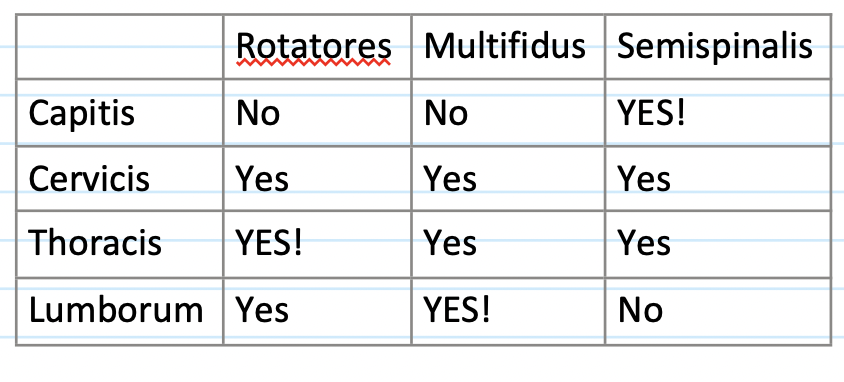
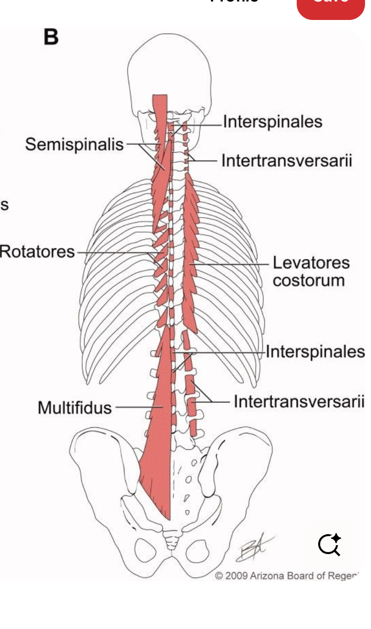
Name the regions of the transversospinalis

Name the regions of the transversospinalis and the erector spinae

Embryological origin and innervation of the transversospinalis
Epimere dorsi rami
Inferior attachment in the semispinalis region
Capitis: upper thoracic and lower cervical TPs (most important)
Cervicis: upper thoracic TPs
Thoracis: lower thoracic TP