week 11 lecture 2
1/34
There's no tags or description
Looks like no tags are added yet.
Name | Mastery | Learn | Test | Matching | Spaced | Call with Kai |
|---|
No analytics yet
Send a link to your students to track their progress
35 Terms
difference between MRI and CT
lack of radiation→ difference between MRI and CT, MRI has none
components of MRI
super conducting magnet
gantry (the giant tube shaped thing that houses everything)
RF coils around the patient (radiofrequency)
gantry contains the magnentic gradient coils
What do we do in MRI for different body parts
use different specialised coils
there are differing MRI scanner designs (e.g. open scanners for claustrophobia)
how are hydrogen atoms used in MRI
body is made up of water (Hydrogen and oxygen)
the behavior of these hydrogen atoms in a magnetic field is what is recorded
MRI uses hydrogen because it’s nucleus has a single proton and is abundant in the body→ when magnet. field is applied, the proton axis all line up, creating a magnetic vector along the MRI scanner
explain the MRI physics
the atoms line up→ radiofreq. pulses from the machine excites then relaxes the hydrogen atoms→ the resultant radio signals are computer processed to form an image
the different types of tissue can be distinguished due to relaxation times→ denser tissue has slower relaxation times
MRI sequences
by changing exam parameters, tissues can take different appearances
e.g. in T1,it is anatomically detatiled, with bright fat and black calcium/fluid
T2: fluid is bright and the muscles are dark
STIR: suitable for brightening fluid
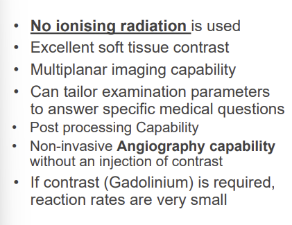
contrast media is used in MRI but not iodine based, it is gadolinium based
MRI advantges
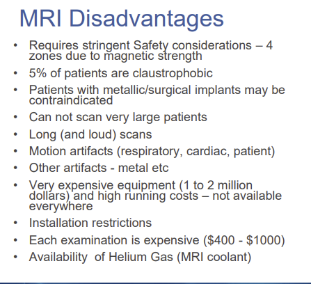
MRI disadvantages
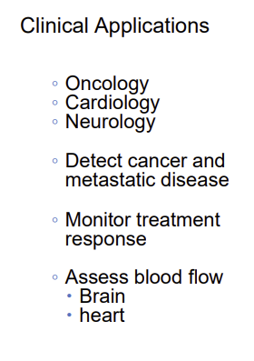
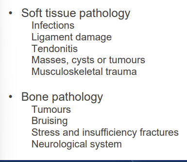
MRI indications (why u would need one)
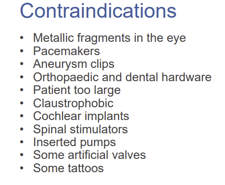
MRI contraindications (why u might not be able to get a MRI)
MRI safety considerations
magnetic strength:
require safety zones around the magnet with safety protocols (make sure we dont transfer metallic things into the area)
things like O2 tanks, mop buckets and crash trolleys have all been pulled into the magnet
patient protection:
need to complete a screenign questionaire
do a metal implant assessment
use ear protection
what is ultrasound
uses non-ionising radiation
the frequencies are above the range of human hearing
we use different frequencies for different body parts, with
- higher FREQ. having better spatial resolution but less tissue depth (they get absorbed quickly) thus they are used for superficial body parts
lower freq. petter penetration but less resolution→ used for deep organs
How does the ultrasound work
when the soundwave meets a interface, it can suffer reflecting or refractions
the reflect/refract varies based on tissue type/density
the images are created by the interpretation of sound reflections, where the image is made up on dots (each dot=echo of structure in patient)
component of ultrasounds
transducer
monitor for display
recording device
housing with controls
what does the transducer do in ultrasound
produces sound waves that interact with body tissue
reflected soundwaves also get sent back to transducer
houses piezoelectric crystal which is the transmitter of pulses and reciever of echos
also converts elect. into mechanical energy to transmit the pulses
converts mechanical energy into electrical. recieved echos
what is echogenicity?
how bright a tissue/structure is
thus how intense the echos are
three types:
anechoic→ no echos (black)
hypoechoic→ low echo, grey
hyperechoic (white, intense echo)
what is echotexture
the description/pattern of echo
can be homogeneous (even pattern)
heterogenous (mixed pattern)
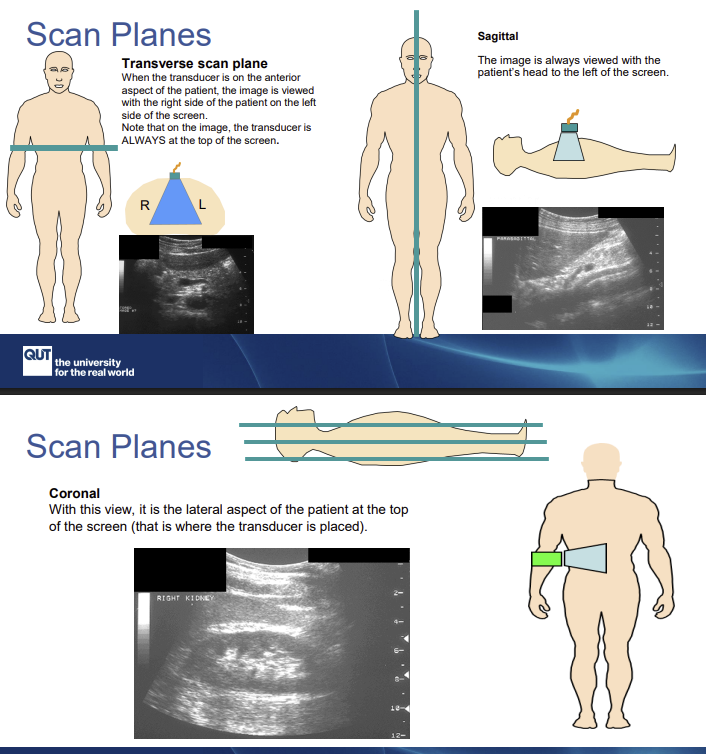
scan planes of sonography
doppler studies in ultrasound
shows directional info about vascular flow
blue→ direction of transducer
red→ away from transducer
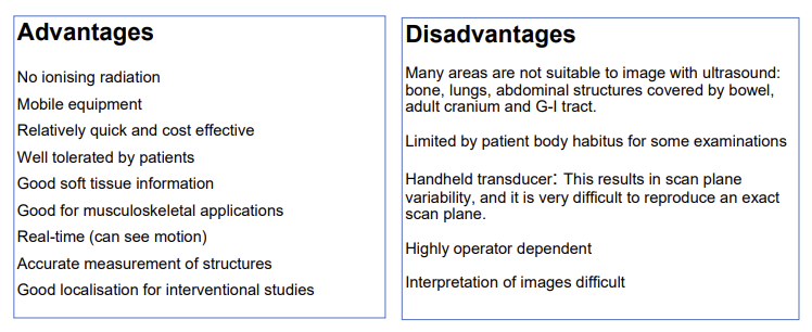
advantages and disadvantages of ultrasound (list 5 for each)
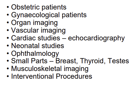
clinical indications for ultrasound
what is nuclear medicine
attaches radioisotopes to compounds. the compounds are then taken up by specifc organs (heart, kidneys) and areas of high metabolic activity (bones, tumours etc)
it is functional imaging, imaging the radiopharmaceutical distribution in a organ. it relied on microcirculation
the resolution and anatomical detail is poor
very sensitive imaging modality because it shows HOW the tissues are working rather than anatomical detail
often provides complementary info to other scans
what is molecular imaging
imaging modality that provides metabolic and functional info
when disease occurs the biochemical activity changes, and molecular imaging detects the celluar changes
list the two scans in nuclear medicine
PET scans
SPECT scans
what is SPECT scan
single photon emission computerized tomography
tracer (radiographic injection) put in, and SPECT machine used to scan specific area of body
tracer highlights blood flow
used to diagnose brain disorder, heart problems, bone disorders, aldo progression of cancer in bones
uses gamma emitting radioisotope for tracer
what is the image receptor used in nuclear medicine
called gamma cameras
it records the image of activity
detects the gamma coming from the body, doesn’t inject/emit anything
PET scans
a molecular imaging technique
uses positron emitting radioisotope (tracer)
uses tracer that emit positrons during decay→ position hits electron in body, they destroy each other and emit 2 photons which are picked up by detector
it demonstrates the metabolism and function of organs/identifies changes
use specific radioactive compounds for specific tissues
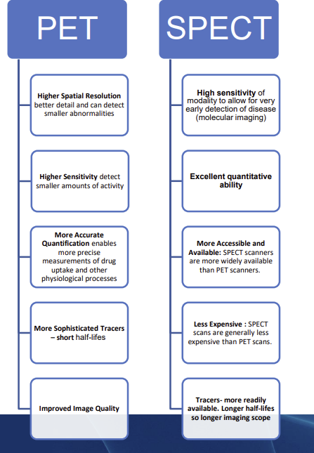
name 5 advantages of PET and SPECT scans
name disadvantages of nuclear medicine
● Low resolution images
● Although highly sensitive, NM is not specific
● NM departments are highly specialised with limited locations outside major population areas.
● Due to the long uptake time the whole procedure takes about 4 hours to complete (SPECT
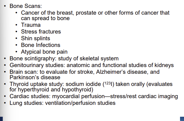
clinical indications for NM
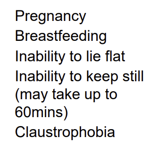
contraindications for NM
list the NM team
nuclear medicine technologist (handling and administers radionuclides)
nuclear medicine physician (interprets the NM procedures)
radiation sfety officer (reviews imaging protocols and dosimetry records
health physicist (calibrates and maintains the equipment)
list two hybrid techniques in moelcular imaging
PET/CT
mMR (PET/MRI)
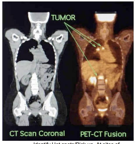
describe PET/CT hybrid technique
provides the detail of disorder and function information (PET)
and detailed image of anatomy (CT)
PET and CT are two completely different imaging systems built into one scanner, but they collect different types of information:
CT → anatomy (structure)
PET → physiology (function/metabolism)
Because they are acquired in the same session, the computer can align (co‑register/fusion) the PET and CT images so they fuse into one image
clinical applications of PET/CT