Brain lab - practical
1/67
There's no tags or description
Looks like no tags are added yet.
Name | Mastery | Learn | Test | Matching | Spaced | Call with Kai |
|---|
No analytics yet
Send a link to your students to track their progress
68 Terms
Neuron
Dendrites
Cell body
Axon hillock
Axon
Myelin Sheath
Node of Ranvier

blue
Dendrites

green
Cell body
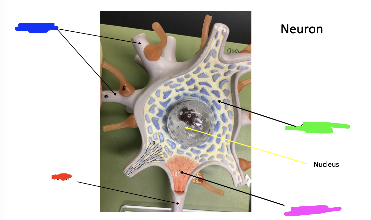
pink
Axon hillock

red
Axon

pink
Myelin Sheath
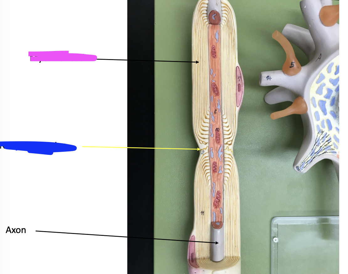
blue
Node of Ranvier
Axonal endings/Synaptic knob/Axon
Mitochondrion
neurotransmitters

blue
Mitochondrion
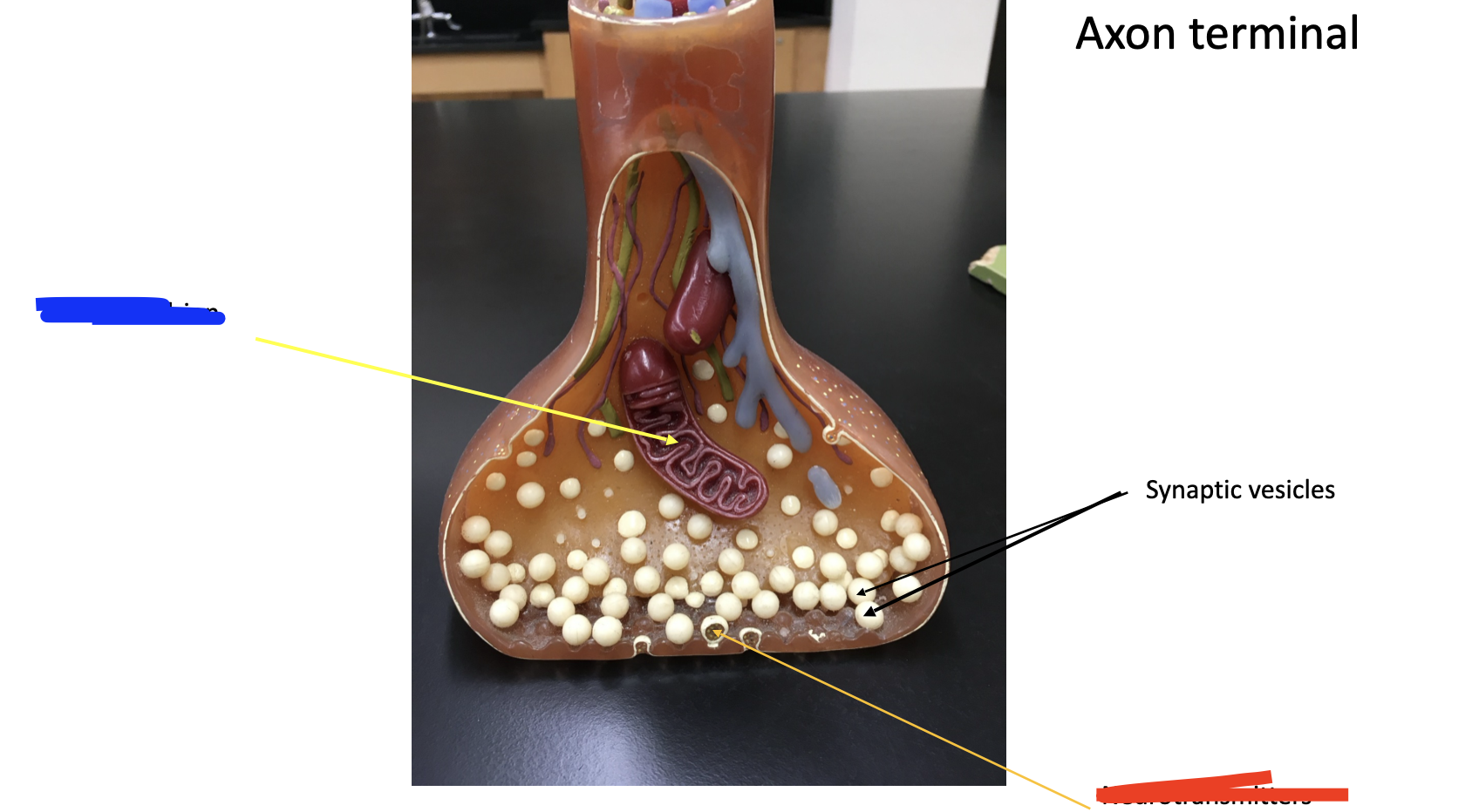
red
neurotransmitters
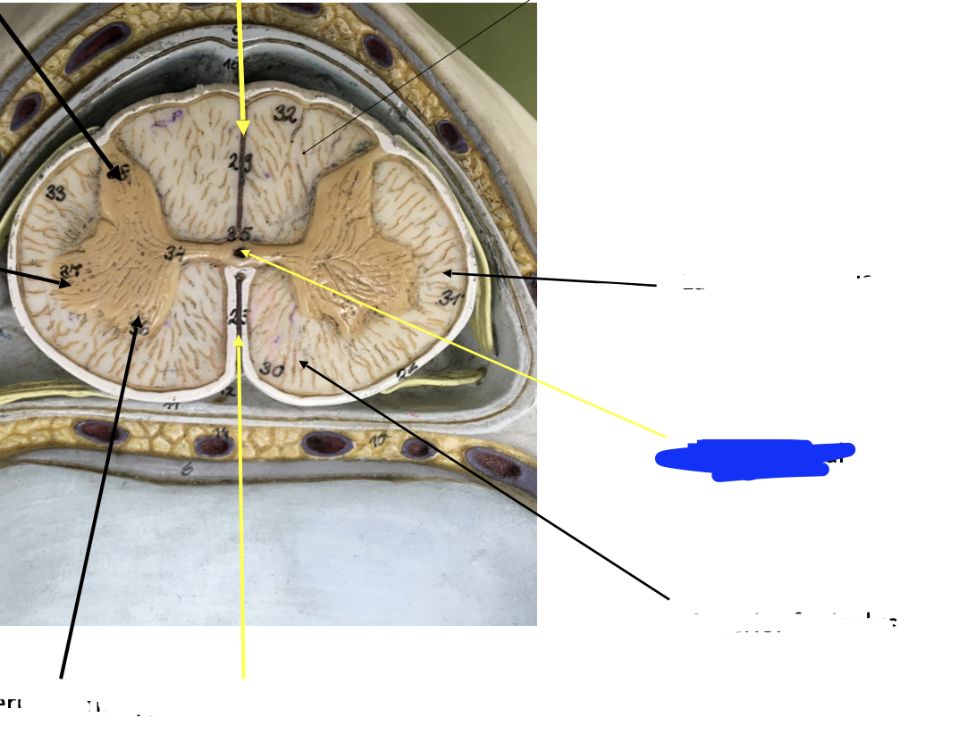
Spinal cord
anterior funiculus
posterior funiculus
lateral funiculus
anterior horn
posterior horn
lateral horn
central canal
posterior median sulcus
anterior median fissure
dorsal root of spinal nerve
dorsal root ganglion
ventral root of spinal nerve

red
anterior funiculus
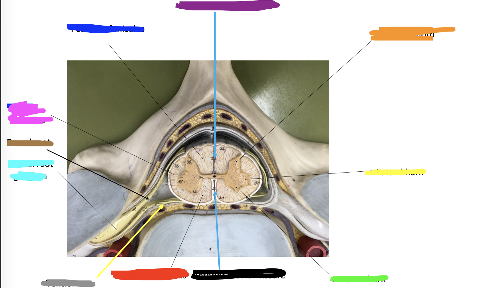
blue
posterior funiculus

pink
lateral funiculus

green
anterior horn

orange
posterior horn

yellow
lateral horn
blue
central canal

dark purple
posterior median sulcus

black
anterior median fissure

brown
dorsal root of spinal nerve

bright green
dorsal root ganglion

gray
ventral root of spinal nerve
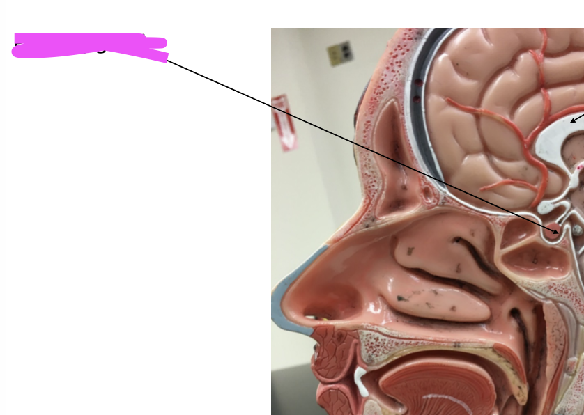
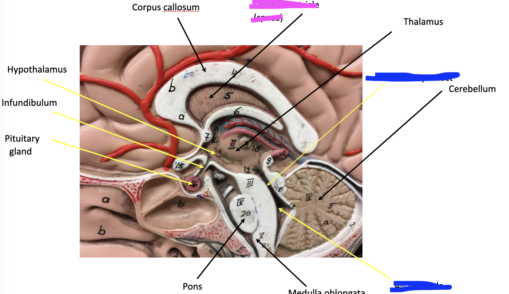
Brain
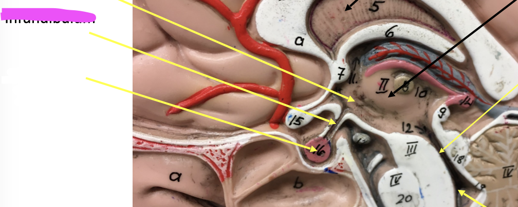
Pituitary gland
Infundibulum
Pink
Pituitary gland
Pink
Infundibulum
What does the Infundibulum connect to?
The pituitary gland and hypothalamus

Gyri (blue)
Elevated tissue ridges
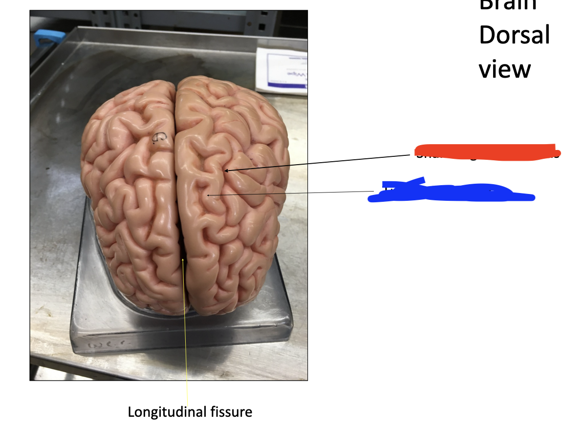
Sulcus (red)
Shallow grooves
Cerebrum
Frontal lobe
Parietal lobe
Temporal lobe
Occipital Lobe
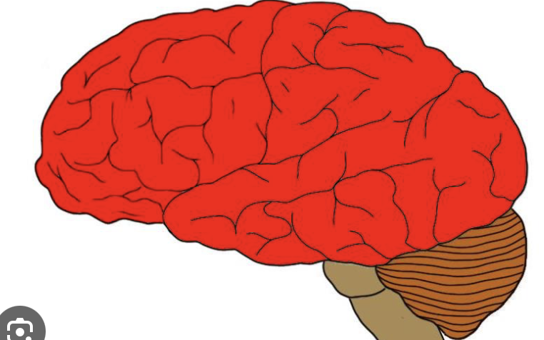
red
Cerebrum
Fissures - Deep grooves
longitudinal fissure
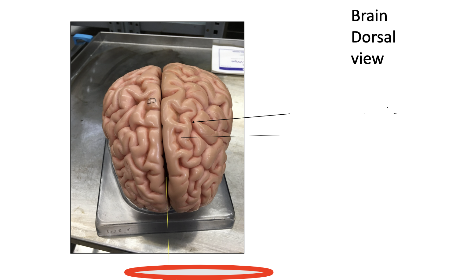
red
longitudinal fissure
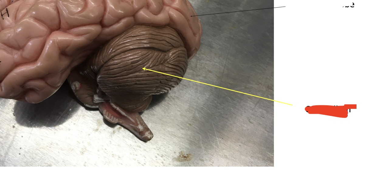
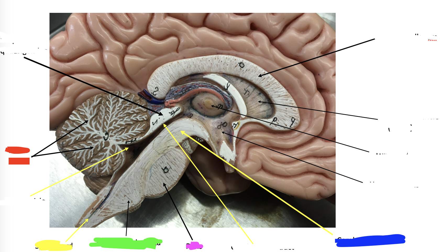
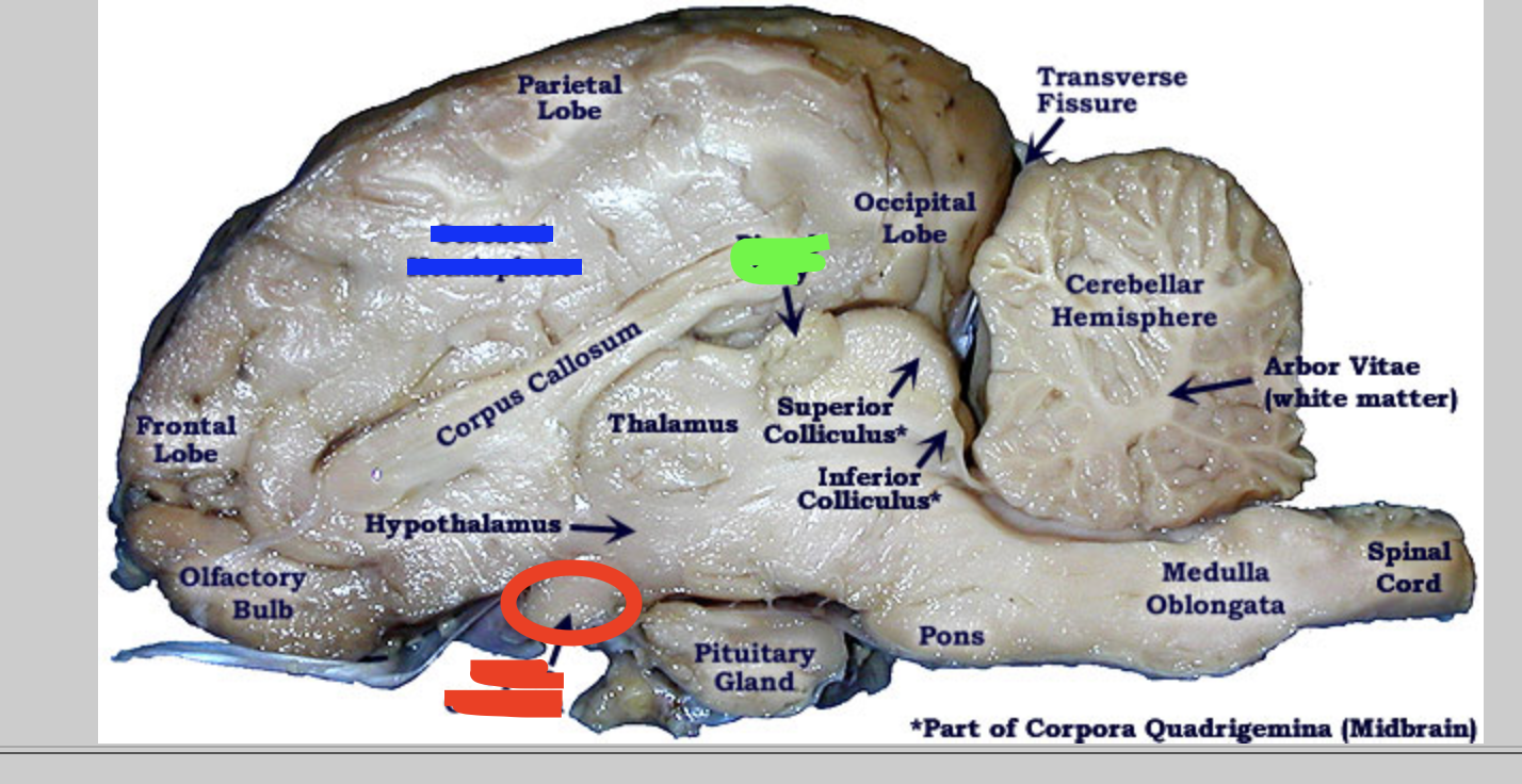
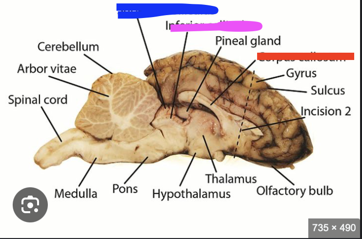
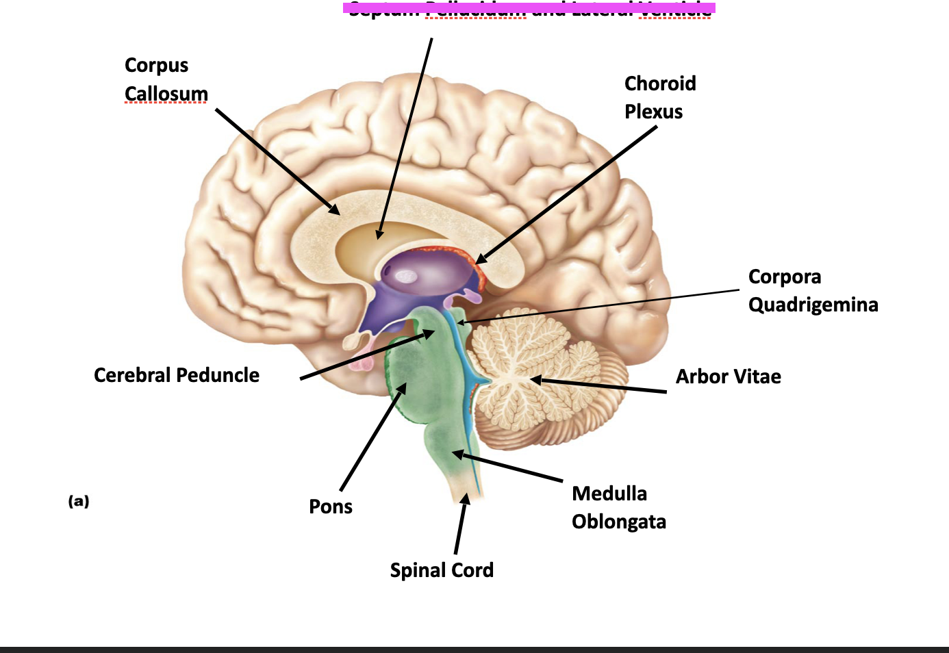
Cerebellum
Arbor vitae
Cerebral peduncles
Pons
Medulla oblongata
Spinal cord
red
Cerebellum

red
Arbor vitae

blue
Cerebral peduncles

pink
Pons

green
Medulla oblongata
yellow
Spinal cord
Cranial nerves
olfactory bulbs and tracts (I) - smell
optic nerves (II) - nerve visions
optic chiasma - off the midbrain
oculomotor nerves (III) - control most of eye movement
trochlear nerves (IV) - control one eye muscle
trigeminal nerves (V) - sensory: face & motor:chewing
abducens nerves (VI) - control one eye muscle

Cranial nerves I - yellow
olfactory bulbs and tracts
smell
I = 1

Cranial nerves II - blue
optic nerves
nerve visions
II = 2

red
optic chiasma - off the midbrain

Cranial nerves III - pink
oculomotor nerves
control most of eye movement
III = 3

Cranial nerves IV - green
trochlear nerves
control one eye muscle
IV = 4
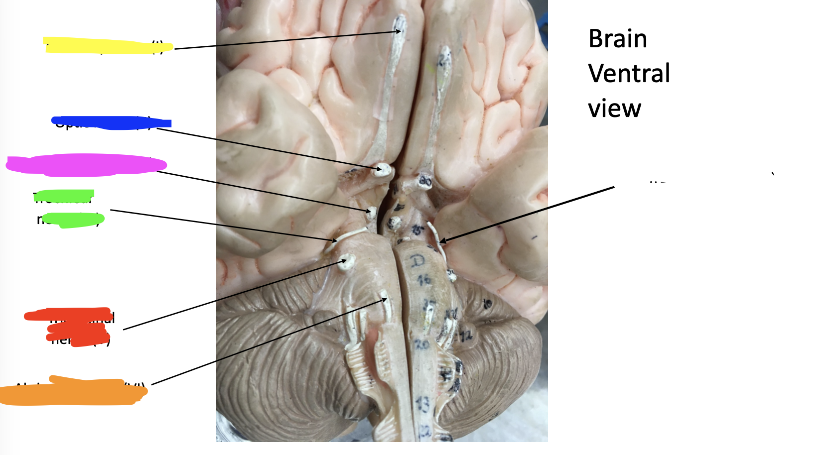
Cranial nerves V - red
trigeminal nerves
sensory: face & motor:chewing
V = 5

Cranial nerves VI - orange
abducens nerves
control one eye muscle
VI = 6
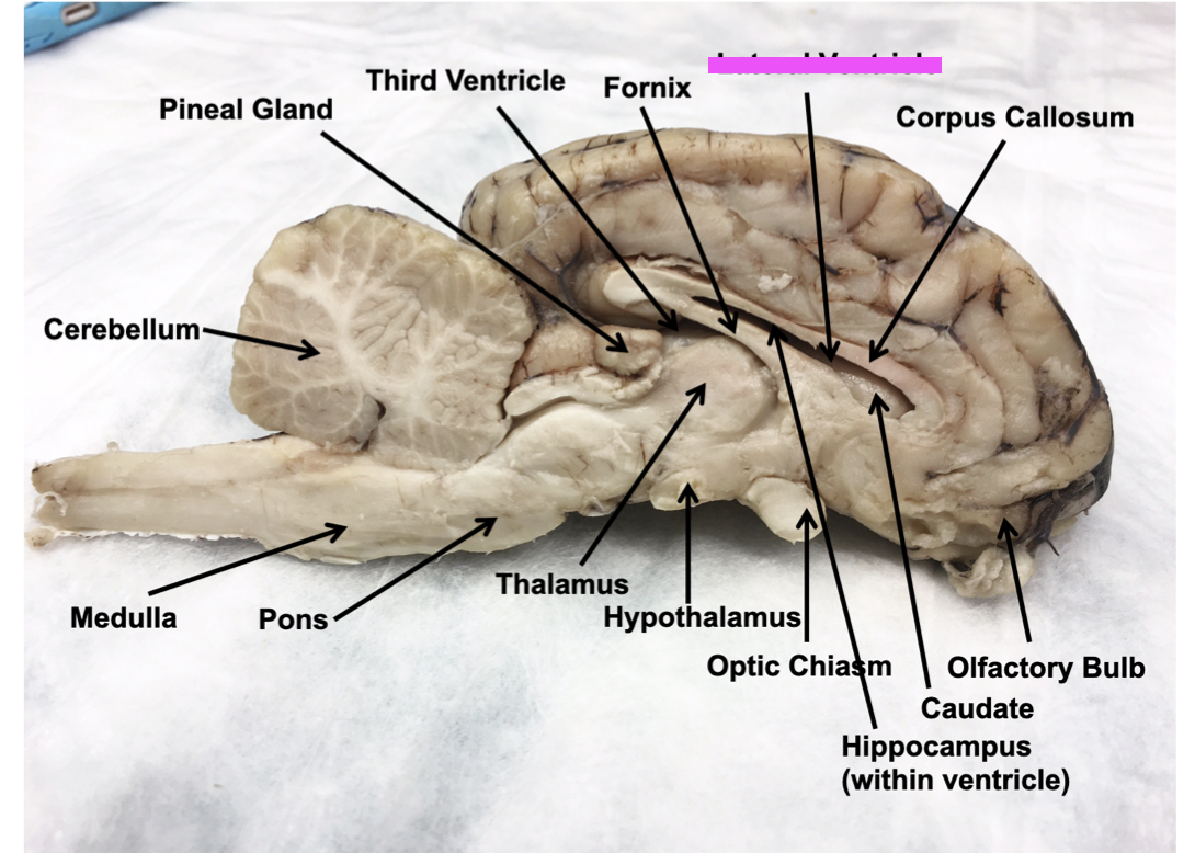
Mid - sagittal view
thalamus
hypothalamus
pineal gland (body) - on sheep brain
corpora quadrigemina - sheep brain
superior colliculi - sheep brain
inferior colliculi - sheep brain
lateral ventricle - sheep brain (space)
septum pellucidum - model (membrane)
Cerebral hemispheres -
corpus callousm
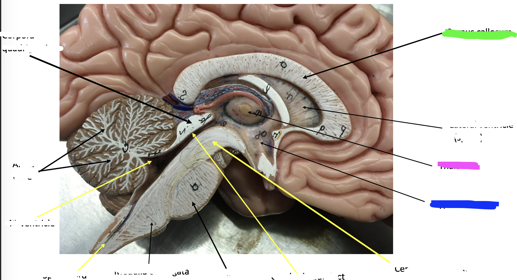
pink
thalamus

blue
hypothalamus
green
pineal gland (body) - on sheep brain

red
corpora quadrigemina - sheep brain

blue
superior colliculi - sheep brain
pink
inferior colliculi - sheep brain
pink
lateral ventricle - sheep brain (space)
pink
septum pellucidum - model (membrane)

blue
Cerebral hemispheres

green
corpus callousm
Meningies
dura mater
dura mater
outermost meninx
2-layered sheet of fibrous connective tissue
below: subdural space
Arachnoid
middle meninx
loose brain covering
does not dip into the sulci below:
subarachnoid space (origin of the arachnoid villi – for reabsorption of CSF)
Pia mater
innermost meninx
delicate tissue
tiny blood vessels
clings to brain
dips into the grooves
What is the corpora quadrigemina comprised of?
super colliculi & inferior colliculi
What is the midbrain comprised of?
cerebral peduncles & corpora quadrigemina. The cerebral aqueduct run through the midbrain
CSF and CFS flow
lateral ventricles
cerebral aqueduct - 4th ventricle
pink
lateral ventricles

blue
cerebral aqueduct - 4th ventricle