NPB 101 MT2 (Neurons+AP) L15-20
1/105
There's no tags or description
Looks like no tags are added yet.
Name | Mastery | Learn | Test | Matching | Spaced | Call with Kai |
|---|
No analytics yet
Send a link to your students to track their progress
106 Terms
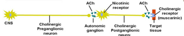
Acetylcholine is in which pathways?
Both the sympathetic and parasympathetic pathways
Autonomic Nervous System
Controls the function of our organs and glands; can be divided into the sympathetic and parasympathetic divisions
Complex network of cells that control the body’s internal state — plays a critical role in maintaining homeostasis
Regulates and supports many different internal processes, often outside of a person’s conscious awareness (Involuntary control)
Ganglion
A cluster of neuronal cell bodies outside of the CNS
Sympathetic pathway
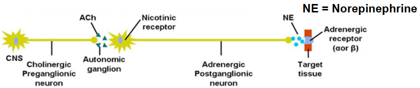
Parasympathetic pathway
Dorsal Root Ganglia
Clusters of neuron cell bodies that transmit signals from CNA to organs
Diffusion
The process of movement of molecules under a concentration gradient
Net diffusion
Difference between two opposing movements
Rate of diffusion through a membrane depends on the following factors:
Magnitude
Permeability
Surface area
Molecular weight
Distance
How does the magnitude of the concentration gradient relate to the rate of diffusion through a membrane?
↑ α ↑ rate of diffusion
How does the permeability of the membrane relate to the rate of diffusion through a membrane?
↑ α ↑ rate of diffusion
How does the surface area of the membrane relate to the rate of diffusion through a membrane?
↑ α ↑ rate of diffusion
How does the molecular weight of the substance relate to the rate of diffusion through a membrane?
↑ α ↓ rate of diffusion
How does the distance (membrane thickness) over which diffusion takes place relate to the rate of diffusion through a membrane?
↑ α ↓ rate of diffusion
Electrochemical gradient
Diffusion down a concentration (chemical) gradient
Movement along an electrical gradient
Movement along an electrical gradient
Electrostatic force (voltage) caused by the separation of electrical charge
Movement along an electrochemical gradient
The combined force of concentration (chemical) and electrical gradients
Neurons
Nerve cells specialized for electrical signaling over long distances (thanks to the long axon)
Soma has a nucleus
Electrically excitable and highly polarized
Membrane potential (mV)
A separation of opposite charges across the plasma membrane
Charge Separation Across a Membrane
Most fluid is electrically neutral
Separated charges form a layer along the plasma membrane
Immediately inside the plasma membrane are negative charge
How does the cell create charge separation?
Establishes and maintains concentration gradients for key ions (Na+, K+).
Ions diffuse through the membrane down their concentration gradients.
Diffusion through the membrane results in charge separation, creating a membrane potential (electrical gradient).
Net diffusion continues until the force exerted by the electrical gradient exactly balances the force exerted by the concentration gradient.
This potential that would exist at this equilibrium is “equilibrium potential.”
Equilibrium potential for K+
Because K+ is more ICF, K+ tends to move out of the cell.
The outside of the cell becomes more positive because K+ moves out.
The membrane is impermeable to the large intracellular protein anion. Therefore, the inside of the cell becomes more negative.
The resulting electrical gradient tends to move K+ into the cell.
No further net movement of K+ occurs when electrical gradient counterbalances concentration gradient.
The equilibrium potential of K+ at -90mV.
Equilibrium potential for Na+
The concentration gradient for Na+ tend to move into the cell.
The inside of the cell becomes more positive because Na+ moves in.
The outside becomes more negative as Na+ moves in, negatively charged ions, mostly Cl- is outside.
Therefore, Na+ move out of the cell.
No further net movement of Na+ occurs when the electrical gradient exactly counterbalances the concentration gradient.
The equilibrium potential for Na+ at +60mV.
Nernst Equation
Describes the equilibrium potential for a particular ion (i)
Ei = RT/zF ln [i]o/[i]i
Where R is the gas constant, T is the temperature in degrees Kelvin, z is the valence of the ionic species, and F is the Faraday constant.
Is sodium more abundant extracellularly or intracellularly?
Extracellularly
Is potassium more abundant extracellularly or intracellularly?
Intracellularly
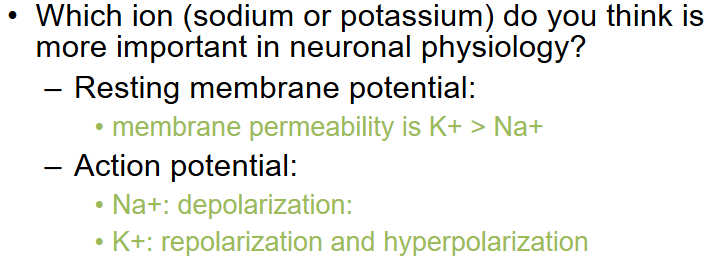
Why is the resting membrane potential closer to the K+ potential than the Na+ potential?
The membrane is 20-30x more permeable to K+ than Na+, so there is a large net diffusion of K+ and a small net diffusion of Na+ (which neutralizes some of the potential created K+)
How do K+ and Na+ penetrate the cell membrane?
Leak channels — permit ions to flow down concentration gradients passively (without ATP use)
Why is Na+ higher outside of the cell and K+ higher inside?
Na/K ATPase — establishes and maintains concentration gradients. Specifically pumps 3 Na+ out of the cell for every 2 K+ pumped into the cell
Resting Membrane Potential
Has a balance of passive leak channels and active Na/K ATPase
Neither K+ nor Na+ is at their equilibrium potentials
Concentration gradients and permeabilities for Na+ and K+ remain constant
What would happen to a cell’s membrane potential if the cell was deprived of ATP?
Na/K+ ATP would not be functional → no membrane potential
Homeostatically Regulated Factors
Nutrients
O2 and CO2
Waste products
pH
Water, Salt, and other electrolytes
Volume and pressure
Temperature
Direct intercellular communication
Gap junctions
Transient direct linkup of cells’ surface market
Endocrine signaling
Acts via hormones and neurohormones secreted into the blood to control processes that rely on duration rather than speed
Main regulatory systems of the body
Nervous system
Endocrine system
Nervous System
Anatomic Arrangement: A “wired” system: a structural arrangement exists between neurons and their target cells, with structural continuity in the system
Type of chemical messenger: neurotransmitters released into the synaptic cleft
Distance of action of the chemical messenger: short distance (diffuses across the synaptic cleft)
Specificity of action on the target cell: dependent on the close anatomic relationship between neurons and their target cells
Speed of response: rapid (milliseconds)
Duration of action: brief (milliseconds)
Major functions: coordinates rapid, precise responses
Endocrine System
Anatomic Arrangement: A “wireless” system: glands are widely dispersed and not structurally related to one another or to their target cells
Type of chemical messenger: hormones released into the blood
Distance of action of the chemical messenger: long distance (carried by the blood)
Specificity of action on the target cell: dependent on the specificity of target cell binding and responsiveness to a particular hormone
Speed of response: slow (minutes to hours)
Duration of action: long (minutes to days or longer)
Major functions: controls activities that require long duration rather than speed
Organization of the nervous system
CNS (brain and spinal cord)
PNS (nerve fibers; has afferent and efferent divisions)
Enteric Nervous System — ENS (nerve network of the digestive tract)
Organization of the Efferent Division of the PNS
Somatic nervous system: fibers of the motor neurons that supply the skeletal muscles
Autonomic nervous system: fibers that innervate smooth muscle, cardiac muscle, and glands
Both receive information from efferent division (PNS)
Somatic Nervous System
Fibers of the motor neurons that supply the skeletal muscles
Receive information from PNS efferent division
Autonomic Nervous System
Fibers that innervate smooth muscle, cardiac muscle, and glands
Receive information from PNS efferent division
Consists of a two-neuron chain
Subdivided into the Sympathetic and Parasympathetic nervous systems
Parasympathetic Nervous System
Division of the autonomic nervous system that maintains resting functions of the internal organs.
“Maintaining homeostasis”
Sympathetic Nervous System
Division of the autonomic nervous system that prepares the body for strenuous physical activity.
“Fight or flight response”
Is eating food a part of the autonomic sympathetic division or parasympathetic division?
Sympathetic
Is digesting food a part of the autonomic sympathetic division or parasympathetic division?
Parasympathetic
Integration of the stress response by the hypothalamus
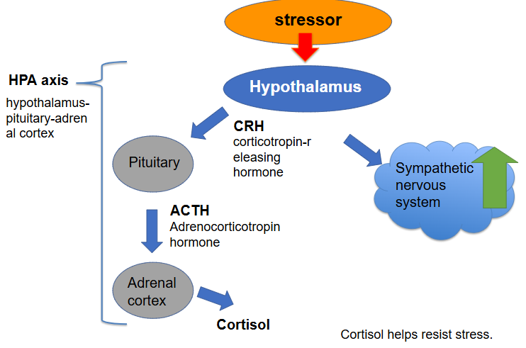
HPA Axis
Hypothalamus-pituitary-adrenal cortex
Stressors activate which system?
Sympathetic
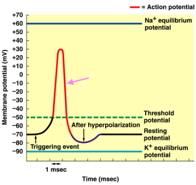
Depolarization
Change in membrane polarization to more positive values than resting membrane potential
Hyperpolarization
Change in membrane polarization to more negative values than resting membrane potential
Repolarization
Change in membrane polarization back to the original polarity
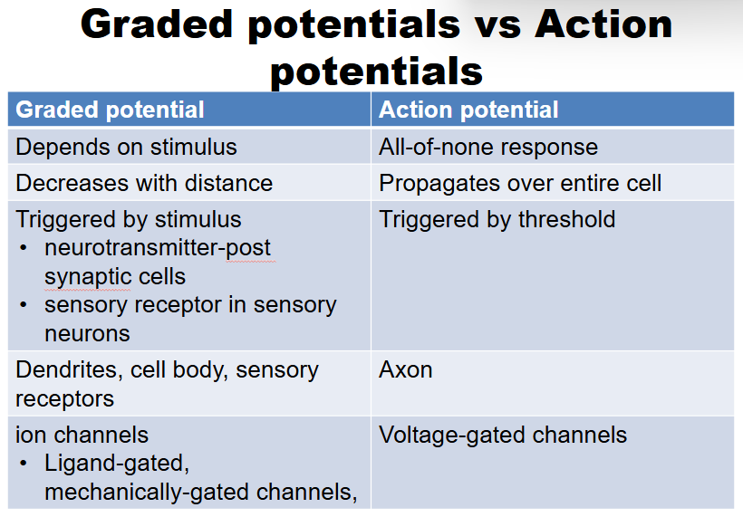
Action Potential
Brief all-or-nothing reversal in membrane potential (spike), lasting on the order of 1 millisecond, that is brought about by rapid changes in membrane permeability to Na+ and K+ ions.
Once initiated in the axon hillock, action potentials are conducted the length of an axon; do not decay with distance
Refractory period ensures one-way propagation of action potentials and limits their frequency (cannot be initiated in a region that has just undergone an AP)
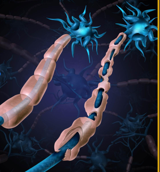
Myelination increases the speed of conduction of AP saltatory conduction
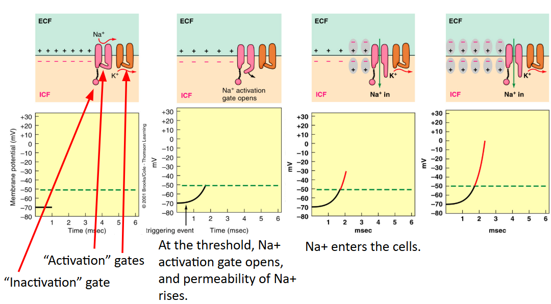
Rising phase of Action Potential
Voltage-gated Na+ channel - opens quickly (< 0.5 ms) in response to depolarization, allowing Na+ to flow down its electrochemical gradient into the cell

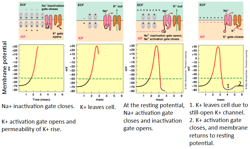
Falling phase of Action Potential
Voltage-gated K+ channel - opens slowly in response to depolarization allowing K+ ions to flow out of the cell down their electrochemical gradient
What are the mechanisms that control these Na+ and K+ specific movements?
Voltage-gated Na+ and K+ channels
Climax of Action Potential
↓ PNa+ ↑PK+
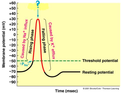
Events Underlying the Rising Phase of the Action Potential
Events Underlying the Falling Phase of the Action Potential
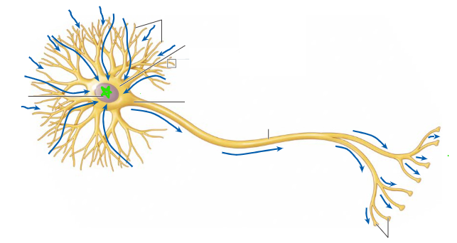
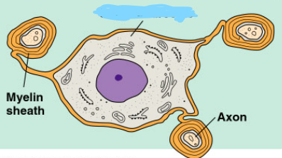
Dendrites
Input zone of neuron; receives incoming signal
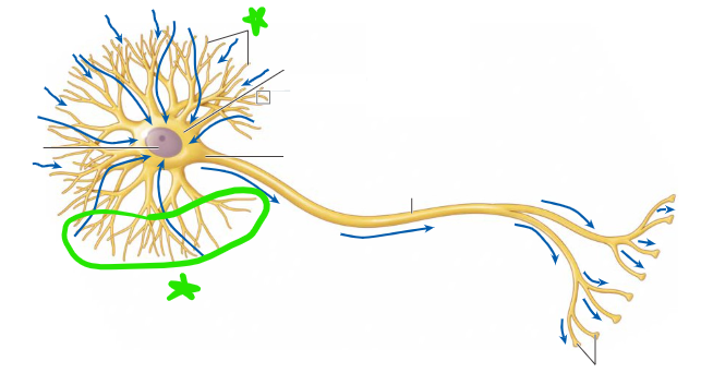
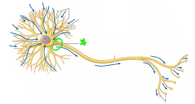
Soma
Cell body of neuron
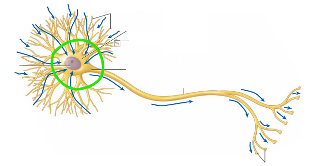
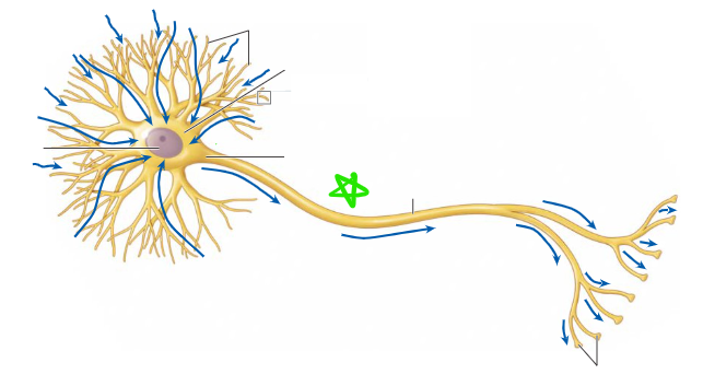
Axon hillock
Trigger zone of neuron, initiates action potentials
Axon
Long tail of neuron
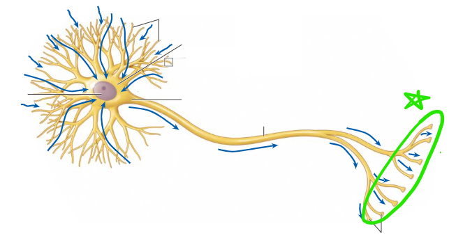
Axon terminals
Output zone of neuron, releases neurotransmitters
Neuron nucleus
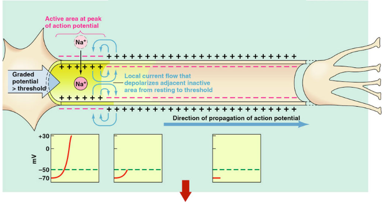
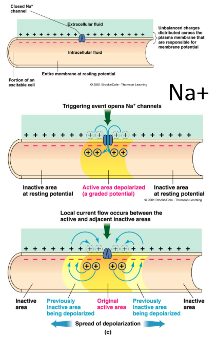
Action potential propagation
Occurs when locally generated depolarizing current spreads to adjacent regions of membrane causing it to depolarize
The original active area returns to resting potential, and the new activate area induces an action potential in the next adjacent inactive area. The cycle repeats itself down the length of the axon.
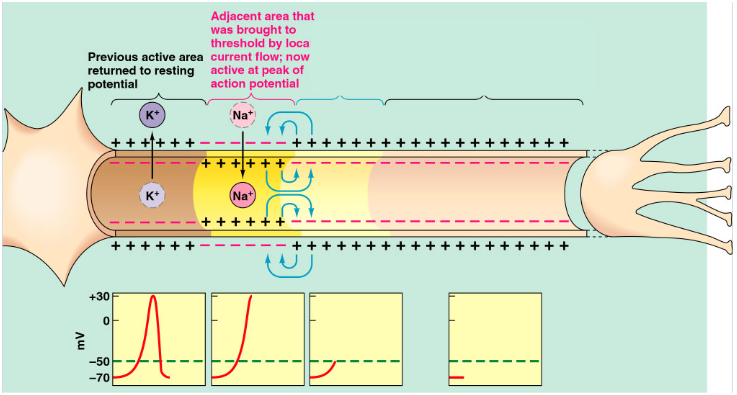
Contiguous conduction
Propagation of action potentials in unmyelinated fibers by spread of locally generated depolarizing current to adjacent regions of membrane, causing it to depolarize
Absolute refractory period
A brief period during a spike
Repolarization: Voltage Gated Na+ channel inactivation gate closes
A second spike cannot be generated
Relative refractory period
A brief period following a spike
Below resting membrane potential: Voltage Gated Na+ channel inactivation gate opens
Capable of opening in response to depolarization
Hyperpolarization: a higher intensity stimulus is needed
Contiguous conduction (of action potentials through a nerve fiber)
Unmyelinated fibers: touching, next to in sequence
Saltatory conduction (of action potentials through a nerve fiber)
Propagation of action potentials in myelinated axons by jumping from node to node, skipping over the myelinated sections of the axon
Faster conduction propagation!
How does the refractory period ensure the one-way communication of action potentials?
Action potential cannot be initiated in a region that has just undergone an action potential
Myelin
A multilayered sheath of plasma membrane, derived from specialized glial cells, that wraps around axonal fibers and acts as an insulator to the flow of current
Nodes of Ranvier
Gaps in myelin insulation containing high densities of voltage-gated Na+ and K+ channels
Schwann cells
Myelin-forming glial cells in the peripheral nervous system
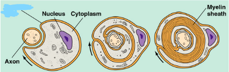
Oligodendrocytes
Myelin-forming glial cells in the central nervous system
Multiple Sclerosis (MS)
1 out of 1000 patients in the US
MS begins between ages 20 and 40
An autoimmune disease (the body’s defense system attacks the myelin sheath.)
Slow transmission of impulse in the affected neurons
Graded potentials
Local changes in membrane potential
Occur in varying grades or degrees of magnitude or strength (size correlates with stimulus)
Spread by passive current flow
Current: any flow of electrical charges
Resistance: hindrance to electrical charge movement
Die out over short distances
Synapse
Junction between two neurons, or between a neuron and a muscle or gland that enables one cell to electrically and/or biochemically influence another cell
Electrical synapse
Neurons connected directly by gap junctions
Chemical synapse
Most synapses in the human nervous system
Chemical messenger transmits information one way across a space separating the two neurons
Gap junctions
Made up of multiple proteins called connexins (a large family of trans-membrane proteins that allow intercellular communication and the transfer of ions and small signaling molecules between cells)
The small diameter of the “tunnel” permits water-soluble particles (such as ions) to pass between cells but blocks the passage of larger molecules.
Convergence
The synaptic input of many neurons into one neuron
Divergence
The synaptic output of one neuron onto many neurons
Synaptic transmission
Primary means of rapid inter-neuronal communication in the brain
Presynaptic axon initiates the signal
Neurotransmitter carries the signal across a synapse; binds to postsynaptic receptors
Postsynaptic (target) cell receives the signal
targets can be a muscle, gland, or another neuron
Postsynaptic targets
Muscles, glands, other neurons
Sequence of events of a Chemical Synapse
AP propagation in presynaptic neuron
Ca+ entry into synaptic knob (terminal button?)
Release of neurotransmitter by exocytosis
Binding of neurotransmitter to postsynaptic receptor
Opening of specific ion channels in subsynaptic membrane
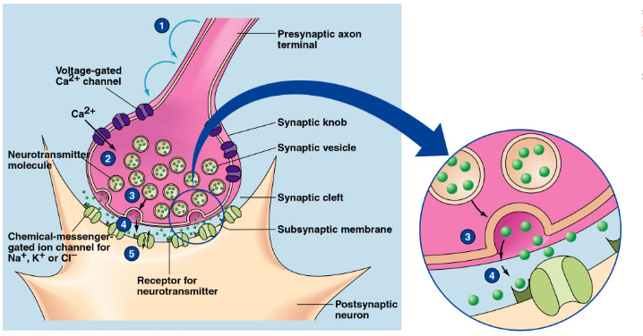
Presynaptic release (chemical synapse)
Voltage-gated Ca++ channels
Synaptic vesicles
Postsynaptic response (chemical synapse)
Postsynaptic receptors
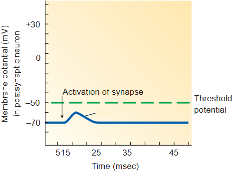
Postsynaptic potential (PSP)
Excitatory postsynaptic potential (EPSP)
Most common excitatory neurotransmitters are glutamate (Glu) and acetylcholine (ACh)
Depolarizing potential that brings mV towards threshold for generation of an action potential
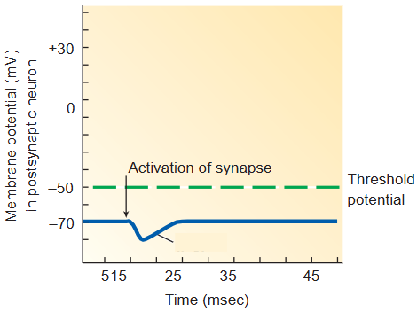
Inhibitory postsynaptic potential (IPSP)
Most common inhibitory neurotransmitters are gamma-amino butyric acid (GABA) and glycine (Gly)
Hyperpolarizing potential that brings mV away from threshold for generation of an action potential
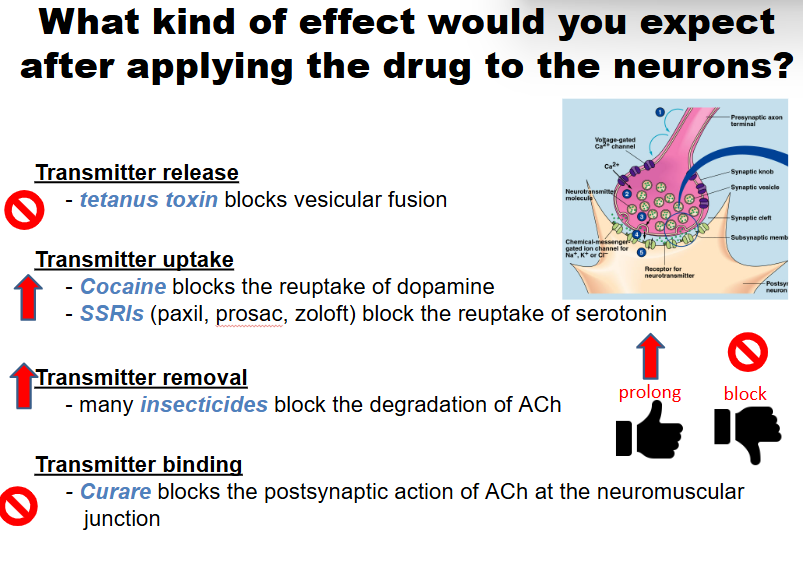
(Neuro)Transmitter removal
Degradation — enzymatic breakdown (ie: AChE)
Transport — active transport back into the presynaptic cell “reuptake”
Diffusion — the transmitter simply diffuses away from the synaptic terminal
Degradation Transmitter removal
Enzymatic breakdown (ex: AChE)
Transport Transmitter removal
Active transport back into the presynaptic cell “reuptake”
Diffusion Transmitter removal
The transmitter simply diffuses away from the synaptic terminal
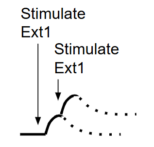
Temporal summation
The additive effect of PSPs (Post-Synaptic Potential) occurring close together in time
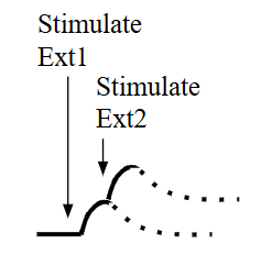
Spatial summation
The additive effect of PSPs (Post-Synaptic Potential) occurring together on nearby parts of the same cell
Cancellation summation
EPSP and IPSP cancel each other out