Neuroanatomy (Exam 4) Main Study Guide SPAUD-210
1/163
There's no tags or description
Looks like no tags are added yet.
Name | Mastery | Learn | Test | Matching | Spaced | Call with Kai |
|---|
No analytics yet
Send a link to your students to track their progress
164 Terms
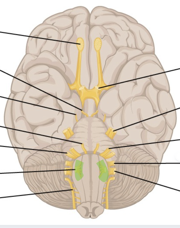
olfactory nerve
CN 1
snakes on bottom of frontal lobe
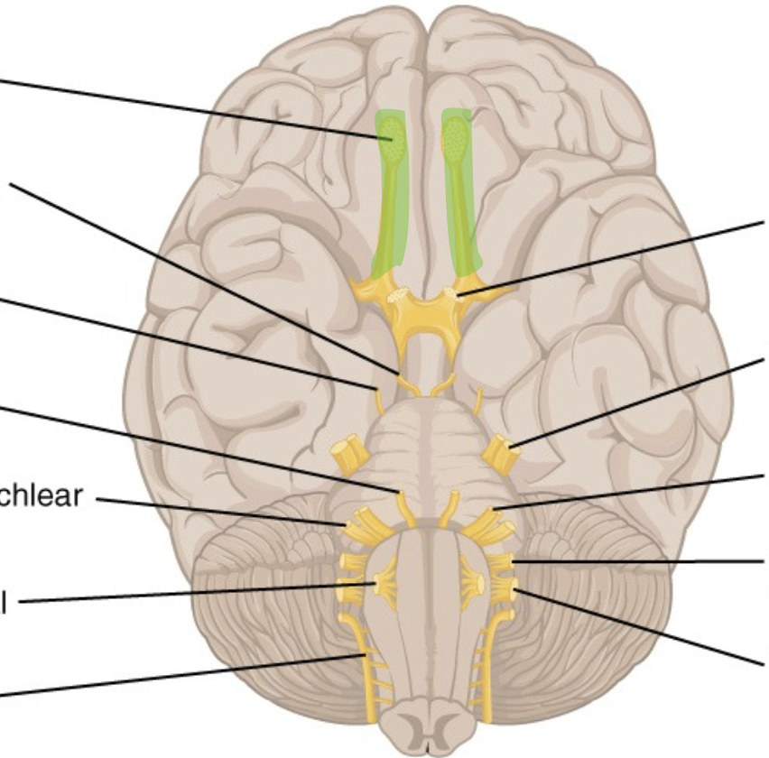
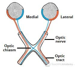
optic nerve
CN 2
snail eyes superior to pons
oculomotor
CN 3
inferior to CN 2, pokes out
troclear
CN 4
lateral on top of pons
trigeminal
CN 5
fat, the middle of pons
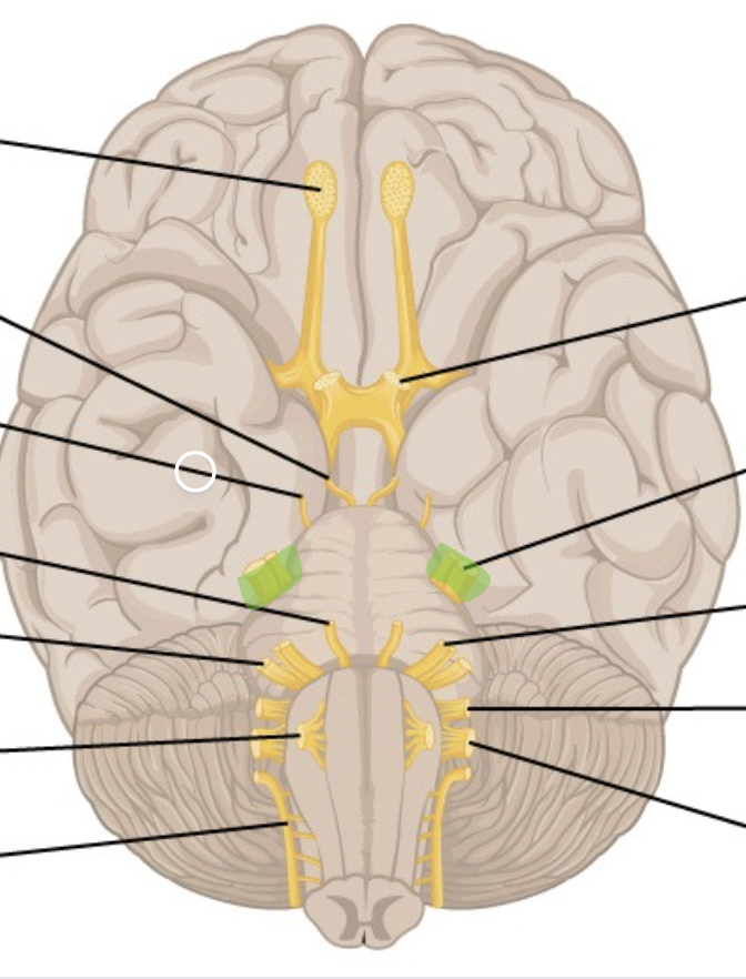
abducens
CN 6
most medial of 6, 7, & 8
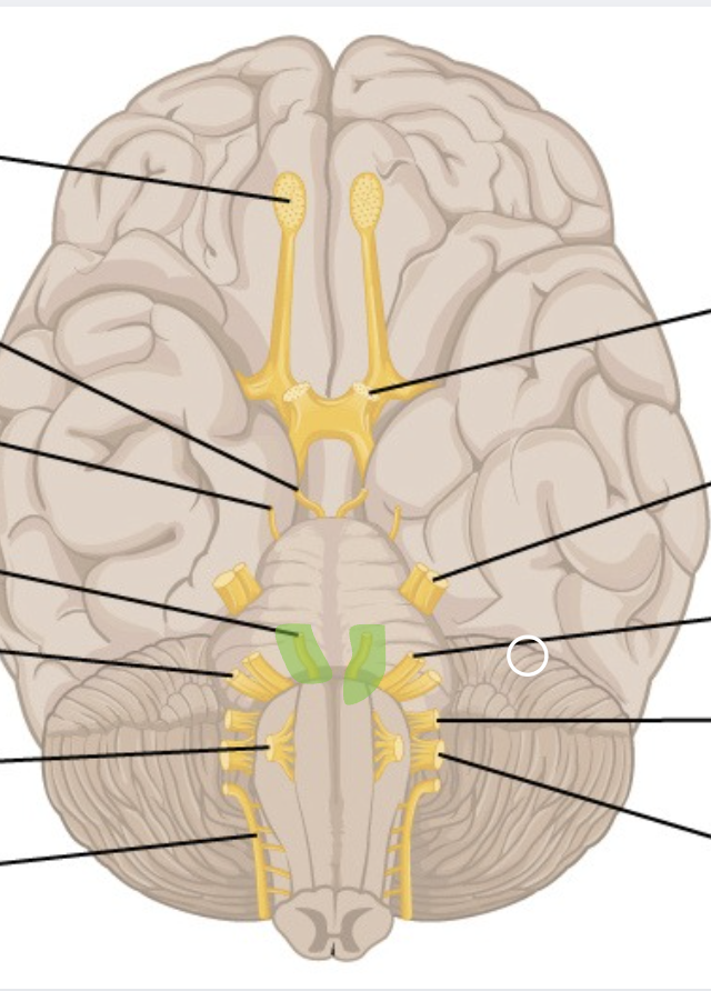
facial
CN 7
lateral to 6, medial to 8
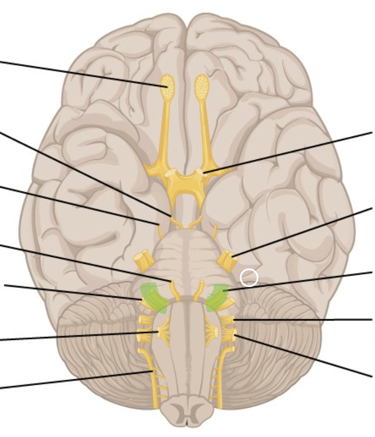
vestibulocochlear
CN 8
most lateral to 6 & 7
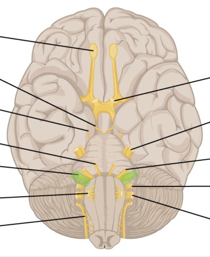
glossopharyngeal
CN 9
most superior to 9 & 10
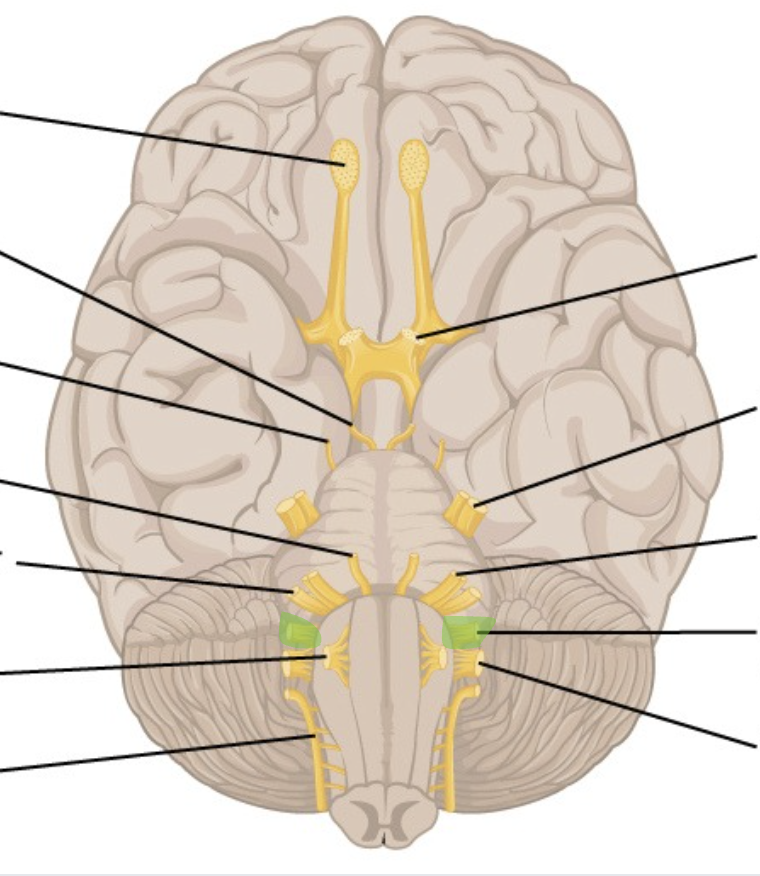
vagus
CN 10
inferior to 9, superior to 11
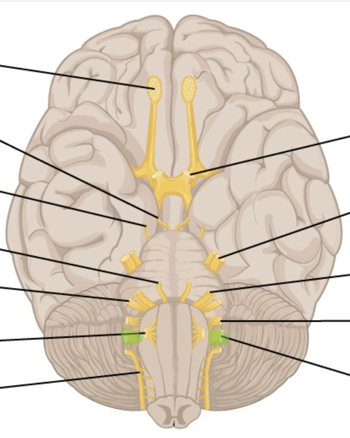
spinal accessory
CN 11
inferior to 9 & 10
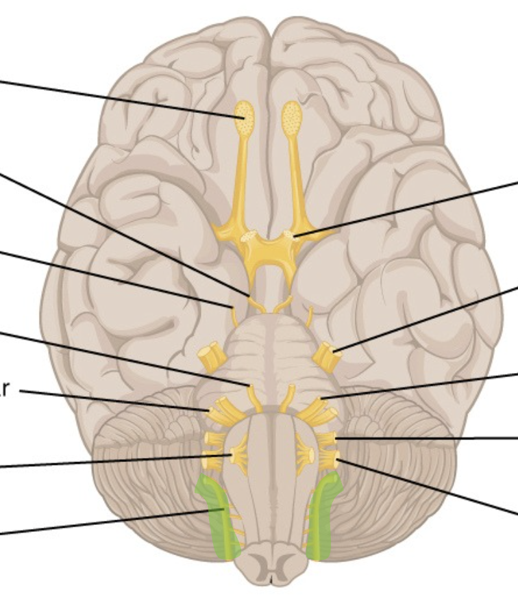
hypoglossal
CN 12
medial to 9, 10, & 11
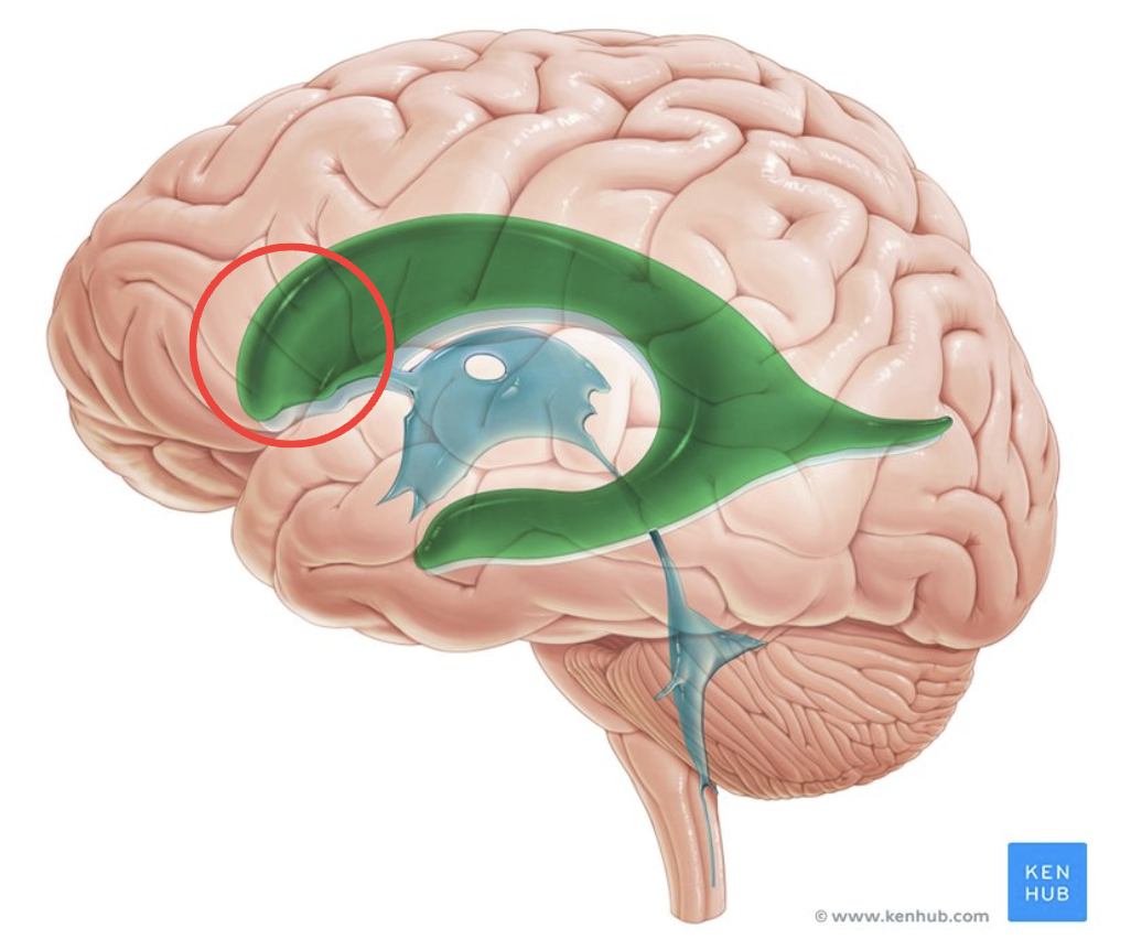
anterior horn of lateral ventricle
front horns, sharp
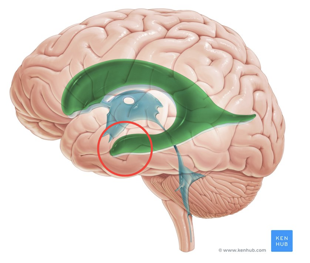
inferior horn of lateral ventricle
bottom one
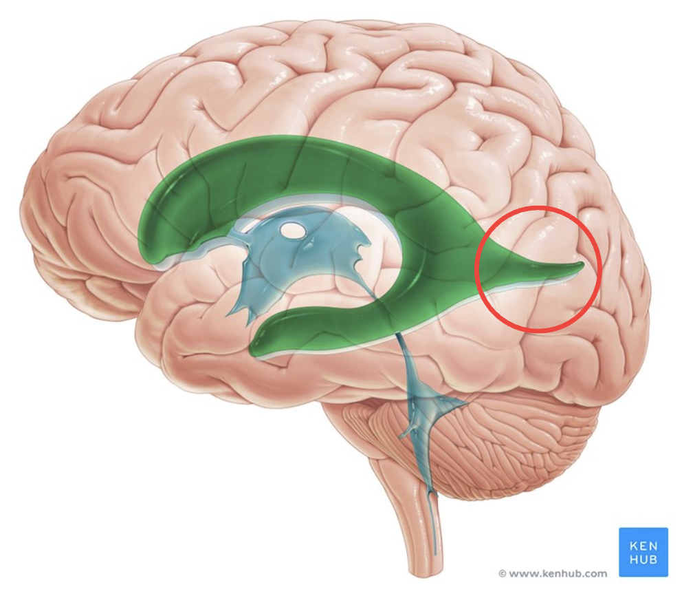
posterior horn of lateral ventricle
back one, pointy
third ventricle
cerebral aqueduct

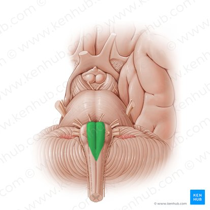
fourth ventricle
triangle shape
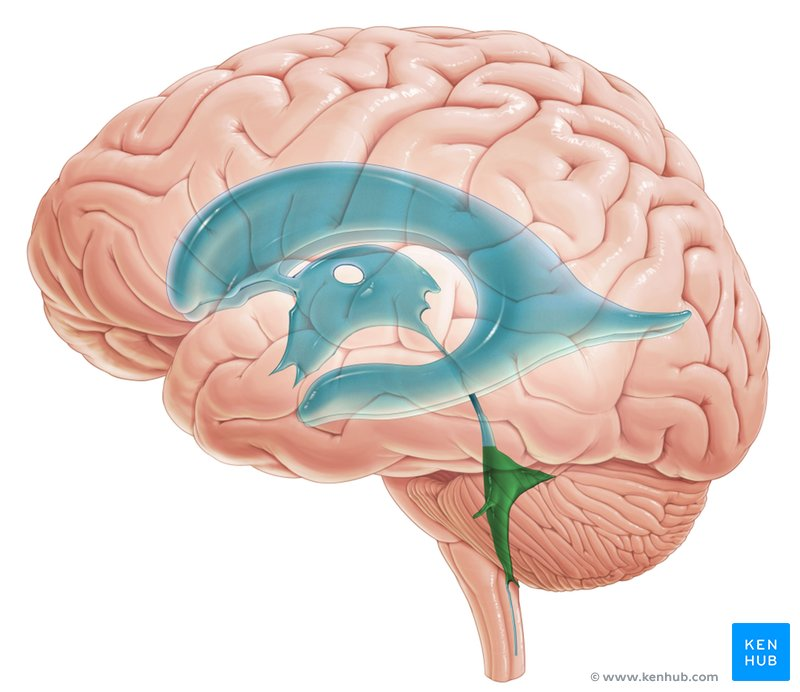
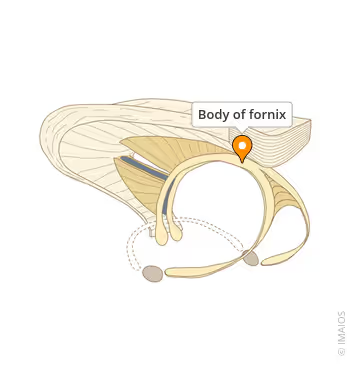
body of fornix
triangle where 3 points connect
choroid plexus
blood vessels in ventricles, on hippocampus
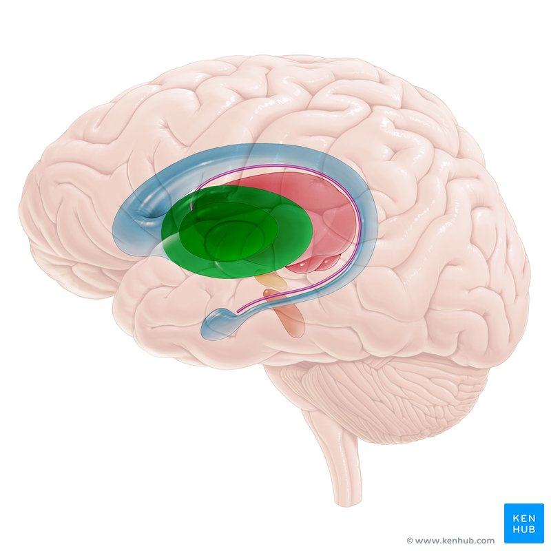
hippocampus
two sides coming off fornix
flocculus
little middle bumps on cerebellum, purple on image
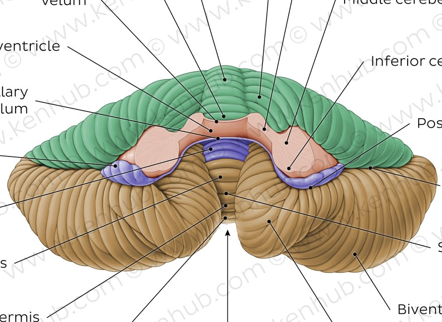
cerebellar tonsil
big bumps on posterior side of cerebellum
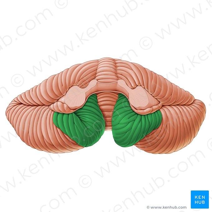
vermis of cerebellum
worm-like line on cerebellum
left island of Reil
also called insular cortex, left shell
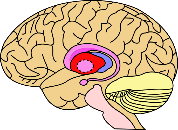
caudate nucleus
top arch
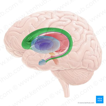
putamen
red on image, below top arch

pallidum
more medial bean, outline & pink
right island of Reil
right side

lentiform nucleus
made up of putamen and pallidum
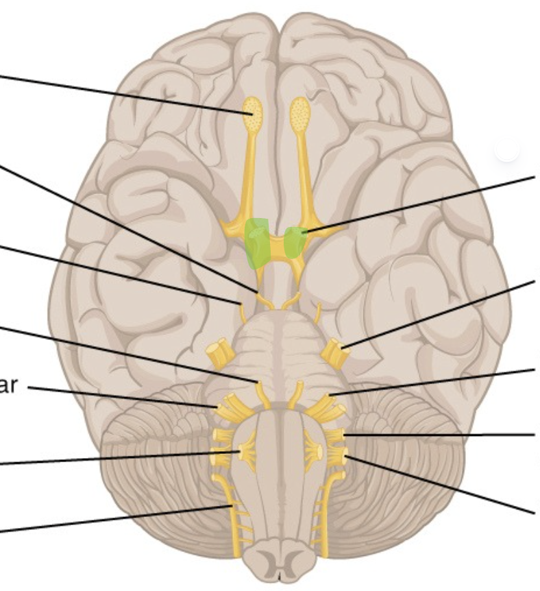
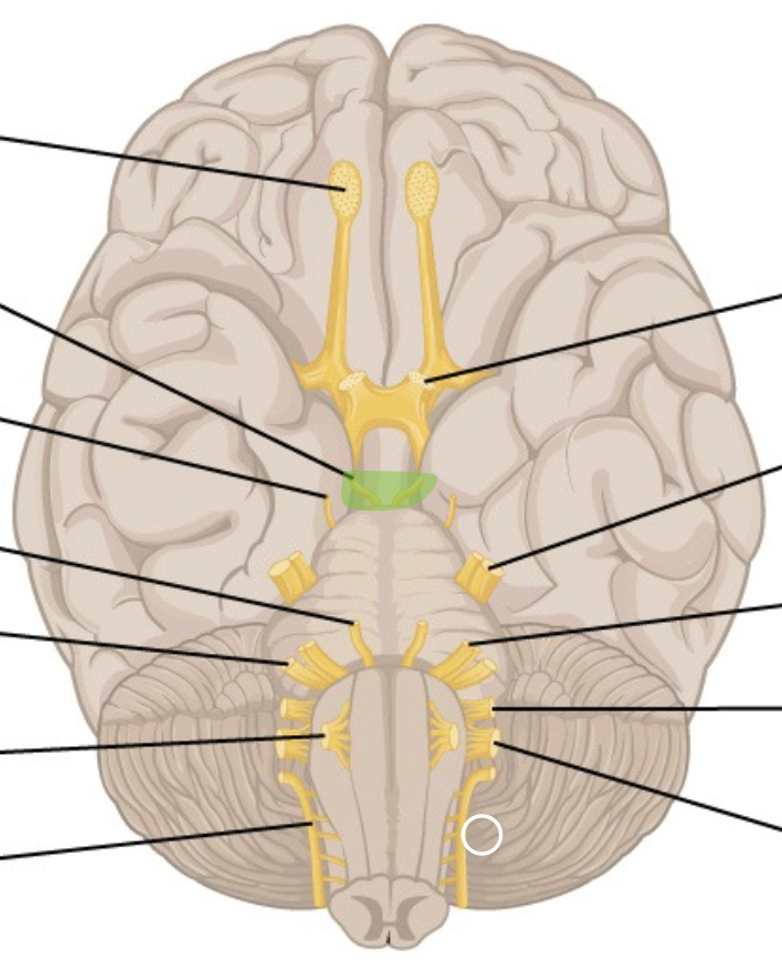
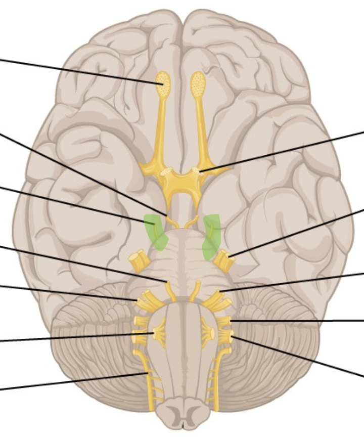
thalamus
oval bulge connected to brainstem

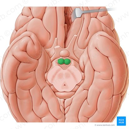
olives
lateral bumps on medulla, green on image
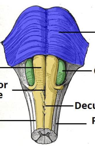
pyramids
medial to olives
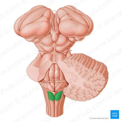
tubercle of nucleus gracilis
y-shaped stems, on medulla, most medial
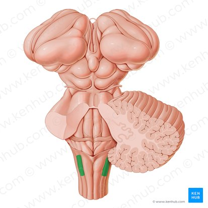
tubercle of nucleus cuneatus
lateral to nucleus gracilis
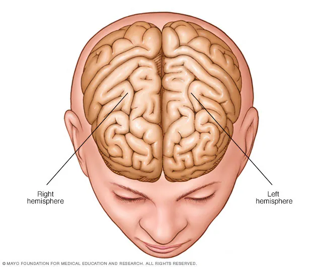
cerebral cortex
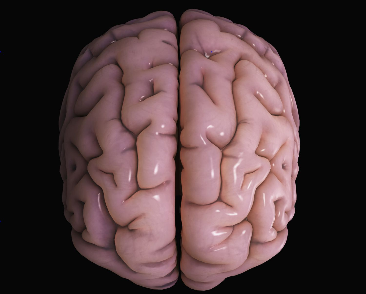
right and left hemispheres
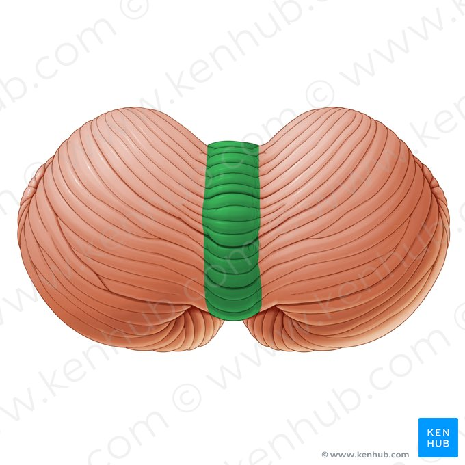
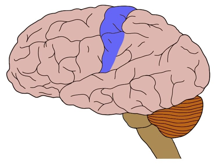
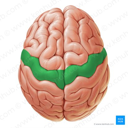
motor area
thin band, medial but slightly toward front

sensory area
next to motor cortex
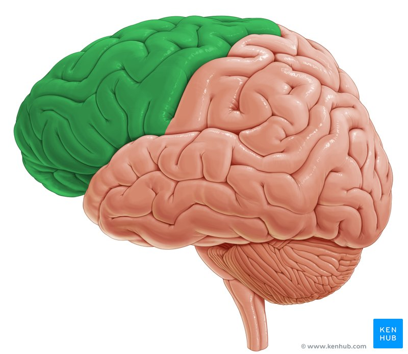
frontal lobe
front
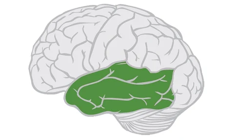
temporal lobe
sides
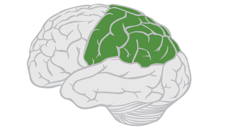
parietal lobe
top
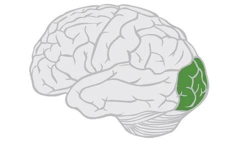
occipital lobe
base/back
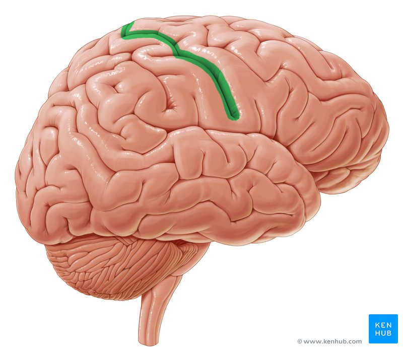
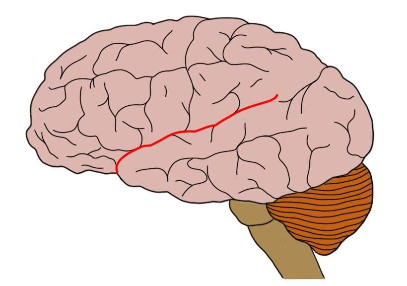
central sulcus
valley between motor and sensory area and between frontal and parietal
precentral gyrus
motor area (peaks)
postcentral gyrus
sensory area (peaks)
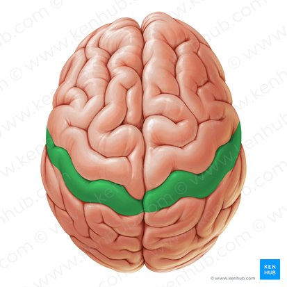
lateral cerebral sulcus (slyvian fissure)
valley near temporal bone
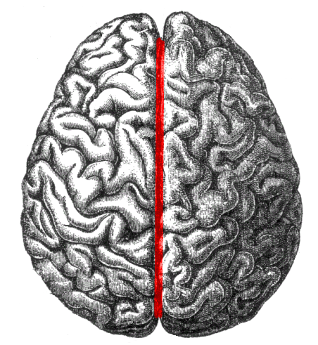
longitudinal cerebral fissure
the way we cut the brain into left and right
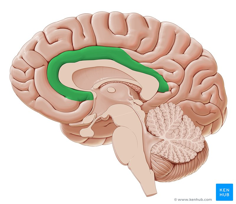
cingulate gyrus
first layer under cerebral cortex
cingulate sulcus
upper line (valley)
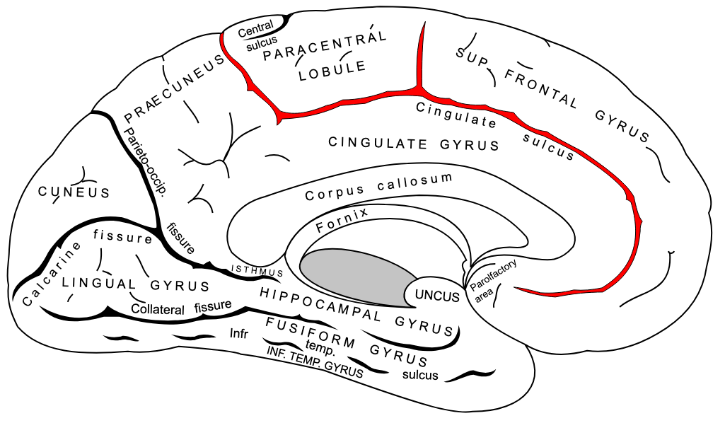
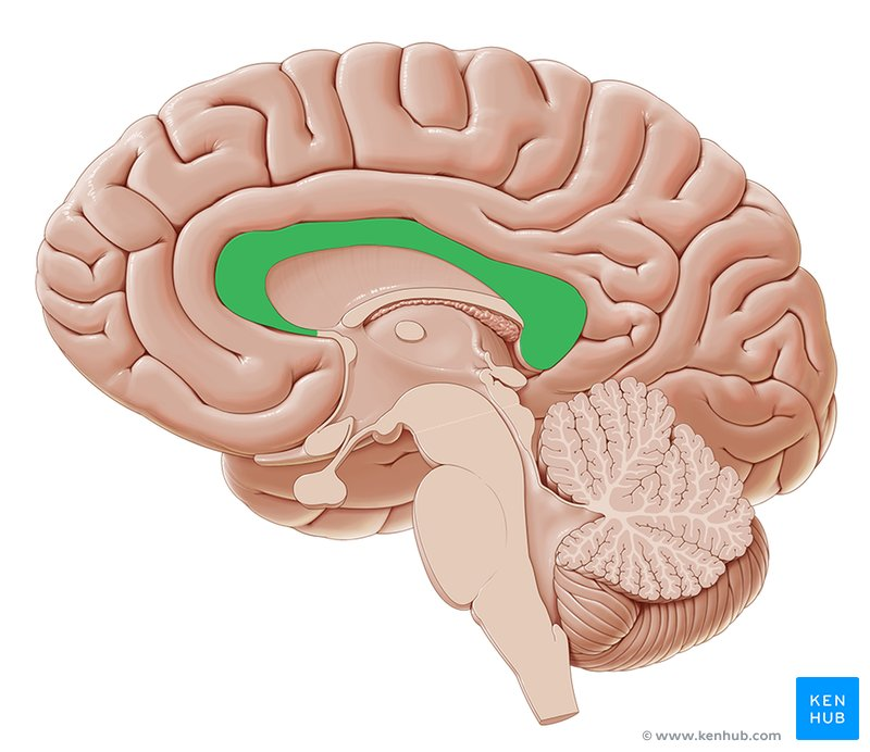
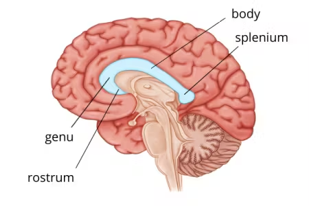
corpus callosum
middle connector between hemispheres
body of corpus callosum
middle part of corpus callosum

genu of corpus callosum
anterior/head part of corpus callosum

splenium of corpus callosum
posterior/back part of corpus callosum
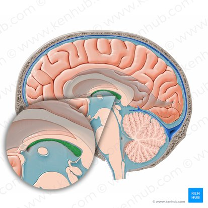
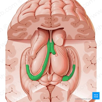
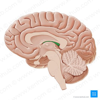
fornix
below corpus callosum, c-shaped curve
septum pellucidum
thin tissue structure, between fornix and corpus callosum body
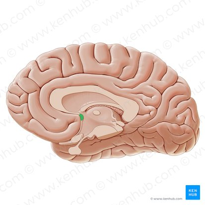
anterior commissure
really small dot, bottom of fornix
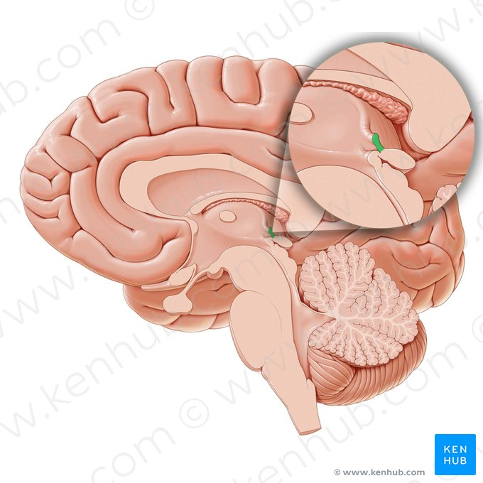
posterior commissure
back dot
thalamus
middle oval shape
optic chiasm
little x from optic nerve
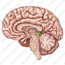
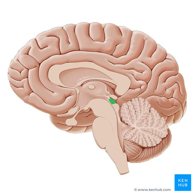
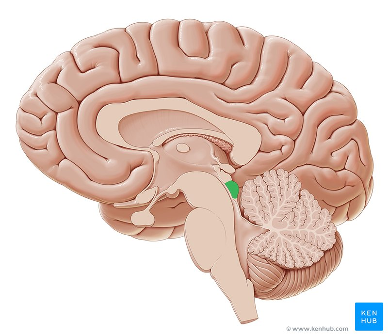
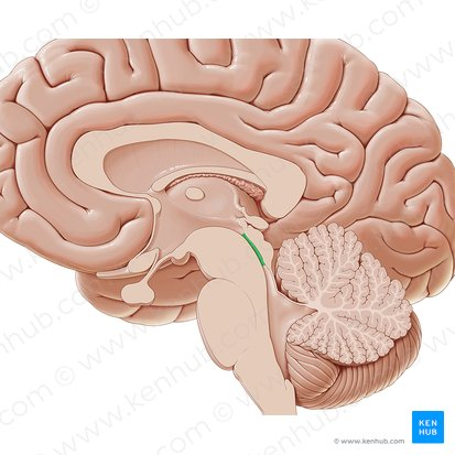
pineal gland
posterior little swoop bulb
superior colliculus
beneath pineal gland
inferior colliculus
beneath superior colliculus
Sylvian (cerebral) aqueduct
valley superior & inferior colliculus and 3rd and 4th ventricle
third ventricle
upper middle ventricle
fourth ventricle
below/connects to 3rd ventricle
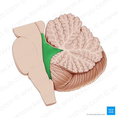
arbor vitae
tree structure on cerebellum, branches are white matter
vermis of cerebellum
worm looking area between parts of cerebellum
medulla oblongata
in-between pons & spinal cord
pons
above medulla, little bubble
cerebral peduncle
above the pons, connected to tube structure
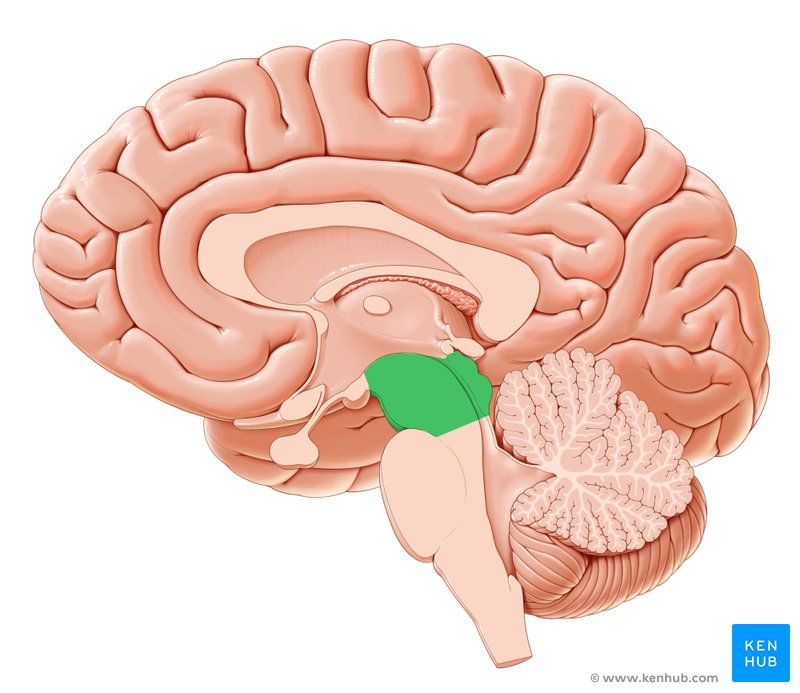
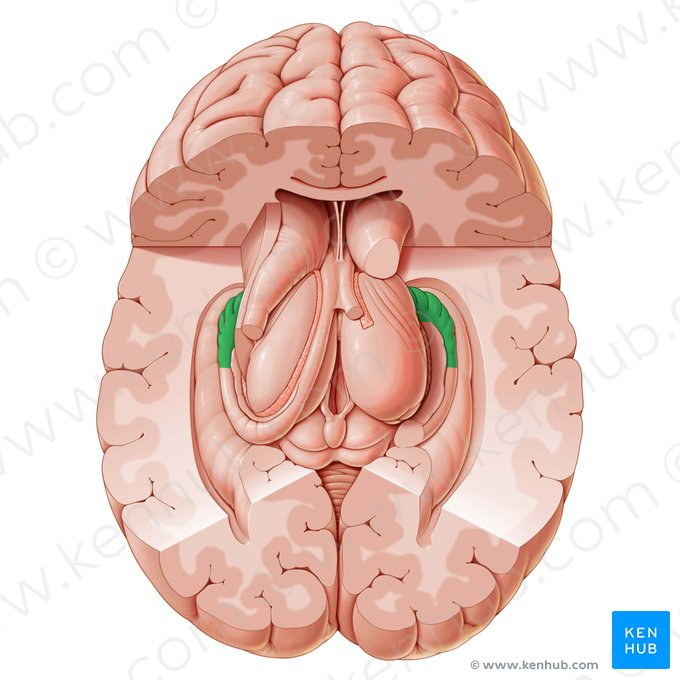
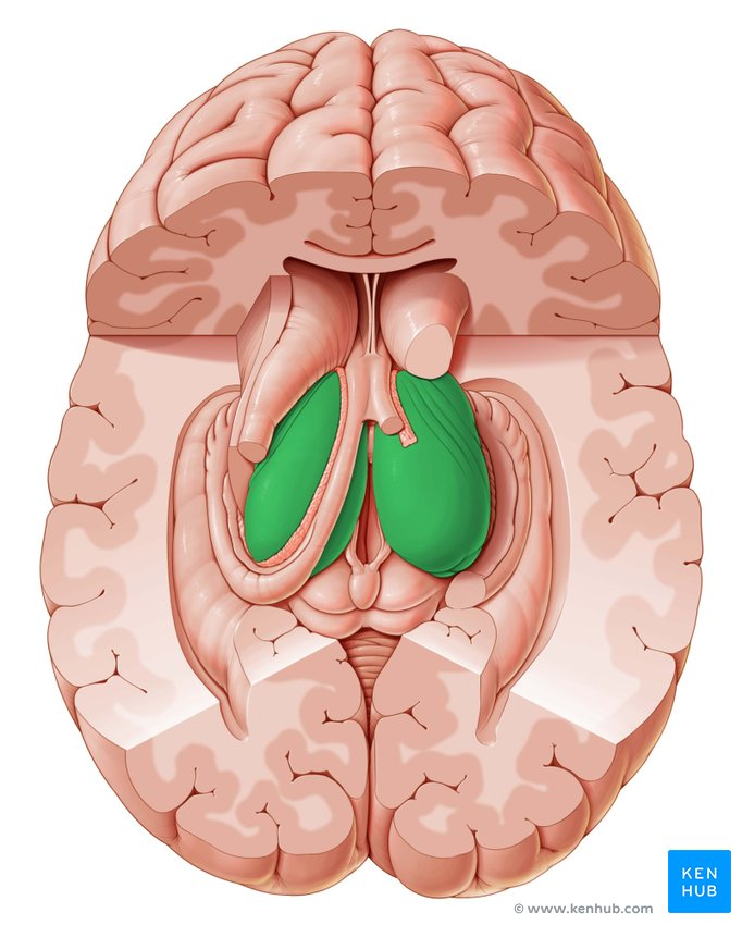
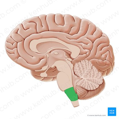
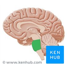
mammillary body
2 little breast like shapes from inferior view
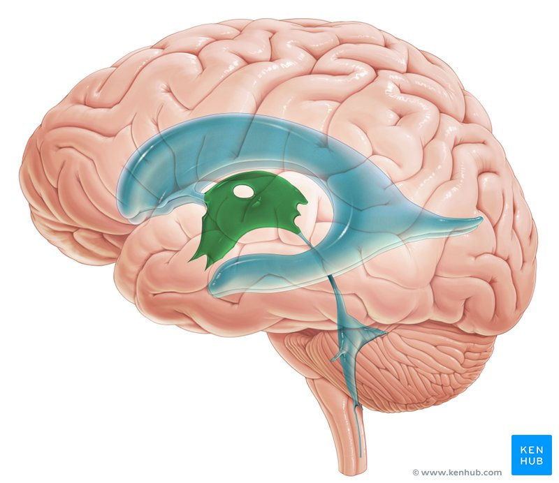
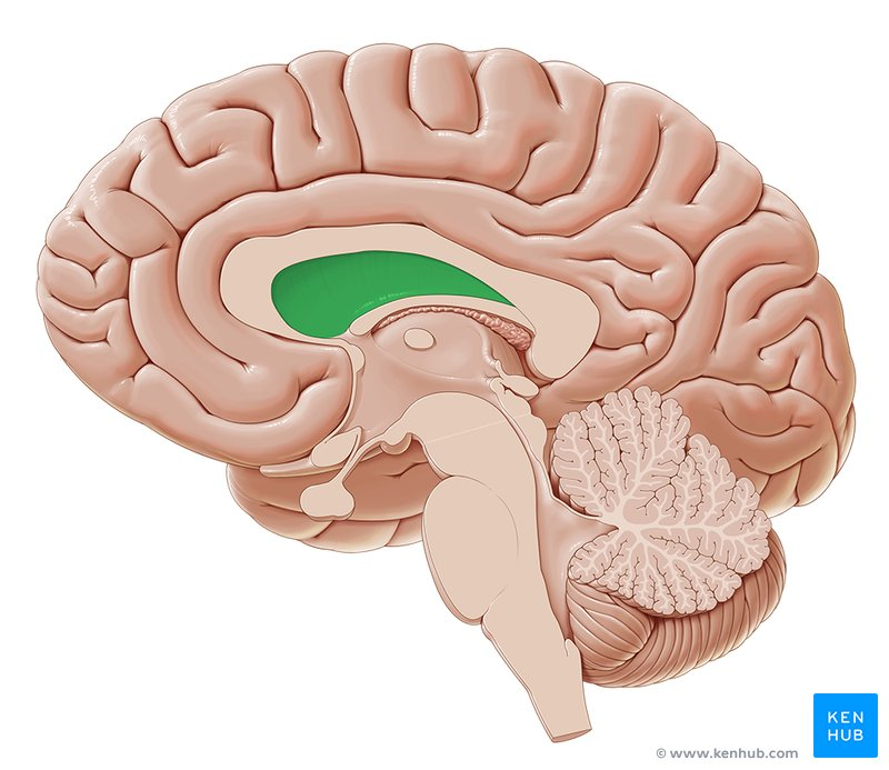
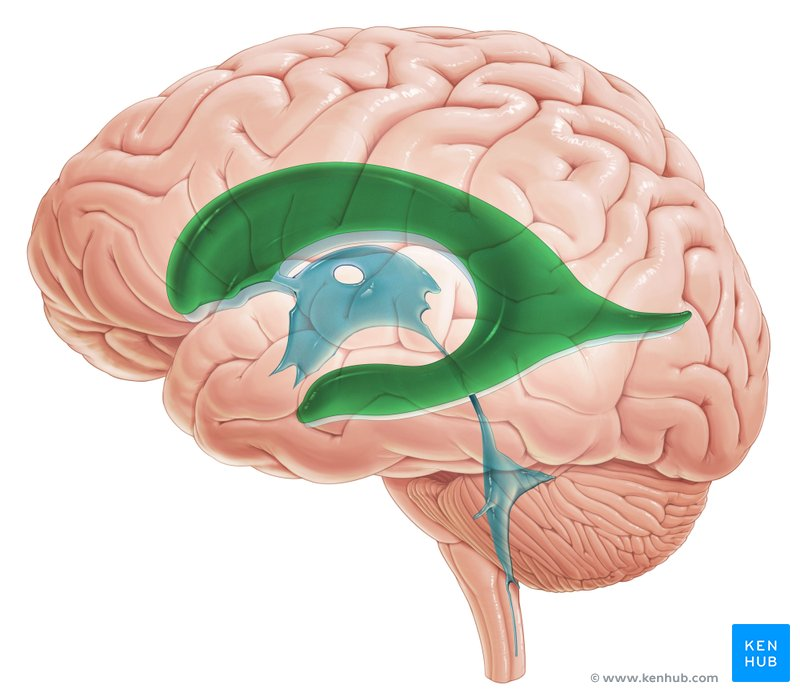
lateral ventricles (first and second)
spaces in corpus callosum
dorsal column
space between dorsal horns
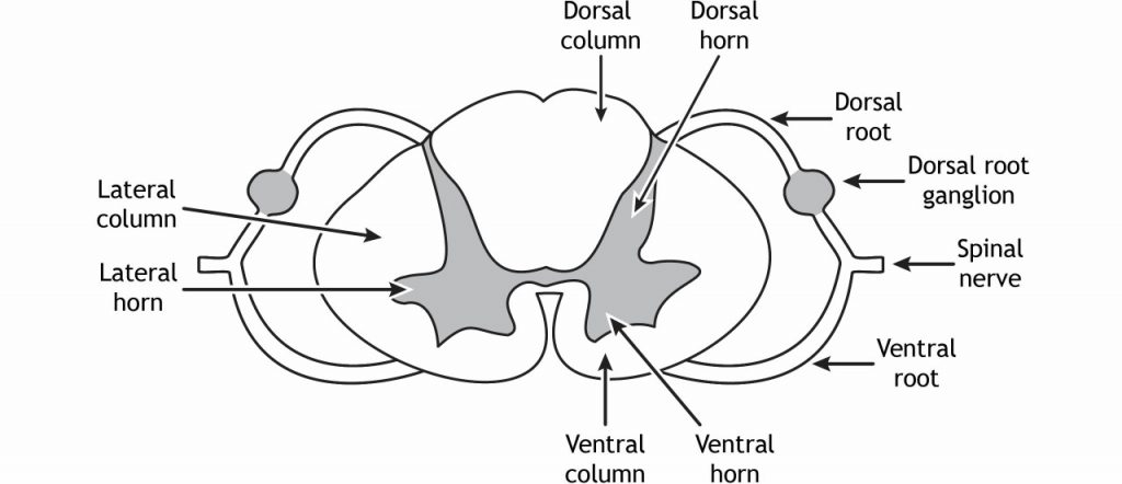
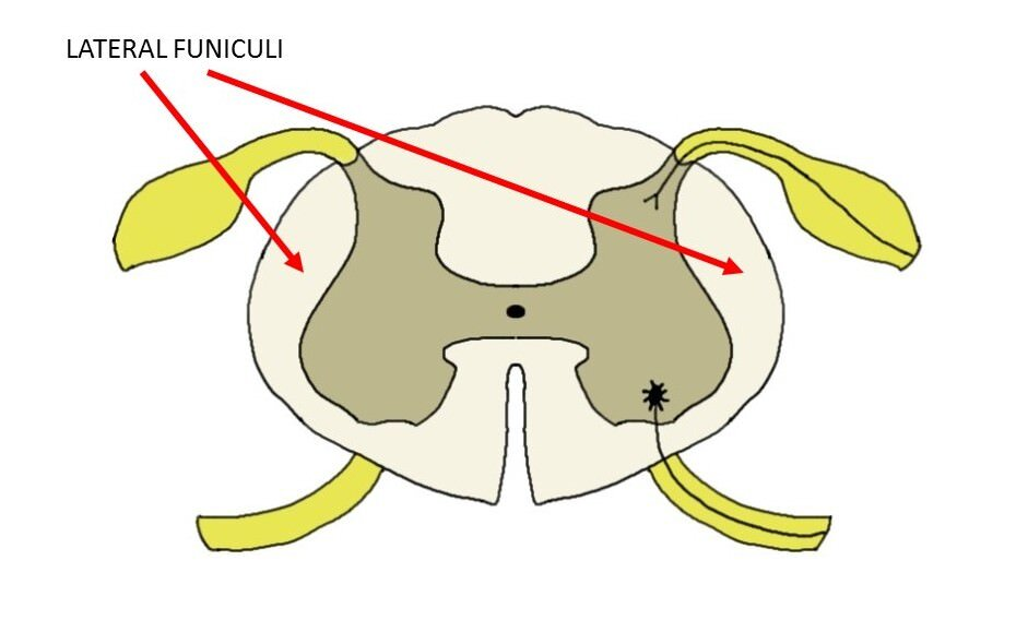
lateral column
side spaces on spinal cord
ventral column
bottom spaces on spinal cord
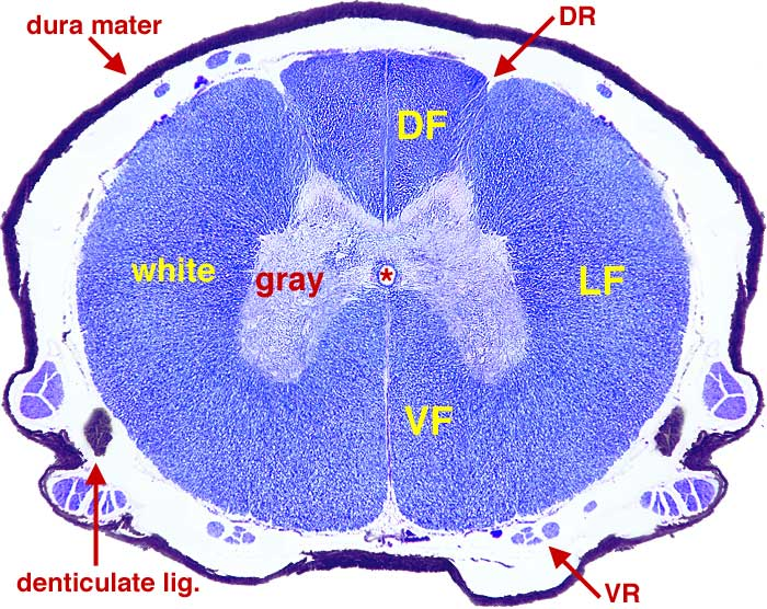
dorsal horn
upper part of butterfly
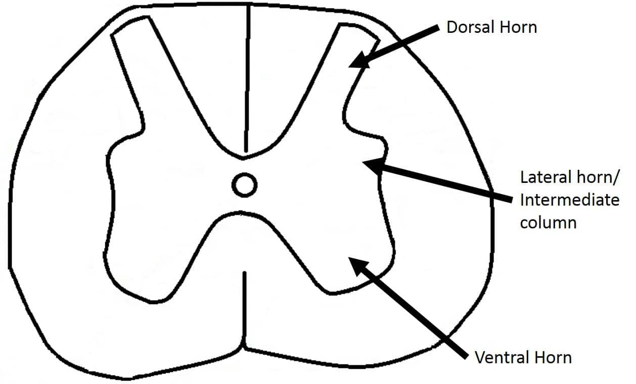
ventral horn
lower part of butterfly

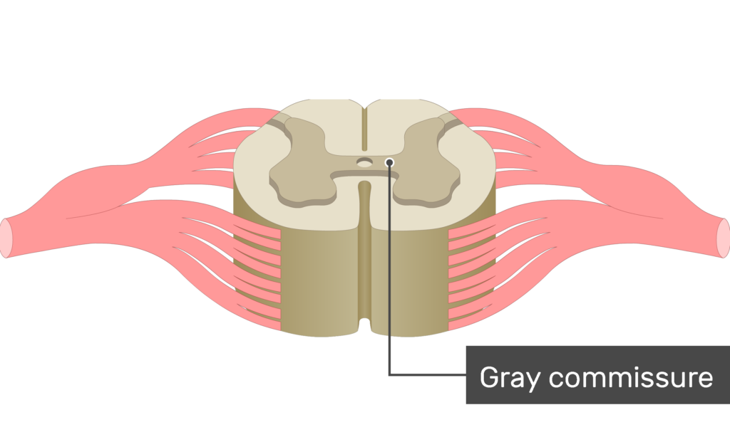
transverse commissure
median line that connects areas of spinal cord
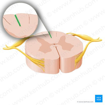
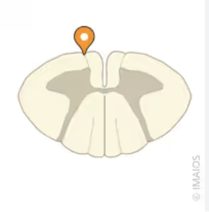
dorsal median sulcus
top, small indentation
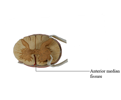
ventral median fissure
back, larger indentation
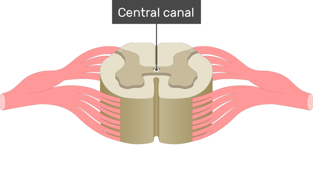
central canal
middle dot on spinal cord, filled with cerebral spinal fluid
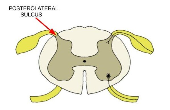
dorsolateral sulcus
bottom dorsal nerve connection
ventrolateral sulcus
bottom ventral nerve connection
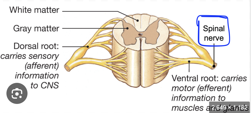
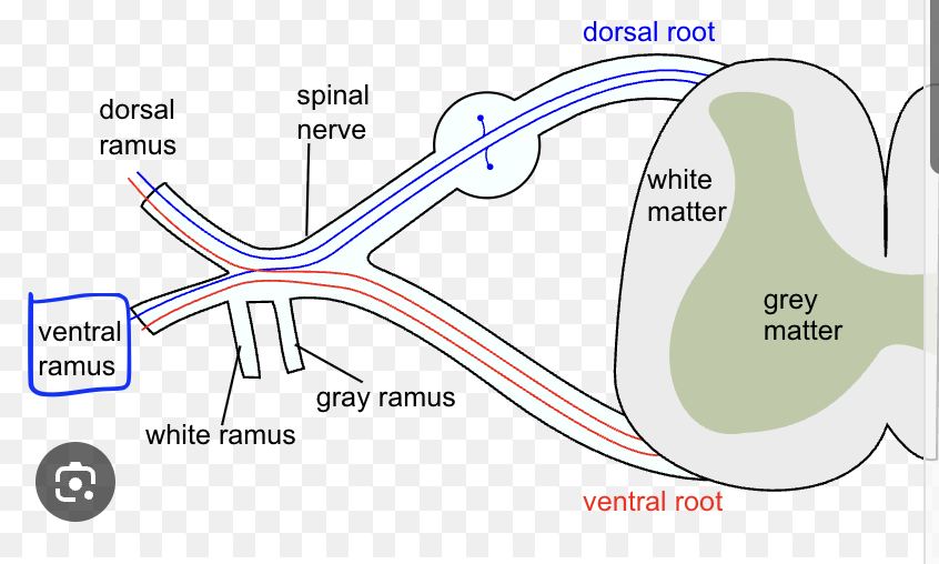
dorsal root
portion that goes to spinal
ventral root
portion that goes to spinal
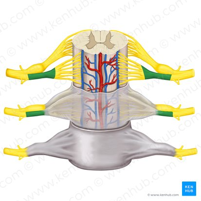
dorsal root ganglion
bubble on dorsal
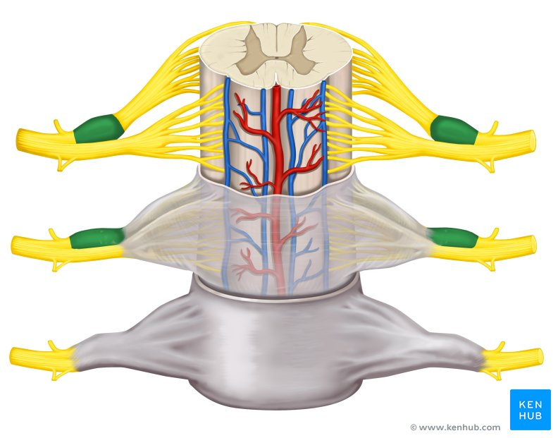
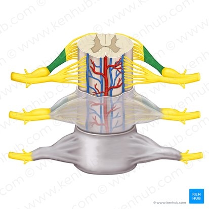
spinal nerve
where roots connect
dorsal ramus
little protrusion, dorsal
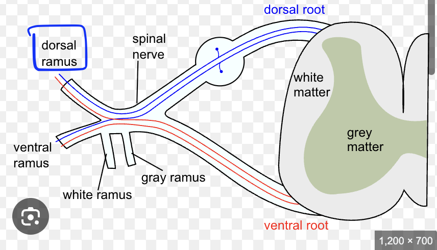
ventral ramus
little protrusion, ventral
billions
how many neurons are in the nervous system?
nervous system
where does the transmitting of info happen?
soma
what is the cell body of a neuron called?
soma
cell body
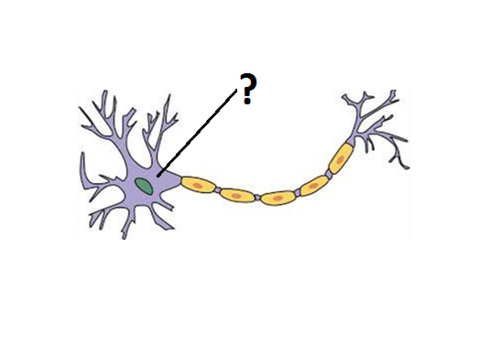
axon

dendrite
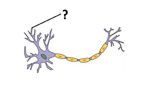
myelin sheath
fatty coverings
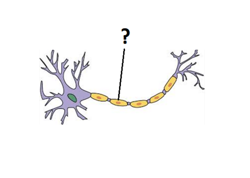
nodes of Ranvier
spots where you don’t have myelin sheath, where impulses jump
away
which way do axons transmit info from the cell body?
toward
which way do dendrites transmit info from the cell body?