BY124L - Exam 3 Models and Specimens
1/148
There's no tags or description
Looks like no tags are added yet.
Name | Mastery | Learn | Test | Matching | Spaced | Call with Kai |
|---|
No analytics yet
Send a link to your students to track their progress
149 Terms
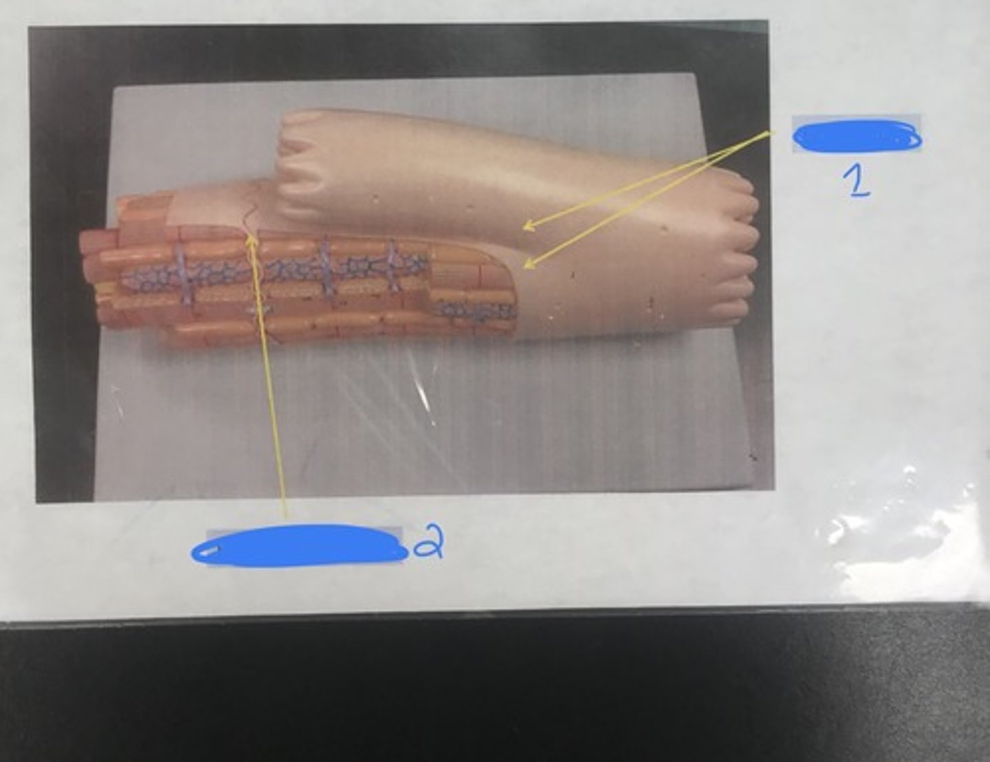
Name: skeletal muscle
For the following model, give the following:
1) name
Name: skeletal muscle (X-sec.)
For the following model, give the following:
1) name
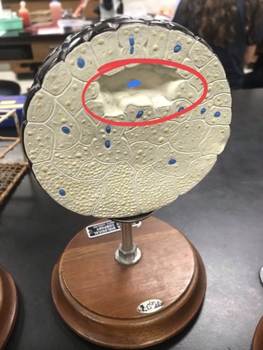
Name: smooth muscle
Structures:
1) nucleus
For the following model, give the following:
1) name
2) structures in numerical order
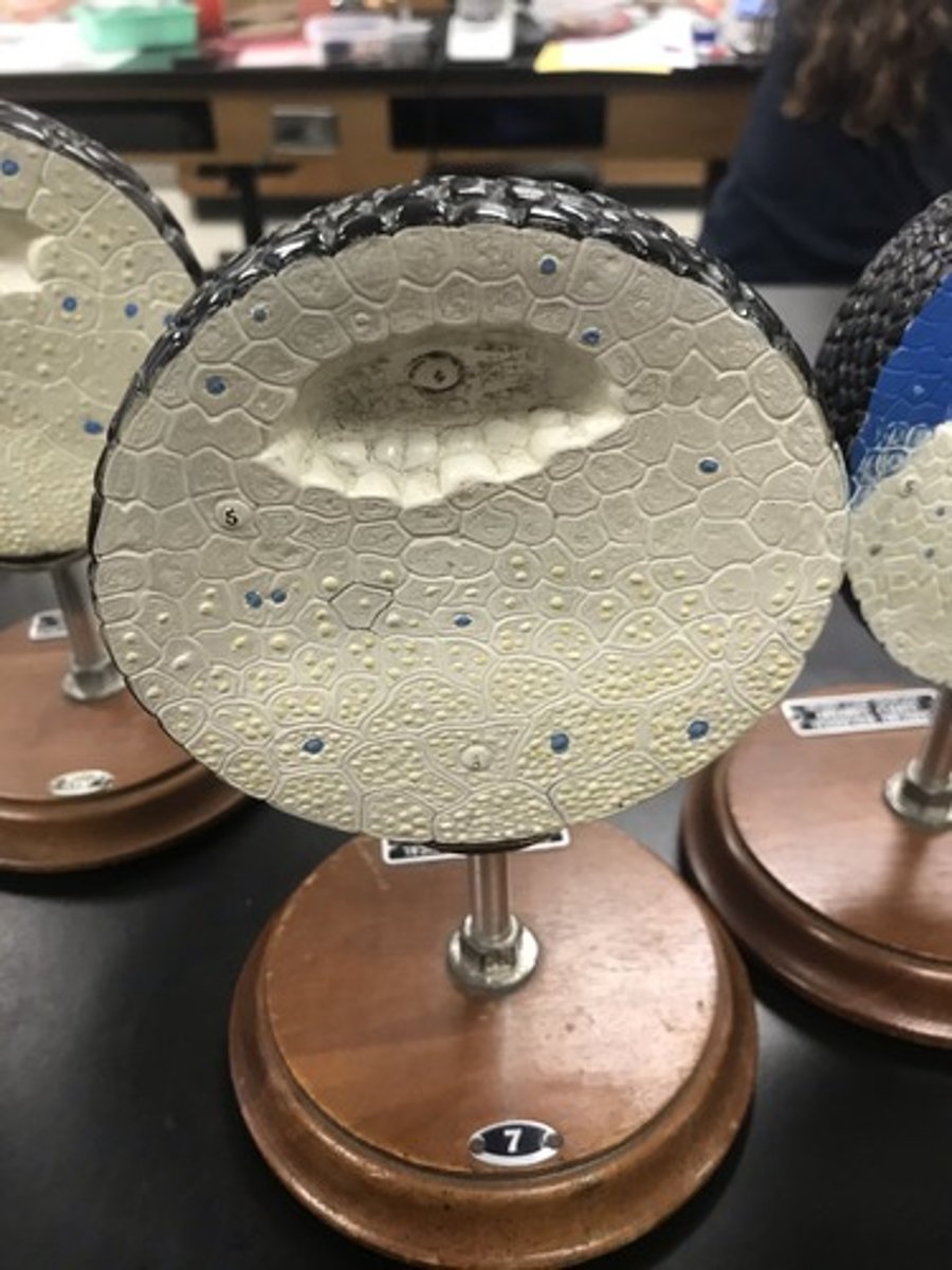
Name: cardiac muscle
Structures:
1) branching
2) intercalated disc
For the following model, give the following:
1) name
2) structures in numerical order
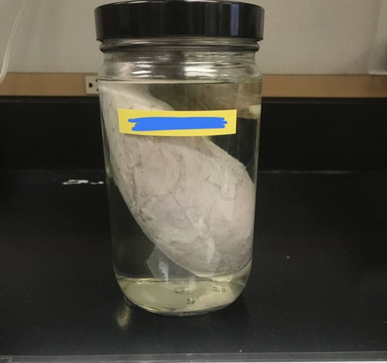
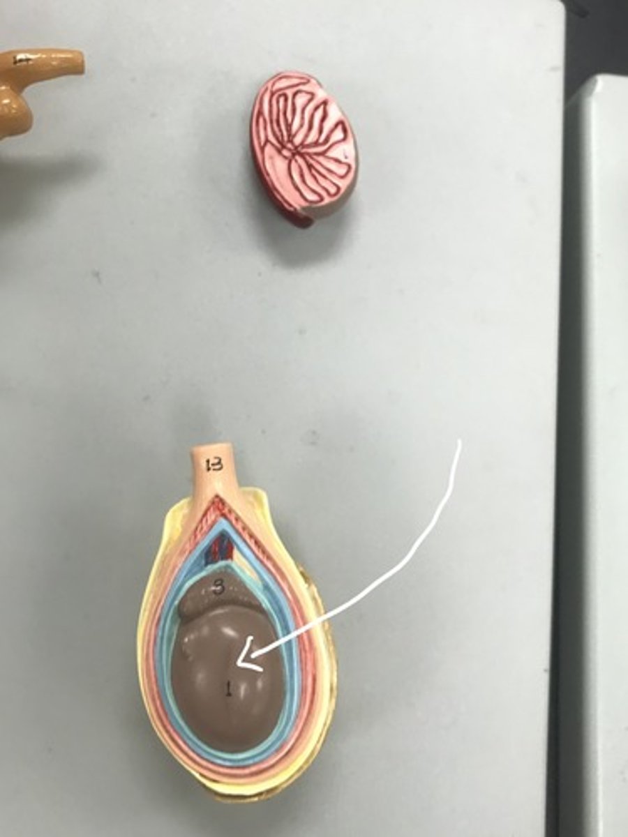
Name: bull testicle
For the following specimen, give the following:
1) name
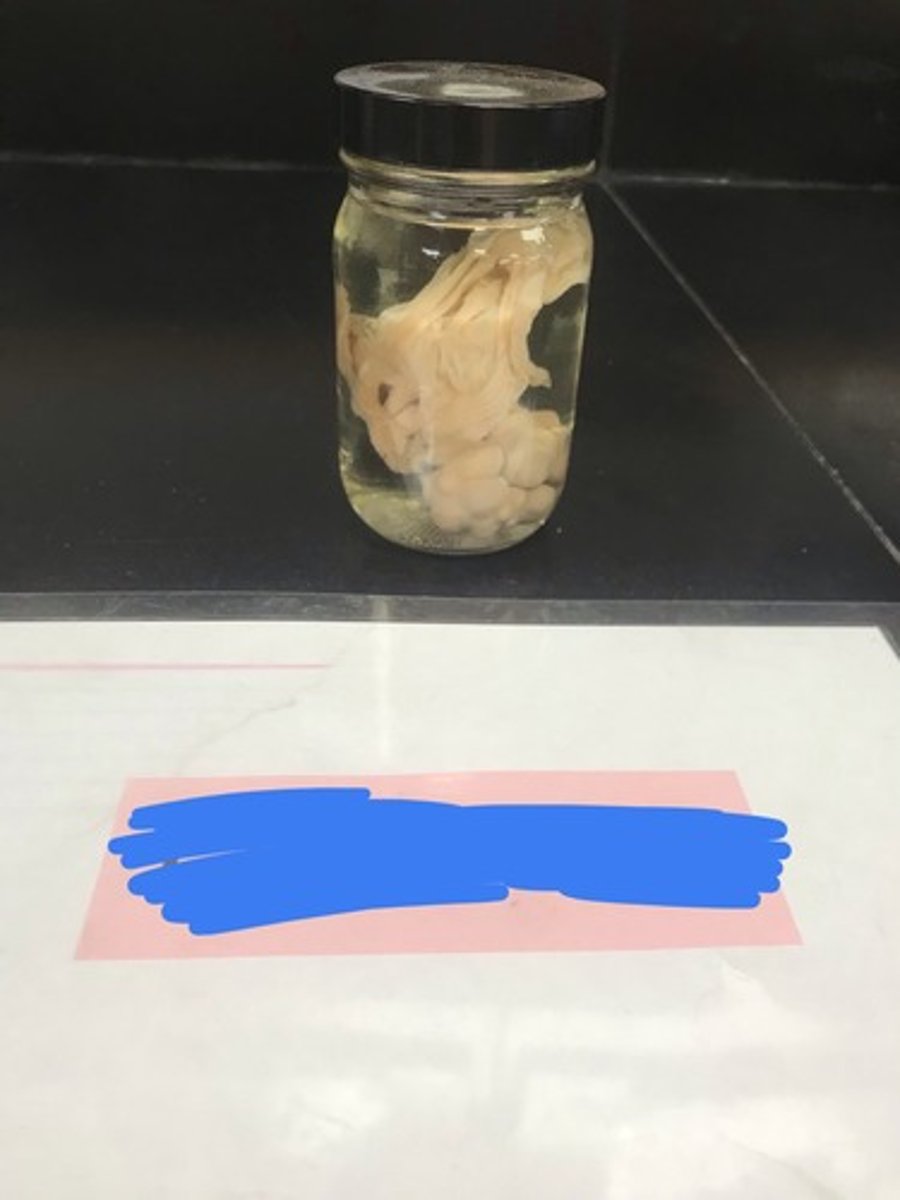
Name: pig ovary
For the following specimen, give the following:
1) name
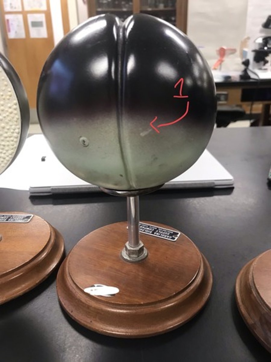
Stage: fertilization
Meoitic Stage: Meiosis II
Structures:
1) animal pole (dark)
2) vegetal pole (light)
For the following model, give the following:
1) stage of embryonic development
2) meiotic stage of the egg before sperm nucleus combines with the egg
3) structures in numerical order
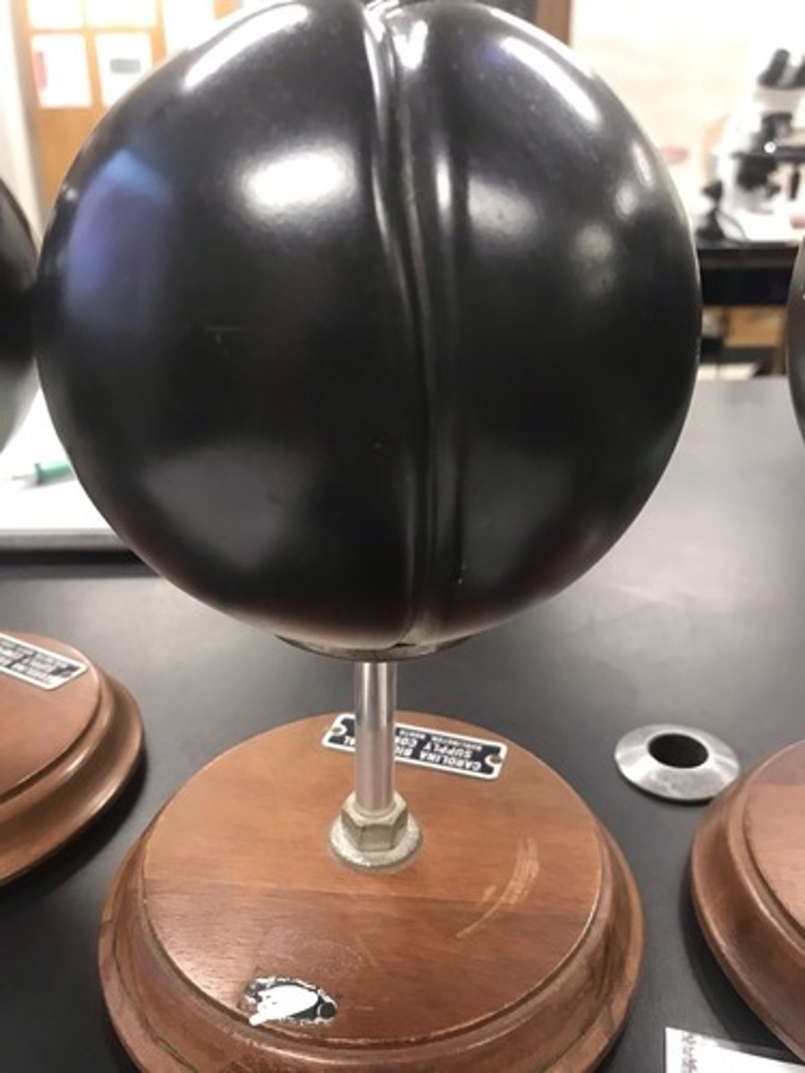
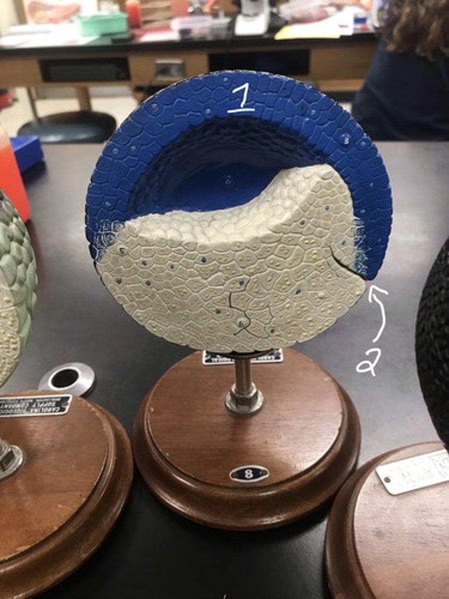
Stage: 1st cleavage
Type: polar/vertical
Structures:
1) gray crescent
For the following model, give the following:
1) stage of embryonic development
2) cleavage type
3) structures in numerical order
Stage: 2nd cleavage
Type: polar/vertical
For the following model, give the following:
1) stage of embryonic development
2) cleavage type
blastomere
Each cell produced from cleavage is known as a...
Stage: 8 cell
For the following model, give the following:
1) stage of embryonic development
Stage: 16 cell
For the following model, give the following:
1) stage of embryonic development
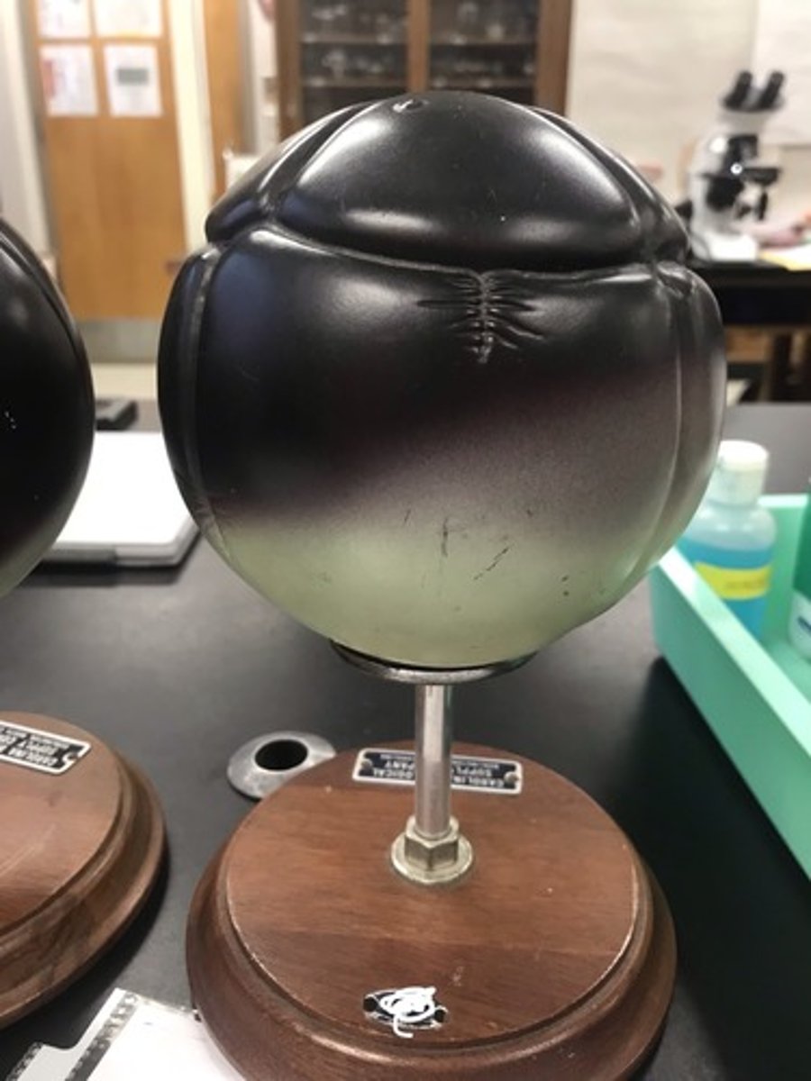
Stage: morula
Structures:
1) blastocoel
For the following model, give the following:
1) stage of embryonic development
2) circled structure
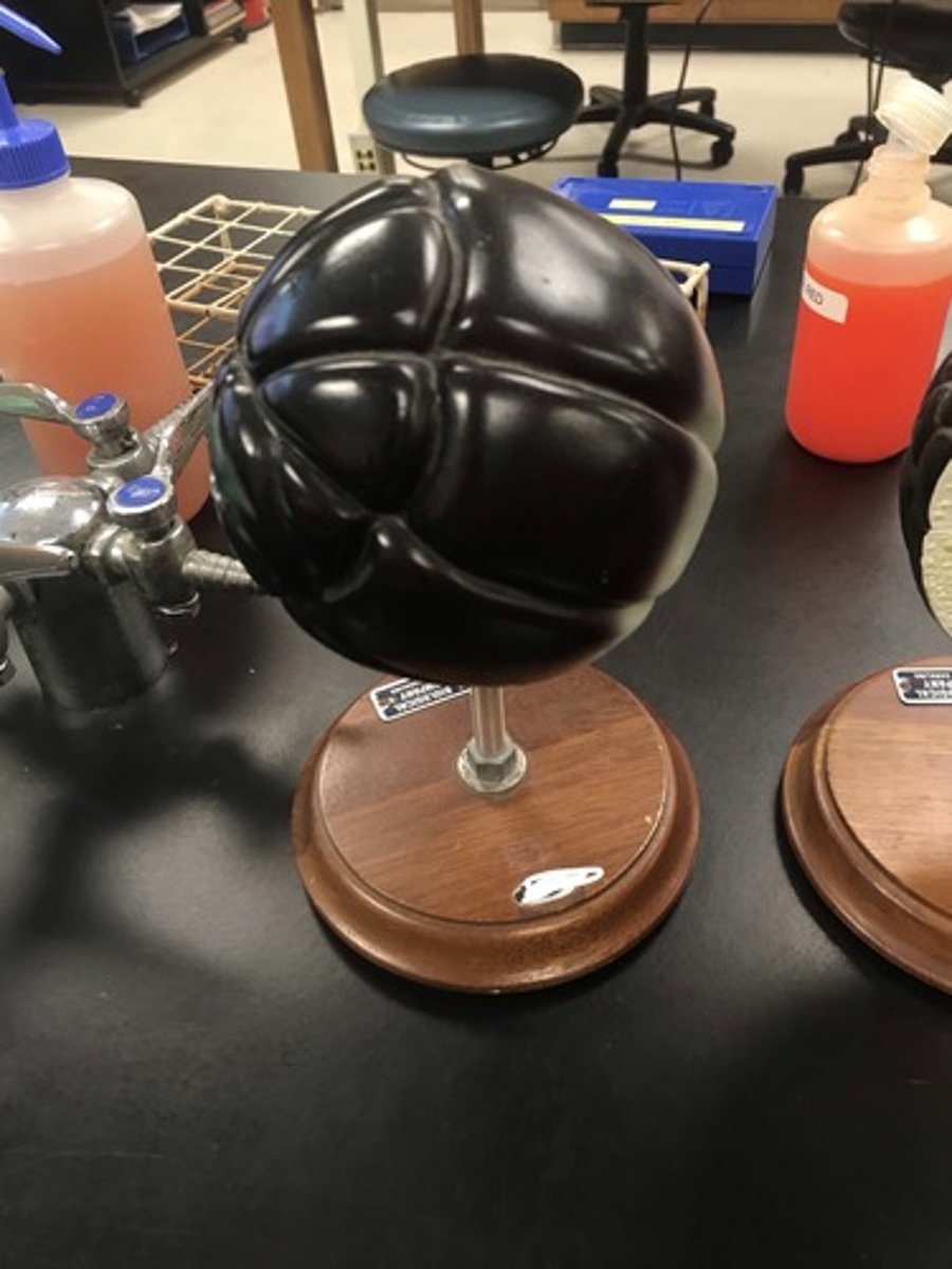
Stage: morula (continued development of blastocoel)
For the following model, give the following:
1) stage of embryonic development
Stage: blastula
Structures:
1) ectoderm
2) dorsal lip
For the following model, give the following:
1) stage of embryonic development
2) structures in numerical order
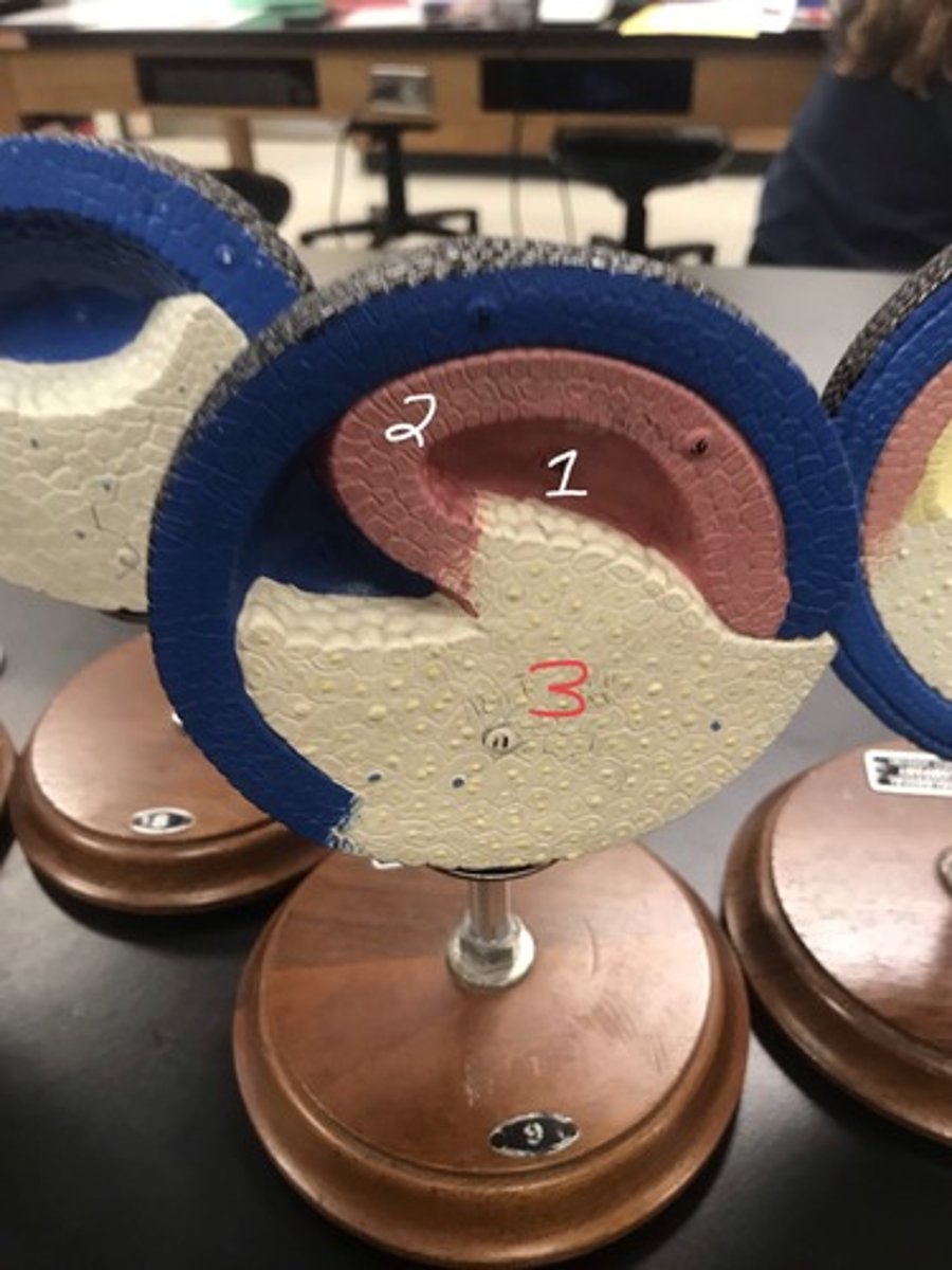
Stage: gastrulation
Structures:
1) archenteron
2) undifferentiated mesoderm
3) yolk cells
For the following model, give the following:
1) stage of embryonic development
2) structures in numerical order
Stage: gastrulation (cont.)
Structures:
1) ectoderm
2) mesoderm
3) endoderm
4) archenteron
5) yolk plug
For the following model, give the following:
1) stage of embryonic development
2) structures in numerical order
Stage: neurulation
Structures:
1) neural plate (gives rise to neural groove and neural crest)
2) to-be notochord
3) shrinking yolk plug
4) blastopore
For the following model, give the following:
1) stage of embryonic development
2) structures in numerical order
Stage: neurulation (cont.)
Structures:
1) neural crest
2) neural groove
3) blastopore (nearly free of yolk)
For the following model, give the following:
1) stage of embryonic development
2) structures in numerical order
Stage: neurulation (cont.)
Structures:
1) neural crests
For the following model, give the following:
1) stage of embryonic development
2) structures in numerical order
Stage: neurulation (cont.)
Structures:
1) oral plate (where ecto- and endoderm fuse to form oral cavity)
For the following model, give the following:
1) stage of embryonic development
2) structures in numerical order
Stage: neurulation (cont.)
Structures:
1) region of brain and spinal cord formation
2) notochord
3) cells that will become heart tissue
4) cells that will become liver
5) to-be hypophysis (pituitary gland)
For the following model, give the following:
1) stage of embryonic development
2) structures in numerical order
Stage: neurulation (cont.)
Structures:
1) mouth
2) to-be pharynx
3) anal opening
For the following model, give the following:
1) stage of embryonic development
2) structures in numerical order
Stage: neurulation (cont.)
Structures:
1) brain
2) somites
For the following model, give the following:
1) stage of embryonic development
2) structures in numerical order
Stage: neurulation (continued embryo elongation)
For the following model, give the following:
1) stage of embryonic development
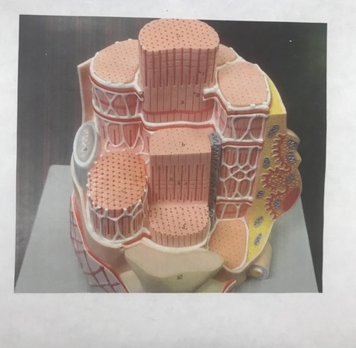
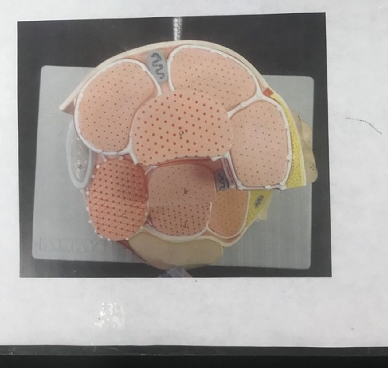
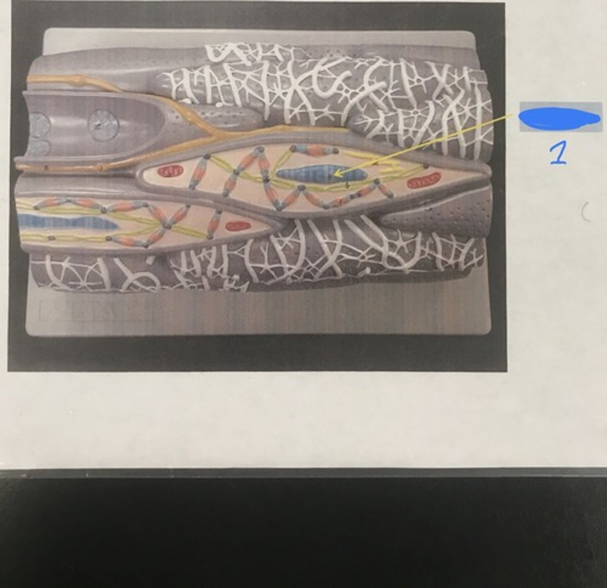
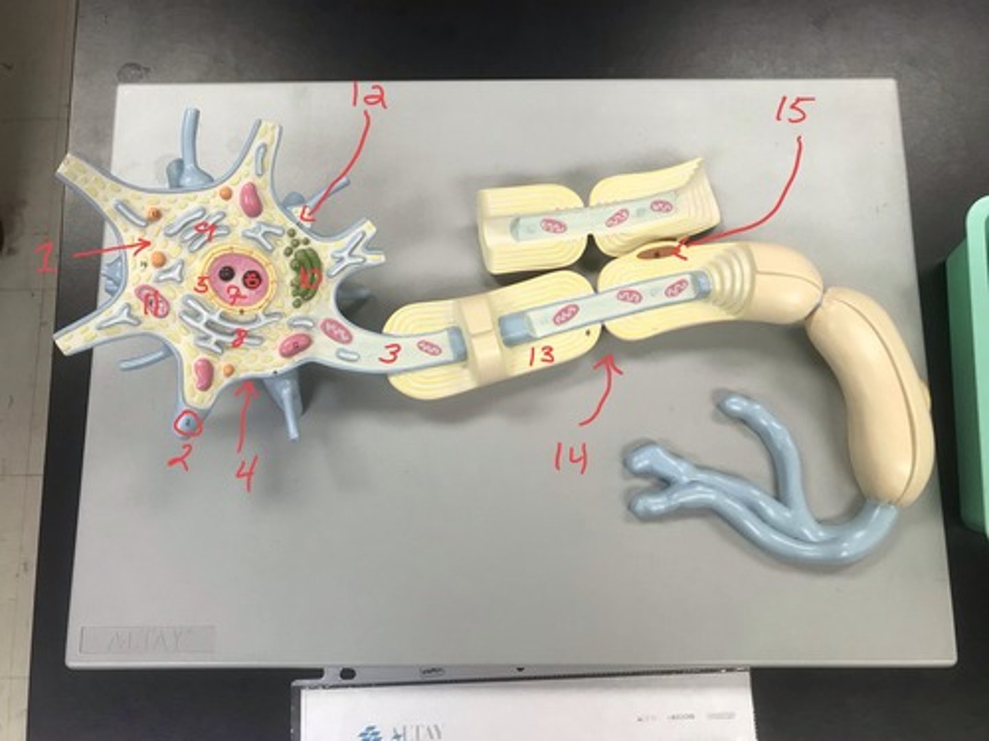
Name: neuron
Structures:
1) cell body
2) dendrite
3) axon
4) cell membrane
5) nuclear membrane
6) nucleolus
7) chromatin
8) rough ER
9) smooth ER
10) golgi complex
11) mitochondrion
12) synapse
13) myelin sheath
14) Ranvier node
15) Schwann cell
For the following model, give the following:
1) name
2) structures in numerical order
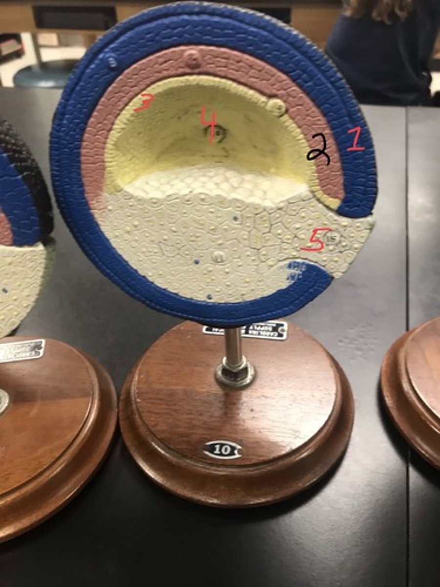
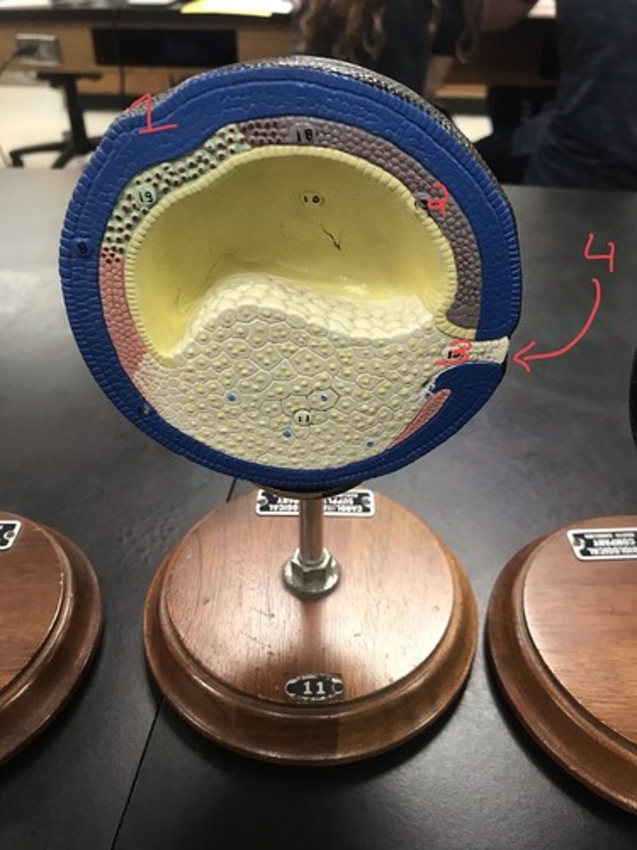
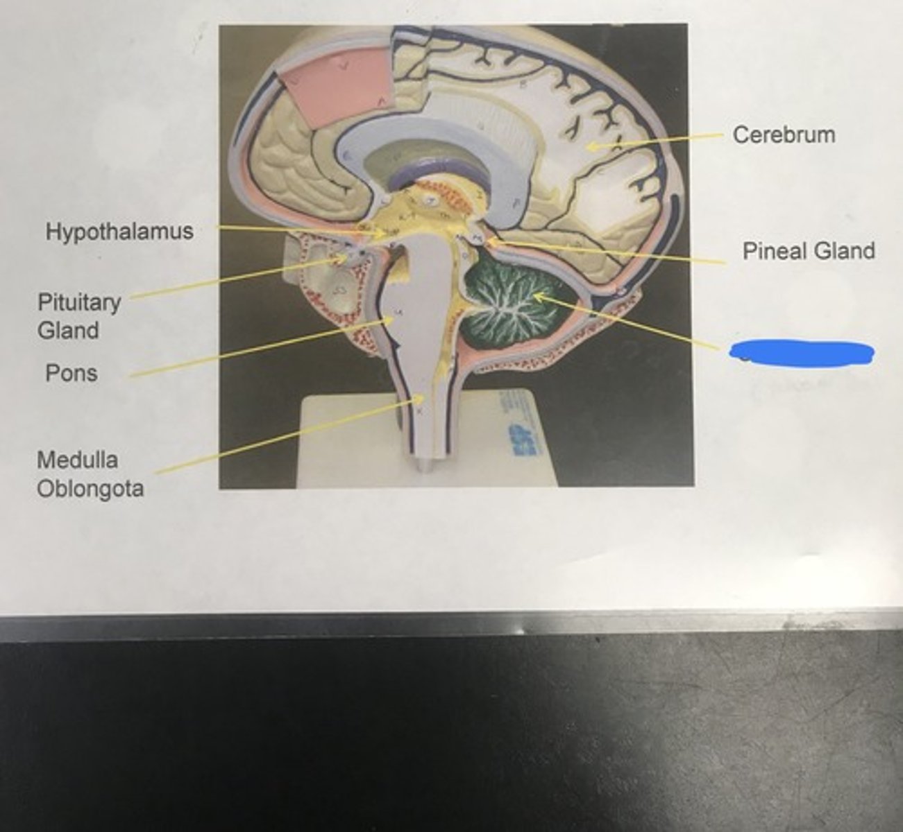
Name: brain (longitudinal view)
Name the model

hypothalamus
- makes oxytocin and ADH, produces releasing-hormones that cause the hypophysis to release its hormones
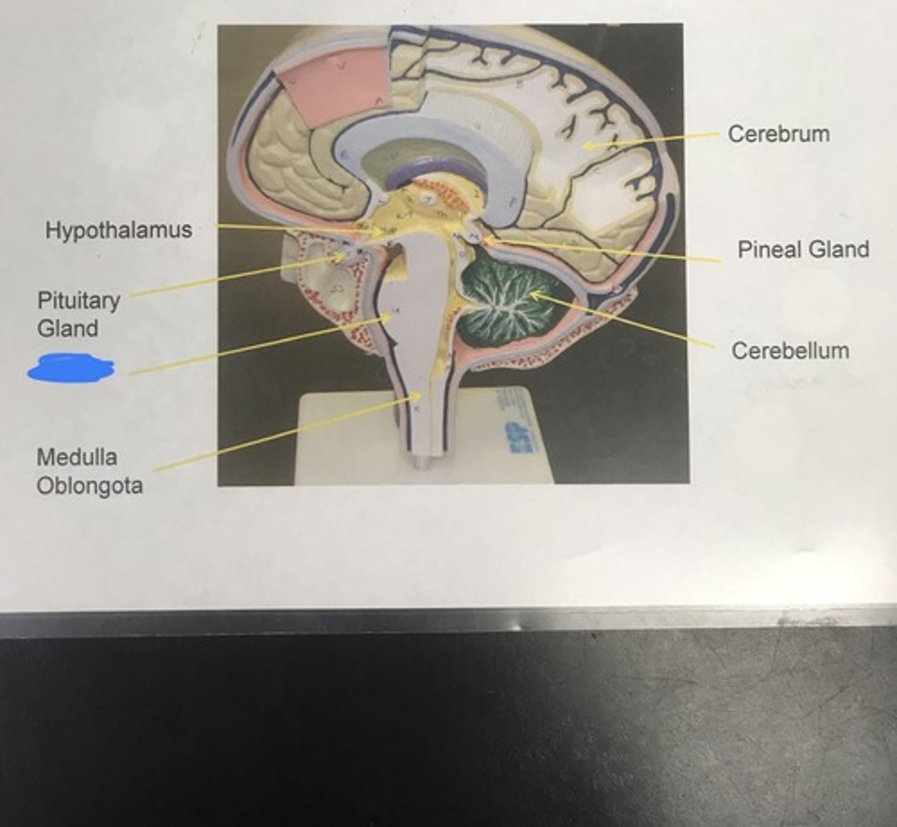
Label the structure covered in blue ink and give a function
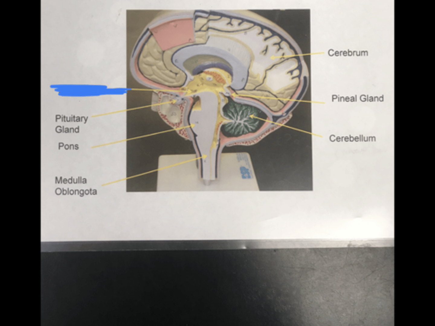
pituitary gland
- manufactures a wide array of hormones (except for posterior pituitary, which stores oxytocin and ADH)
Label the structure covered in blue ink and give a function
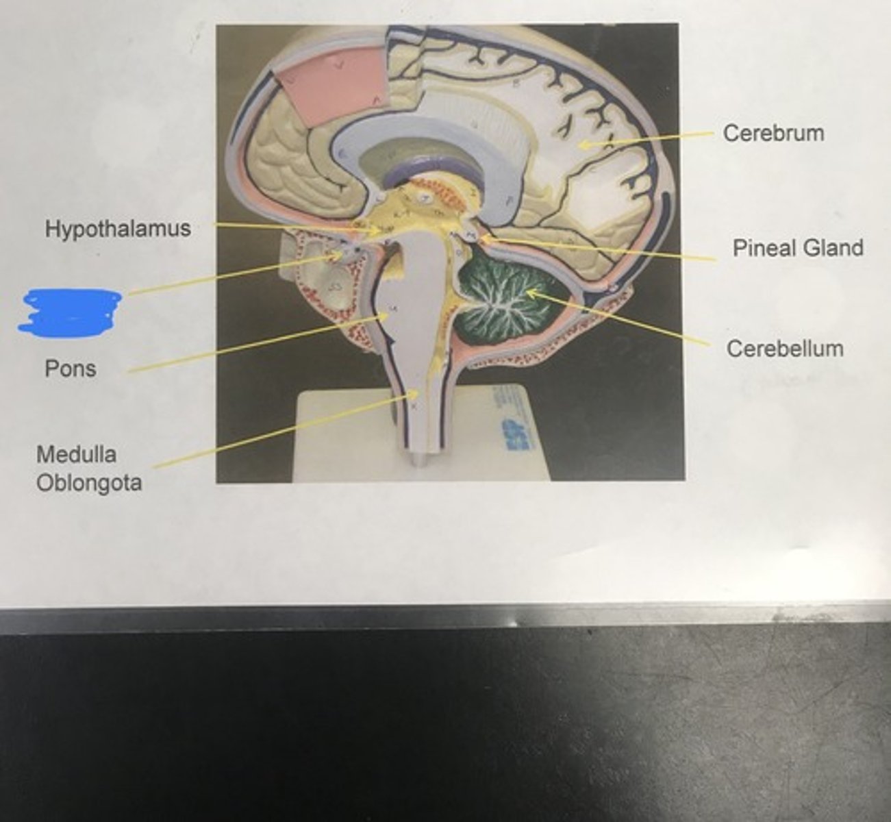
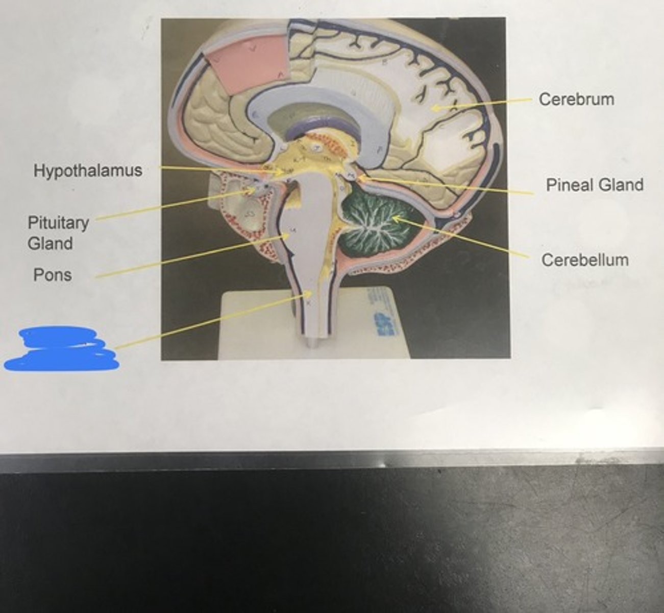
pons
- helps medulla oblongata with its functions
Label the structure covered in blue ink and give a function
medulla oblongata
- controls autonomic/homeostatic functions including:
1) swallowing
2) breathing
3) digestion
4) heart rate
5) vomiting
Label the structure covered in blue ink and give a function
brainstem
medulla oblongata + pons =
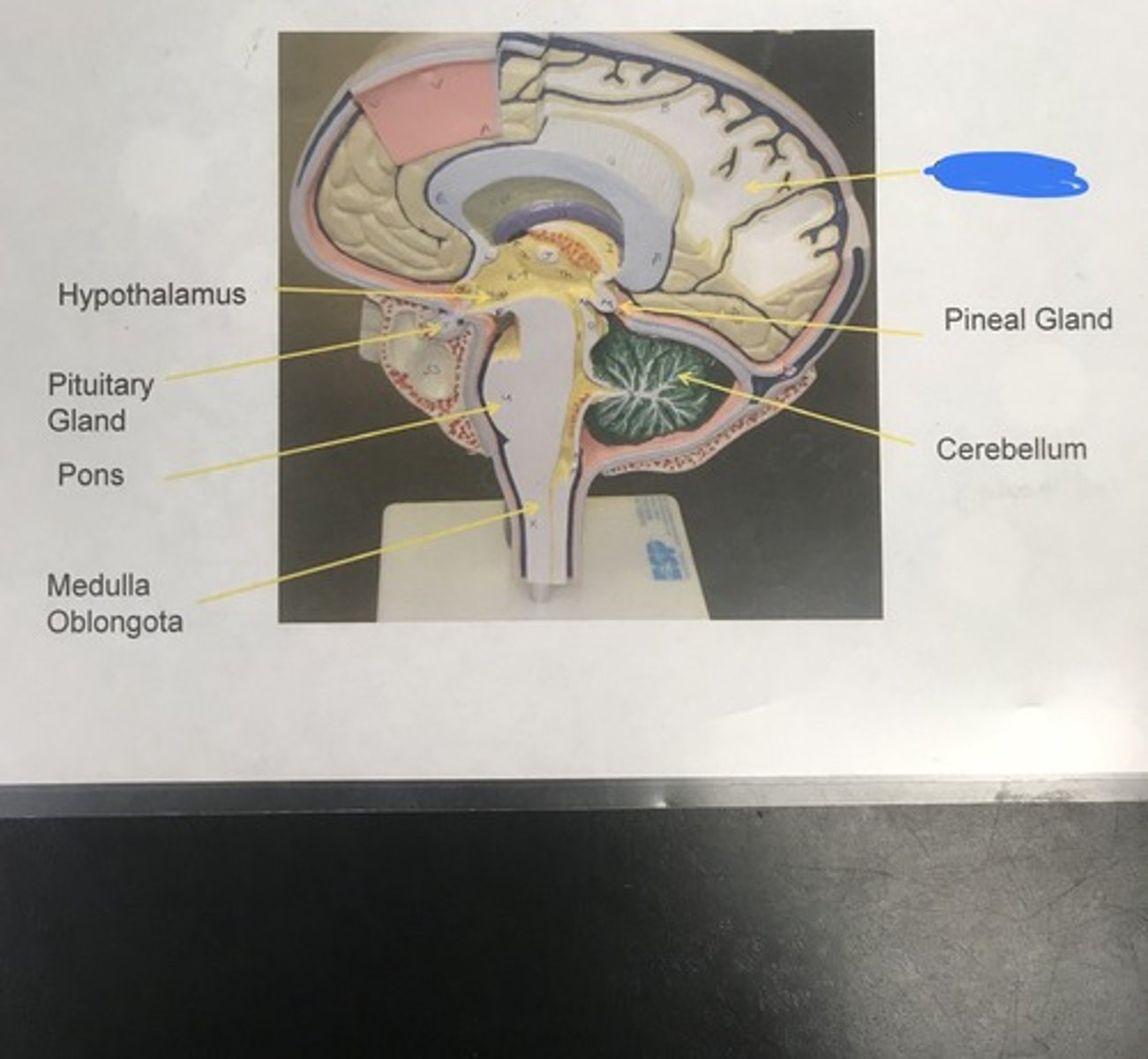
cerebrum (all of the brain's lobes combined)
Label the structure covered in blue ink
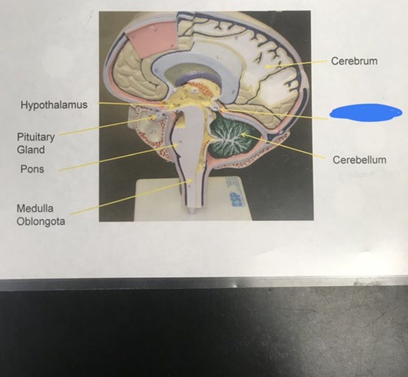
pineal gland
- manufactures melatonin, an important hormone in the management of biological rhythms
Label the structure covered in blue ink and give a function
cerebellum
- coordinating movement and balance
Label the structure covered in blue ink and give a function
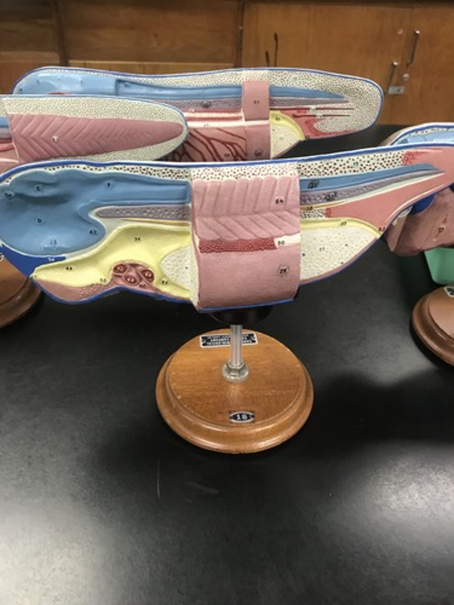
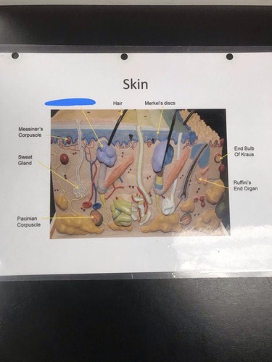
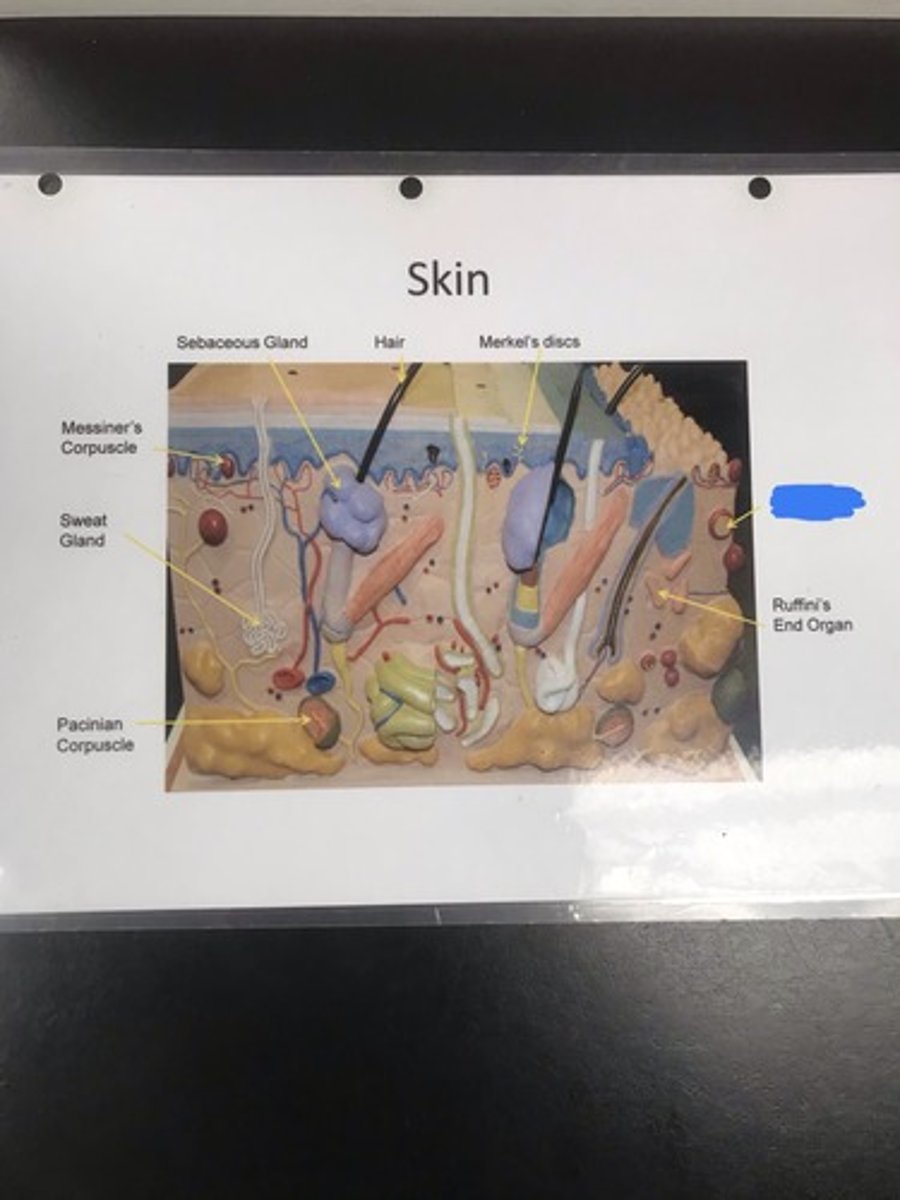
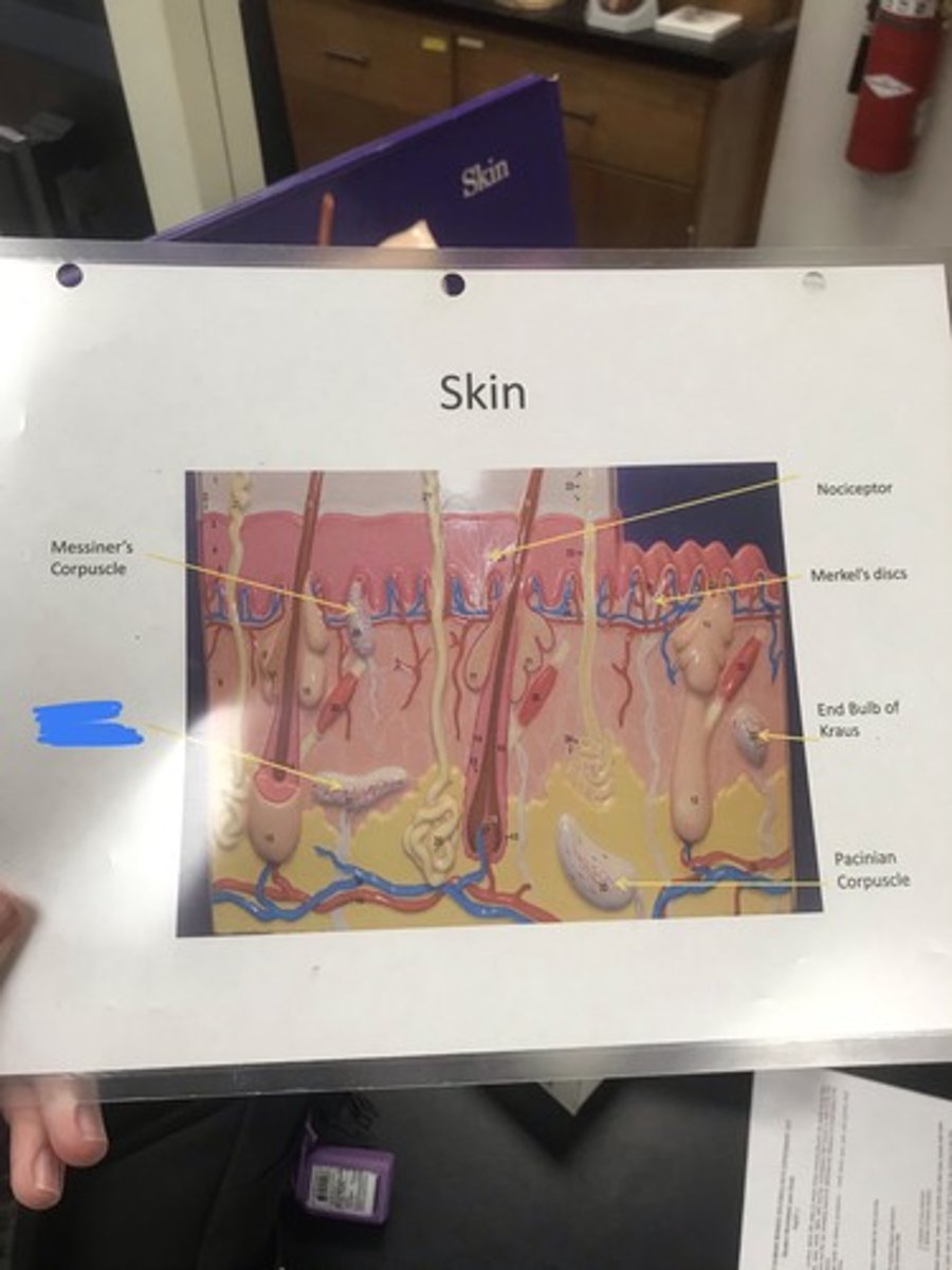
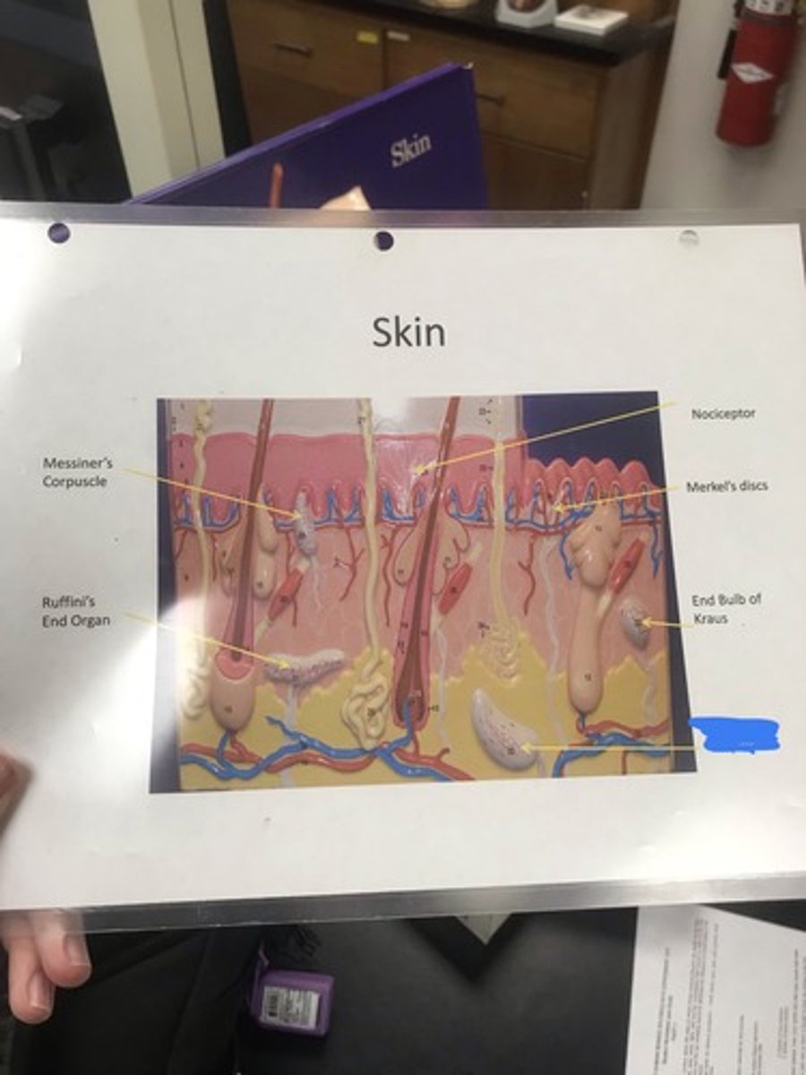
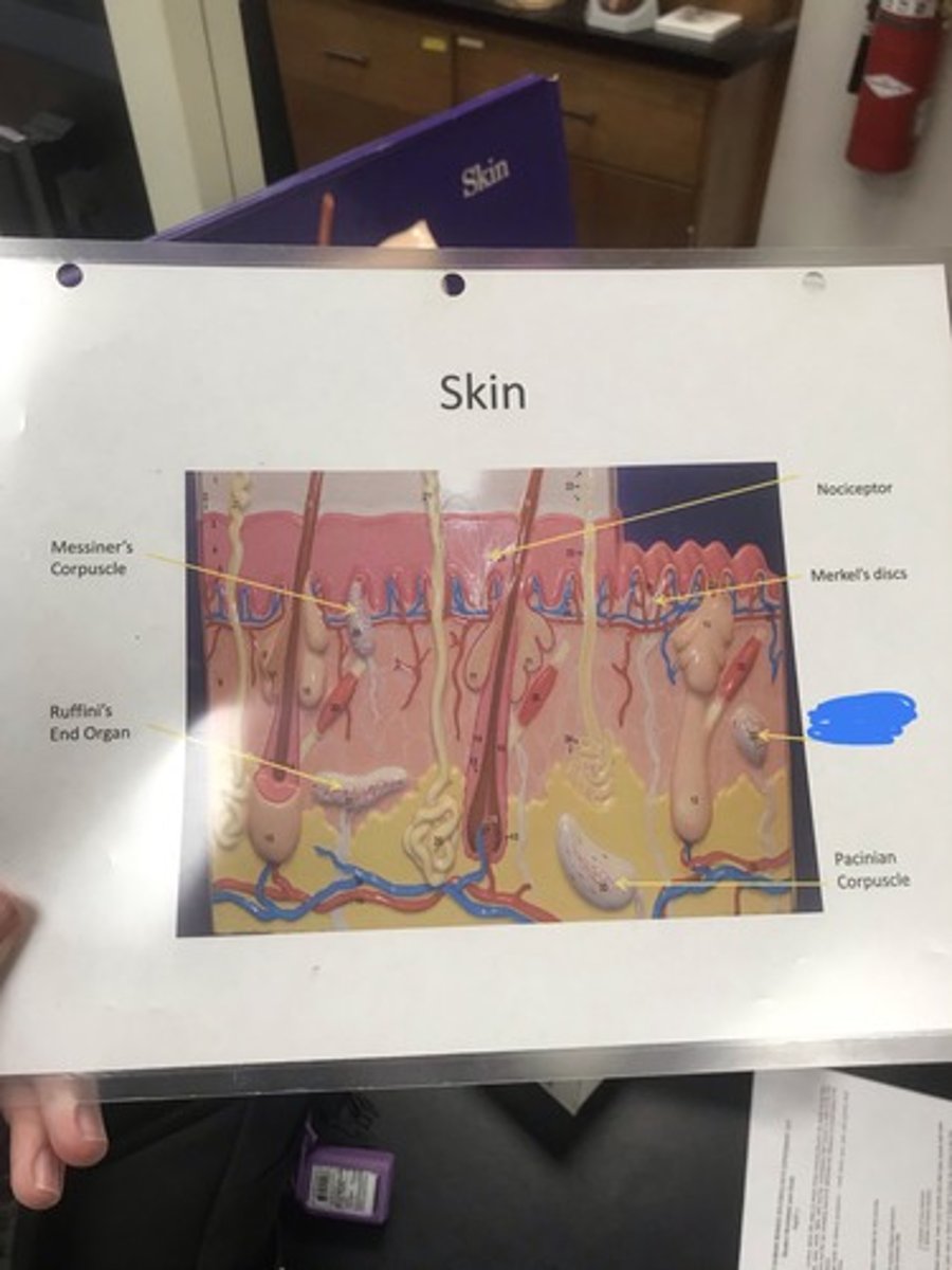
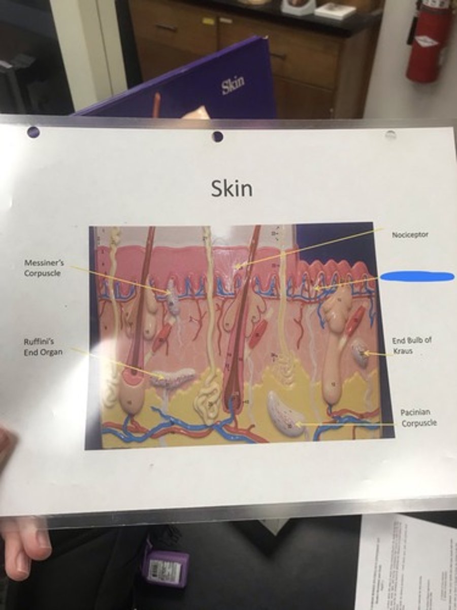
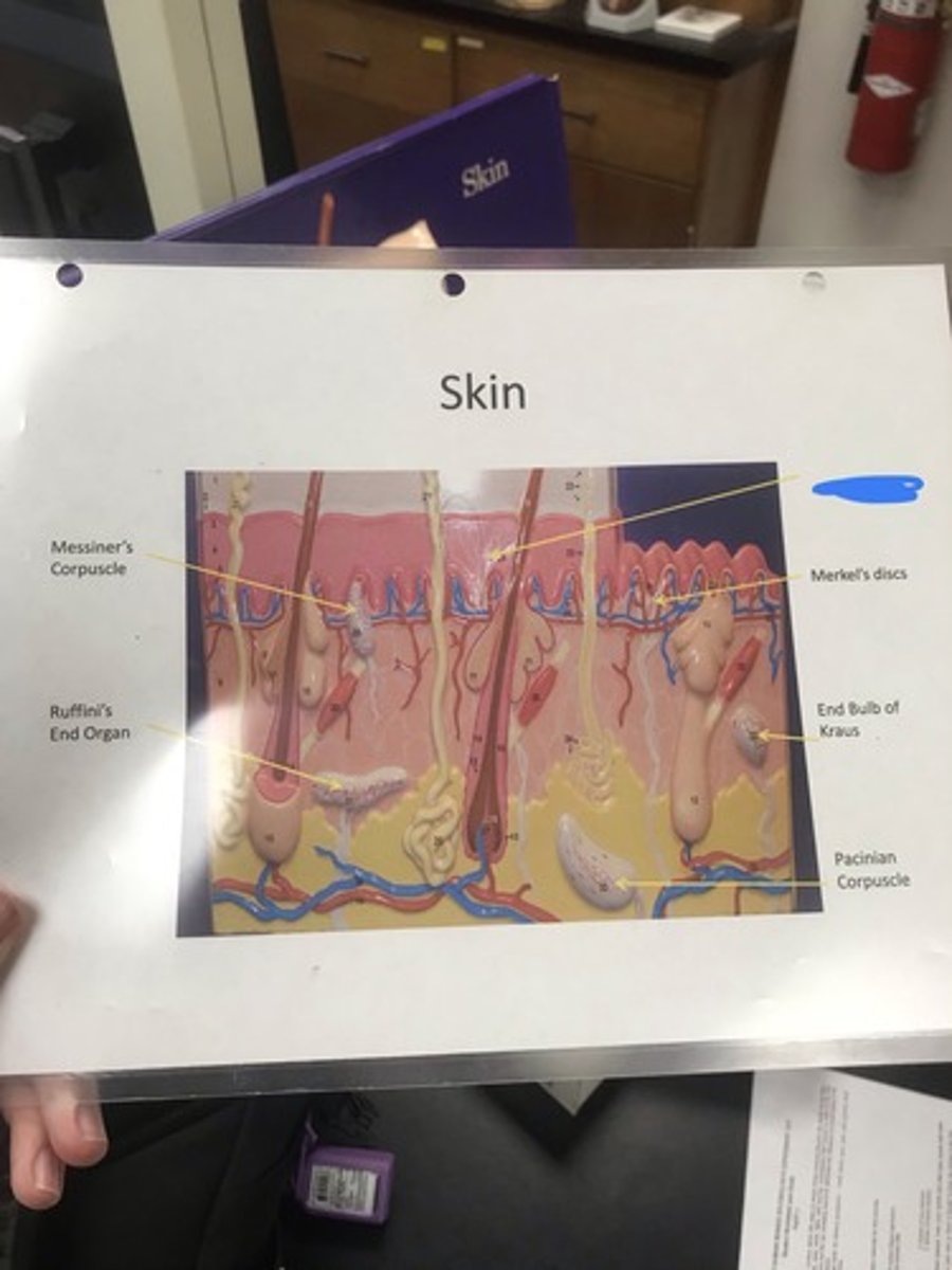
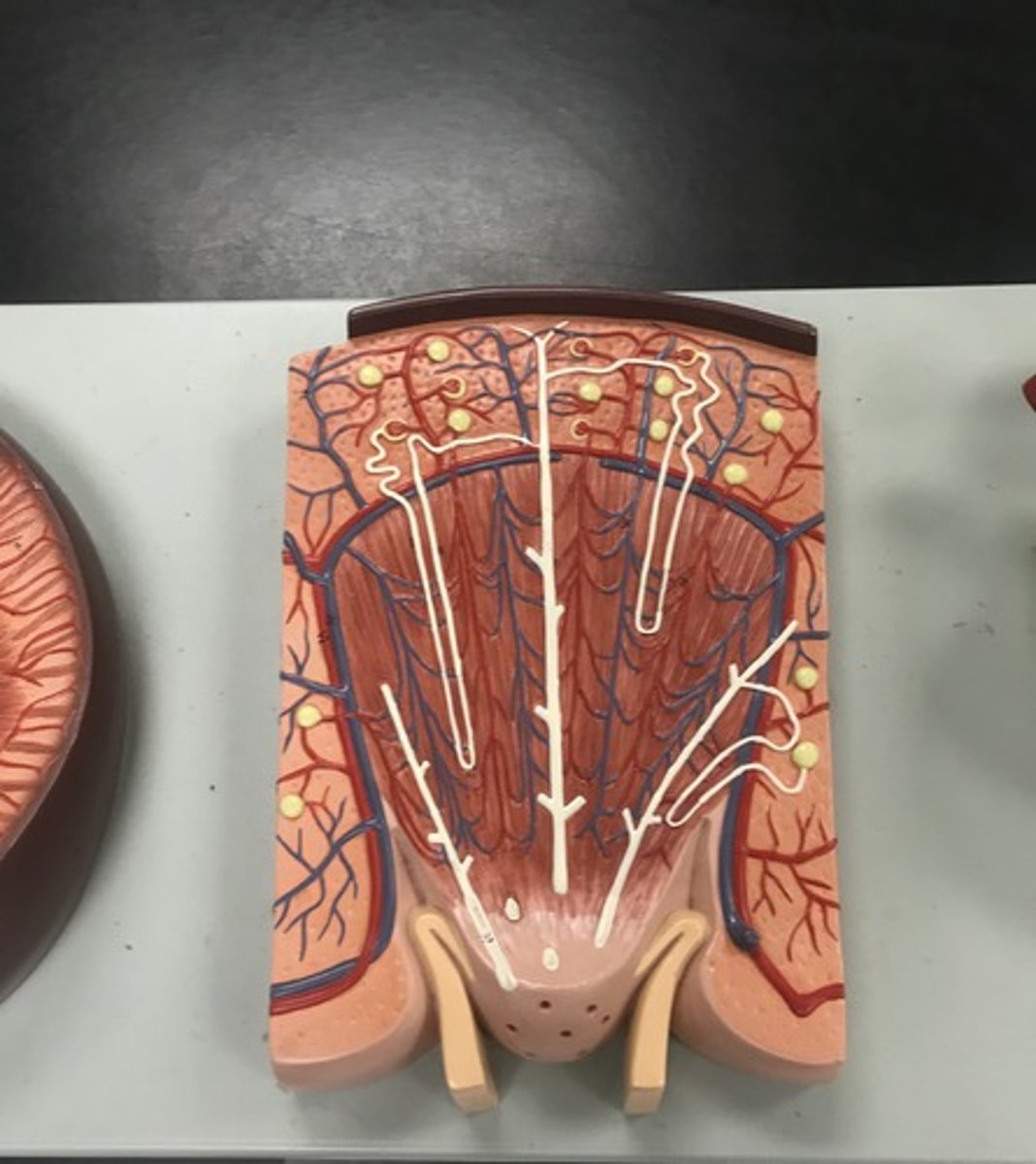
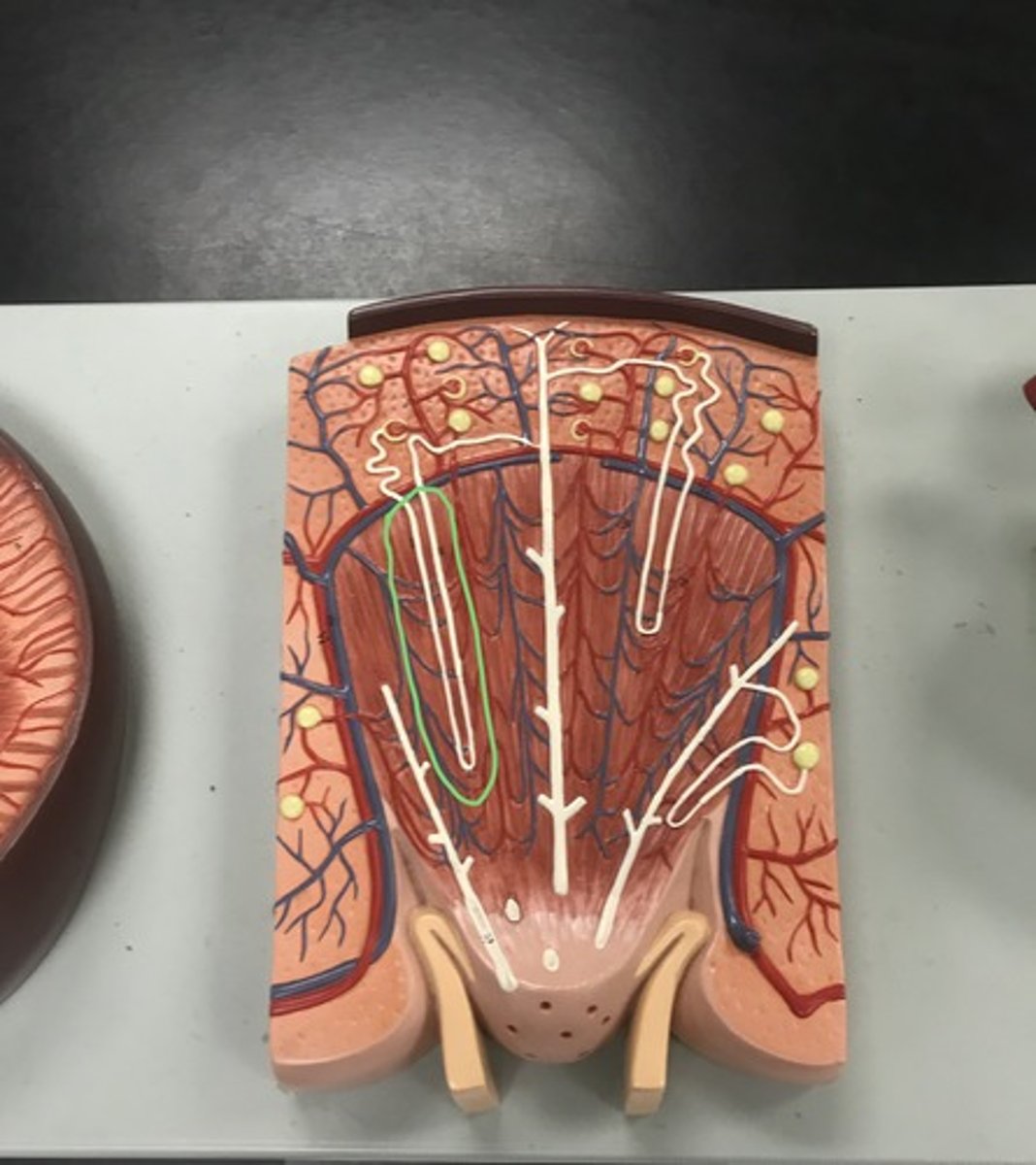
skin
Name the model
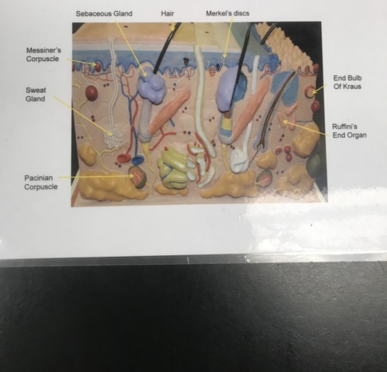
Merkel's discs/tactile discs
- allow for discriminative touch
Label the structure covered in blue ink and give a function
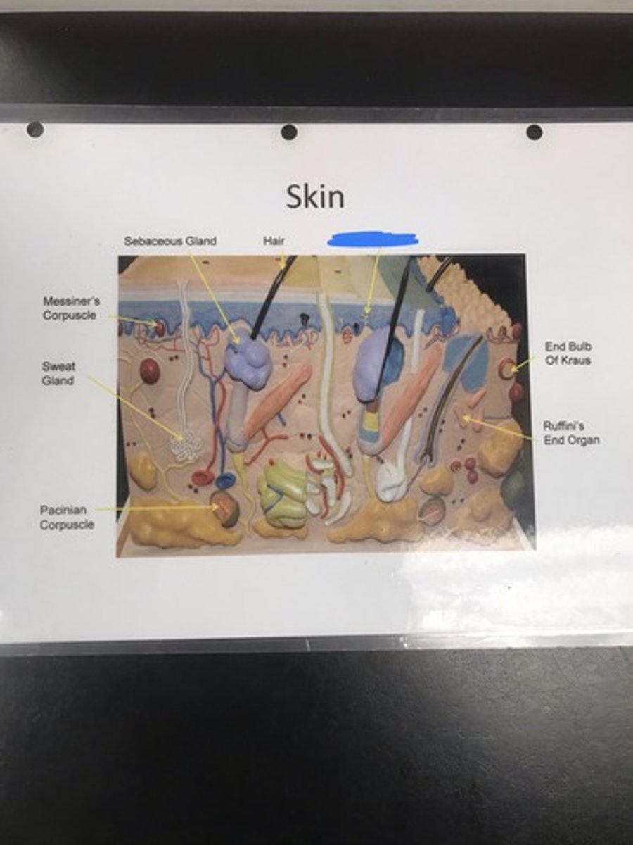
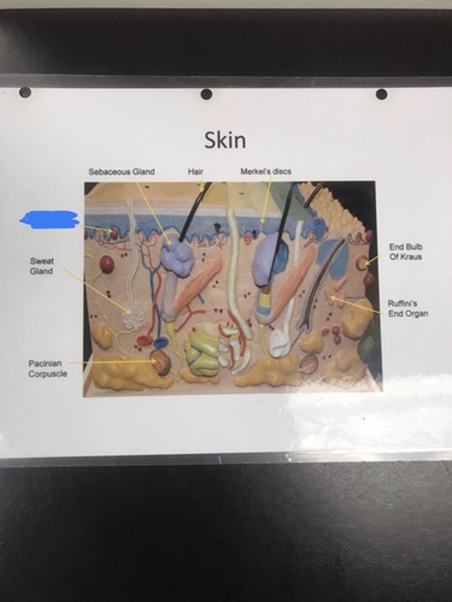
hair
Label the structure covered in blue ink
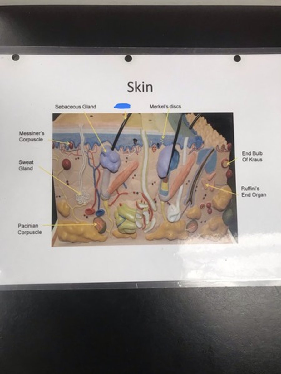
sebaceous gland
- secretes sebum into hair shaft to waterproof and lubricate skin and hair
Label the structure covered in blue ink and give a function
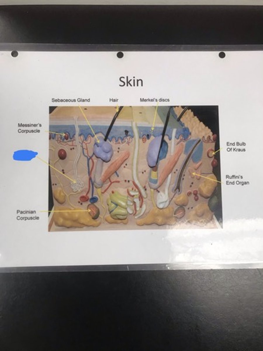
Meissner's corpuscle
- allow for discriminative touch
Label the structure covered in blue ink and give a function
sweat gland
- secretes sweat
Label the structure covered in blue ink and give a function
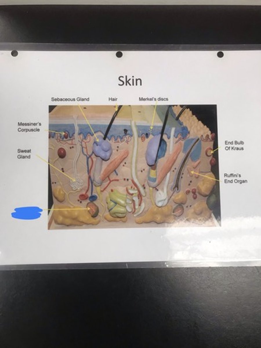
Pacinian corpuscle
- detecting pressure/sustained touch
Label the structure covered in blue ink and give a function
Ruffini's end organ
- heat sensation
Label the structure covered in blue ink and give a function
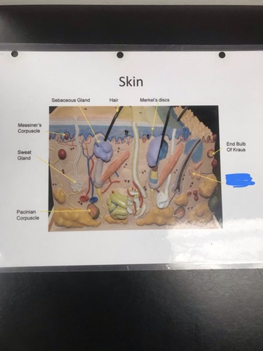
End bulb of Kraus
- cold sensation
Label the structure covered in blue ink and give a function
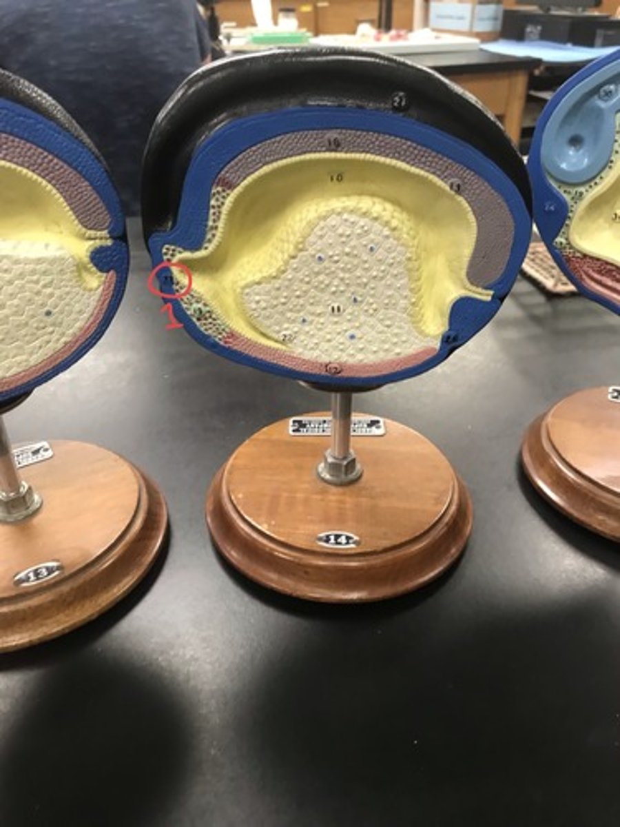
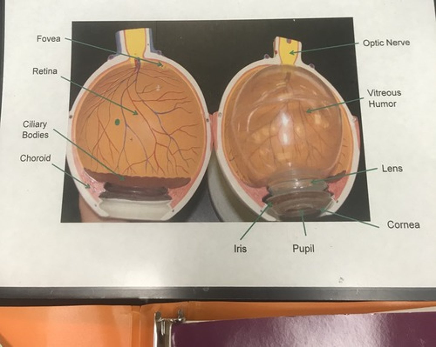
eye
Name the model
fovea
- concentrated area of cones that gives us sharp sight in the center of our visual field
Label the structure covered in blue ink and give a function

retina
- innermost eyeball layer that contains rods and cones
Label the structure covered in blue ink and give a function

ciliary bodies
Label the structure covered in blue ink
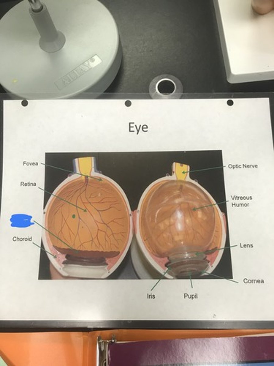
choroid
- pigmented inner eye layer responsible for "night shine" in nocturnal mammals
Label the structure covered in blue ink and give a function

iris
- pigmented muscle that surrounds the pupil
Label the structure covered in blue ink and give a function
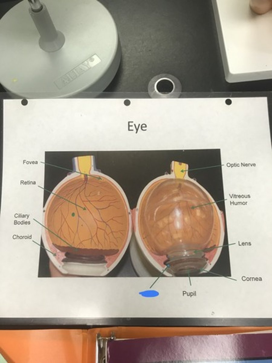
vitreous humor
- jellylike material that occupies the area behind the lens; most of eye volume
Label the structure covered in blue ink and give a function

optic nerve
Label the structure covered in blue ink

lens
- hardened structure behind cornea that helps focus light onto the retina
Label the structure covered in blue ink and give a function

cornea
- front of the eye where sclera becomes transparent
Label the structure covered in blue ink and define it
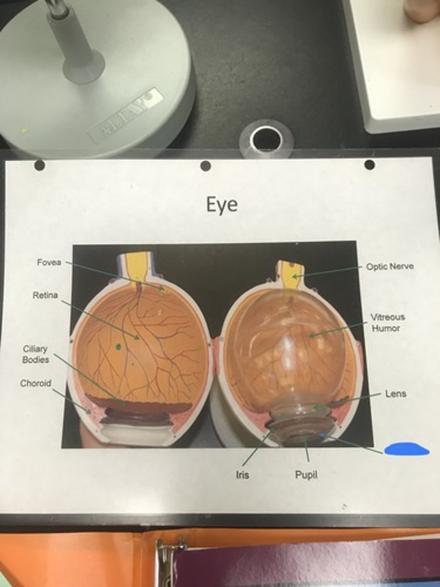
pupil
- hole in center of iris that allows light into the eye
Label the structure covered in blue ink and give a function

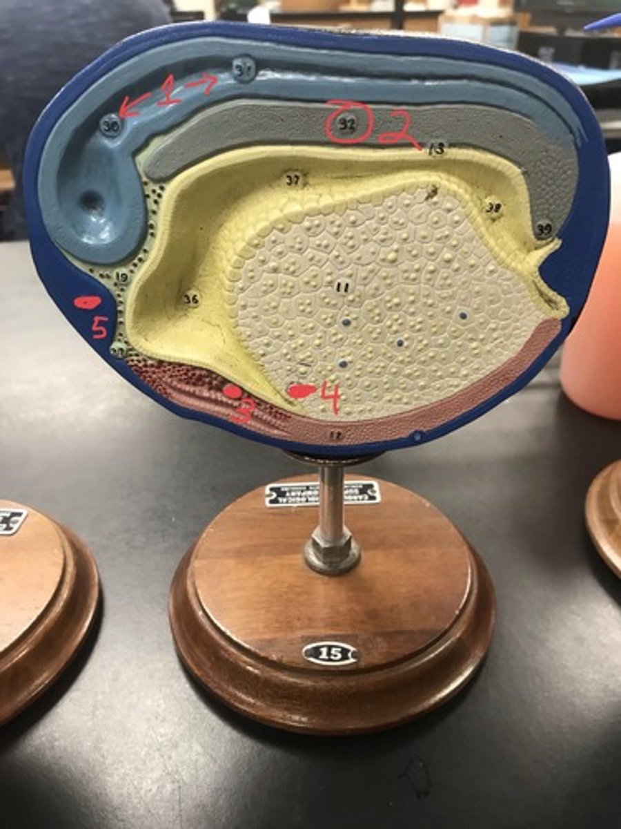
ear
Name the model
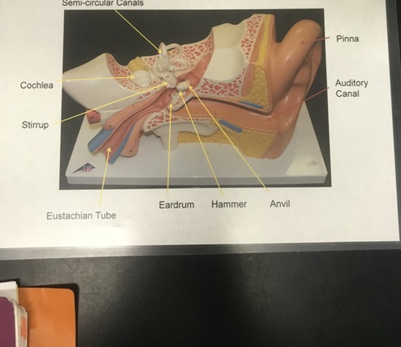
semi-circular canals ("pretzel rings"; contain ciliated epithelium)
- detect position of the head, giving our sense of balance
Label the structure covered in blue ink and give a function
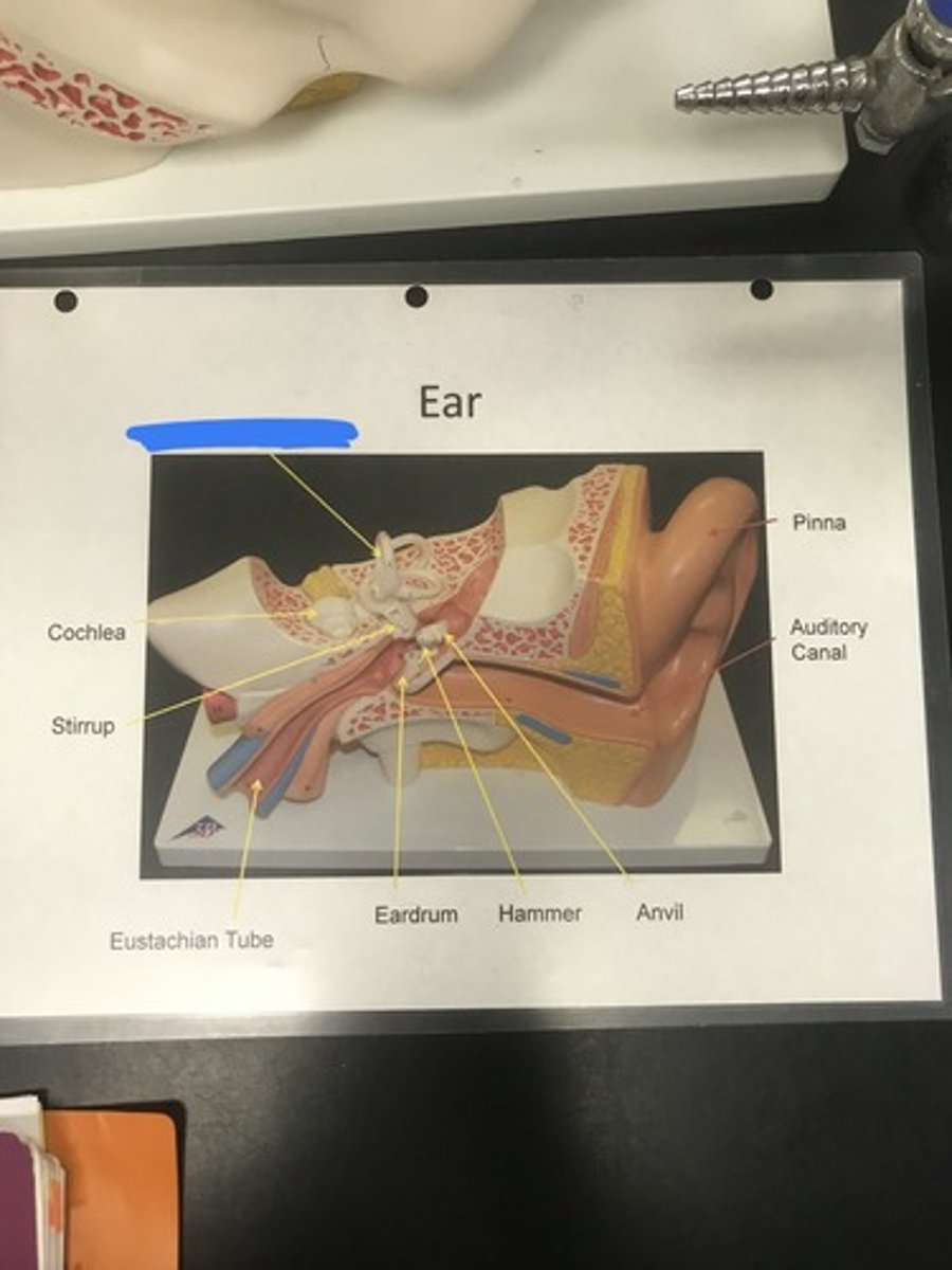
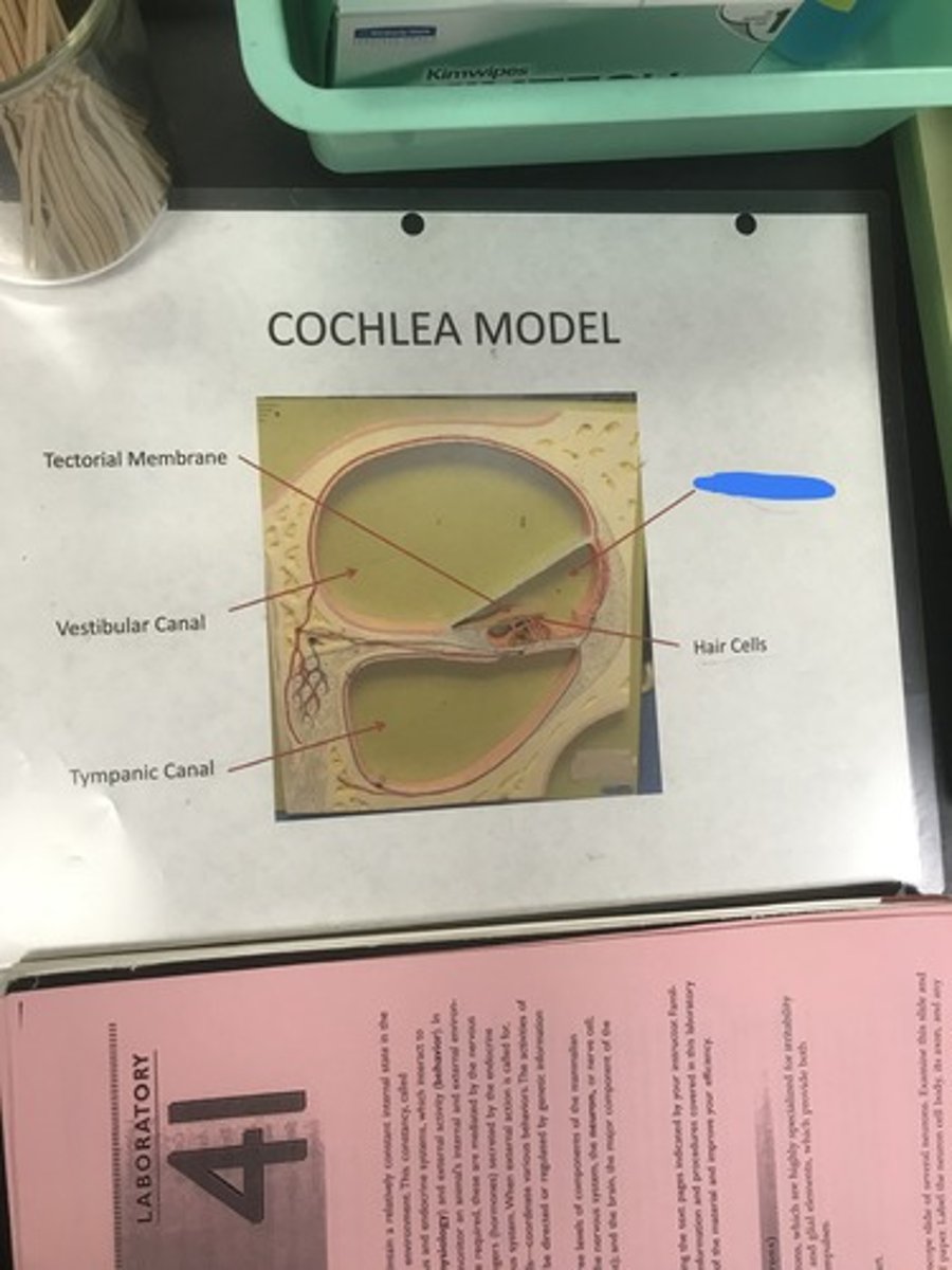
cochlea
- snail-shaped structure that contains hair cells used for sound detection
Label the structure covered in blue ink and give a function

stirrup (stapes)
- 3rd bone of middle ear that attaches to the oval window
Label the structure covered in blue ink and define it
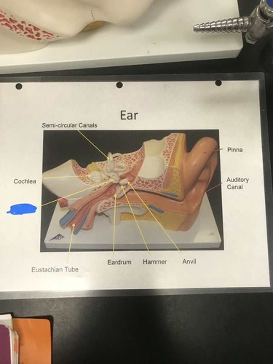
Eustachian tube
- connects middle ear with pharynx, allowing for equalized air pressure of the middle ear with the atmosphere
Label the structure covered in blue ink and give a function
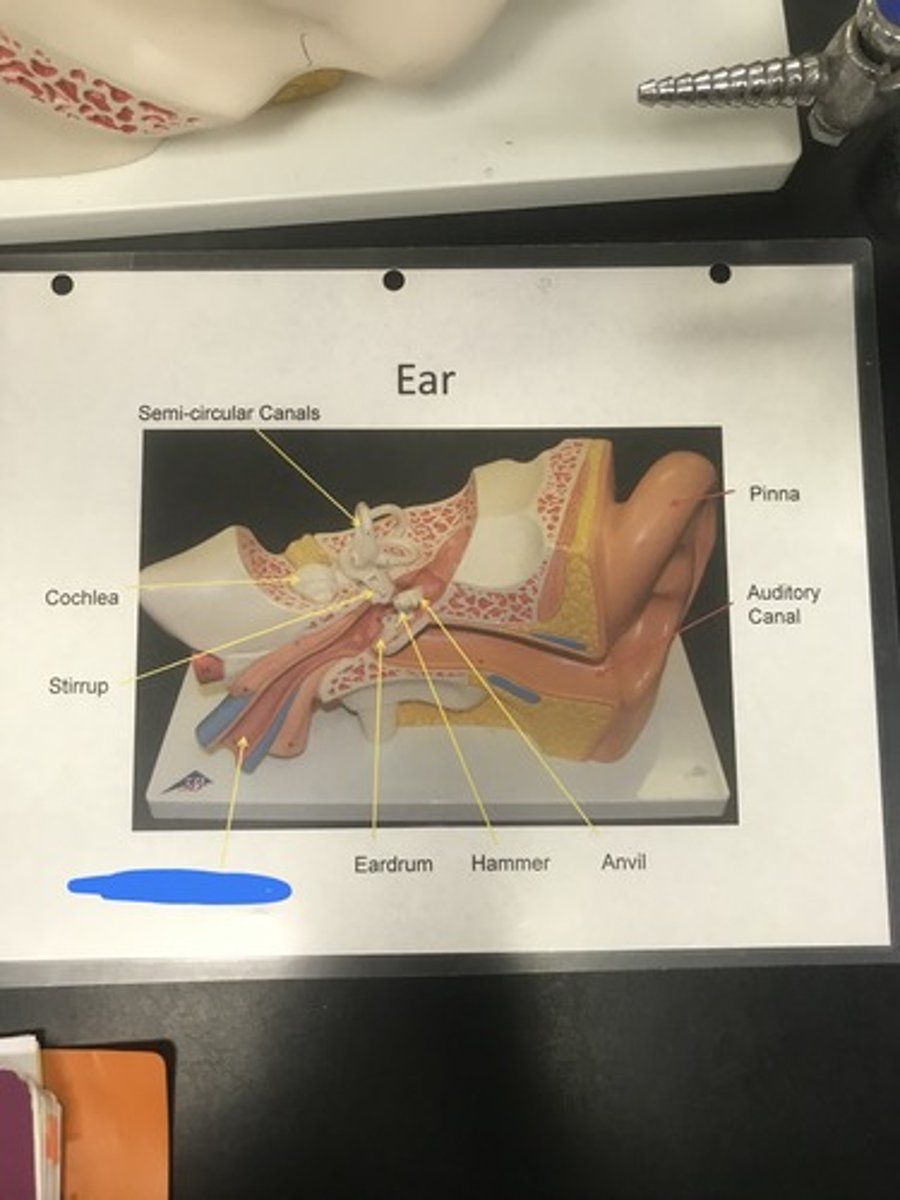
eardrum/tympanic membrane
- membrane that vibrates in response to sound waves
Label the structure covered in blue ink and define it

hammer (malleus)
- 1st bone of middle ear; directly attaches to eardrum
Label the structure covered in blue ink and define it
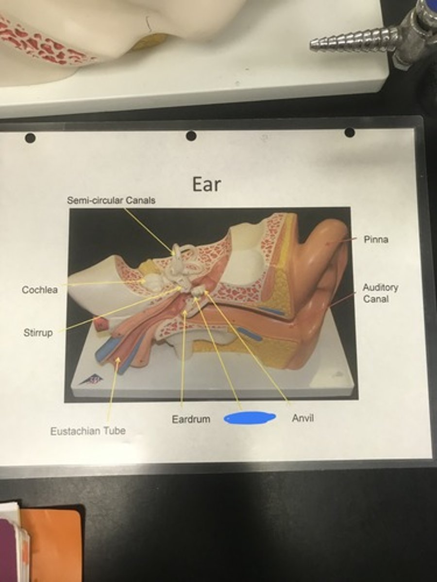
anvil (incus)
- 2nd bone of middle ear b/w hammer and stirrup
Label the structure covered in blue ink and define it
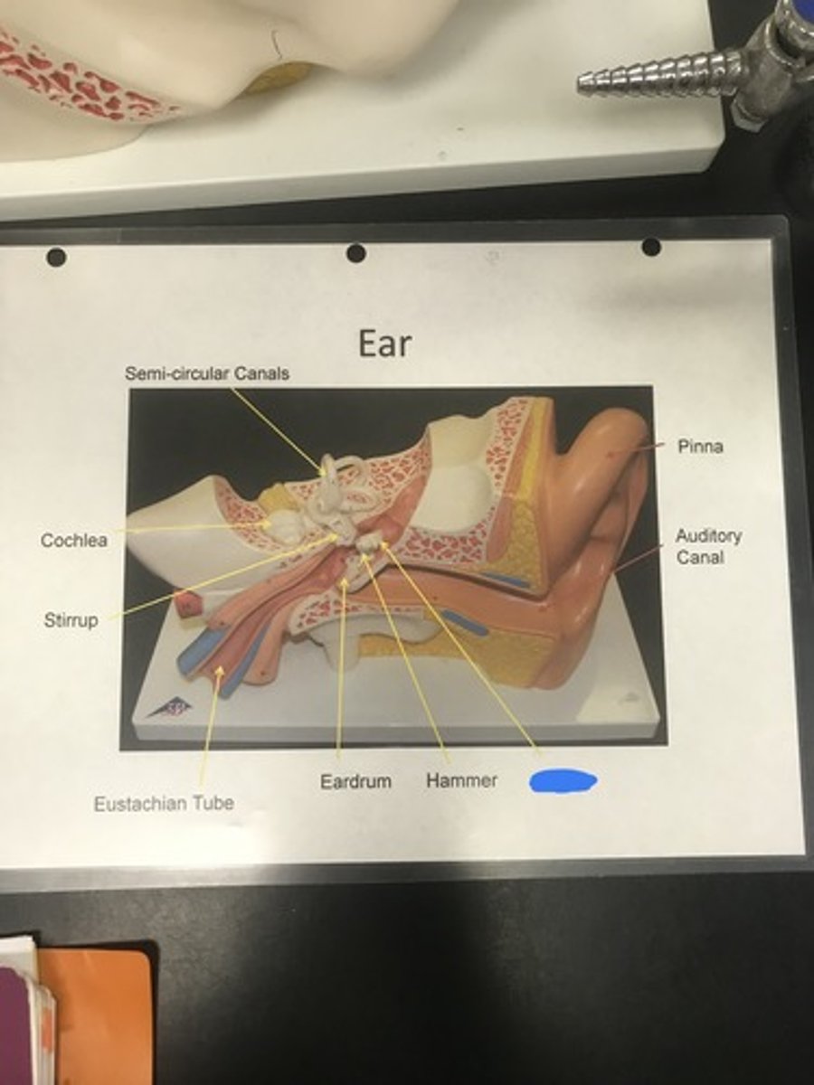
auditory canal
- passage from outside to eardrum
Label the structure covered in blue ink and give a function
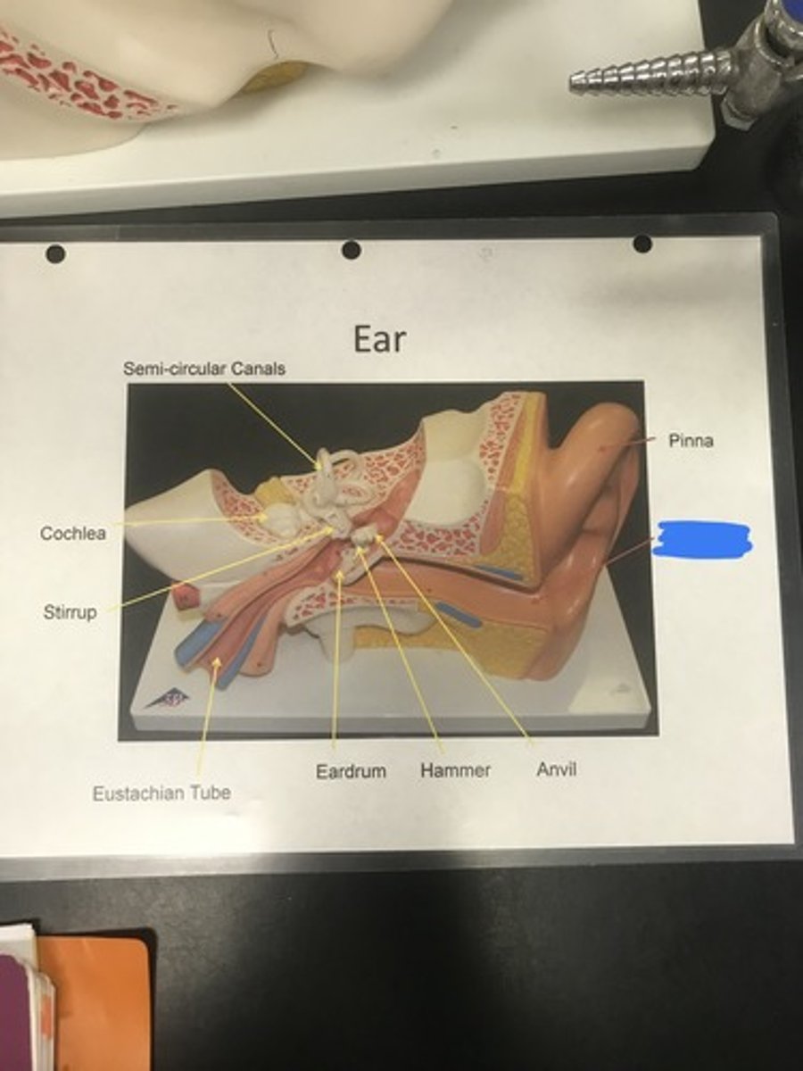
pinna
- external ear surface that funnels sound
Label the structure covered in blue ink and give a function

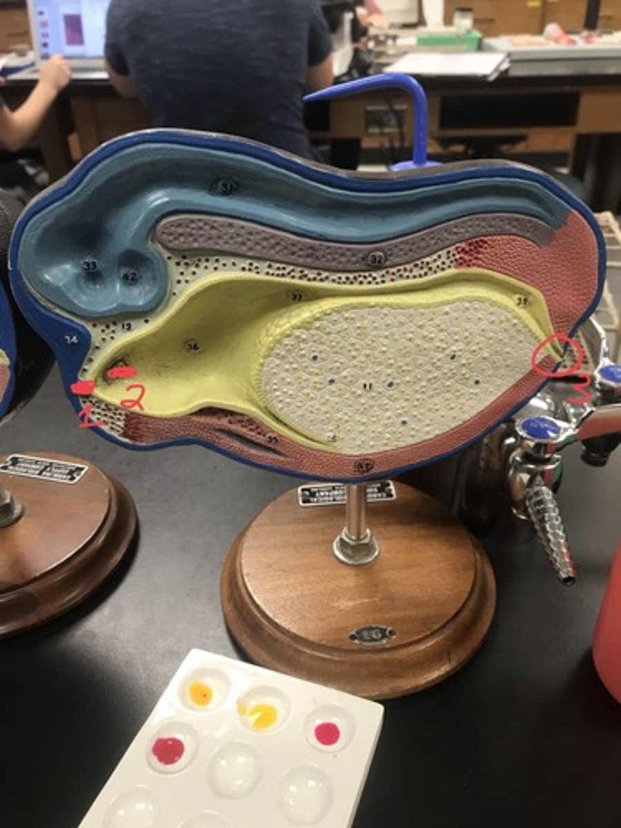
cochlea
Name the model
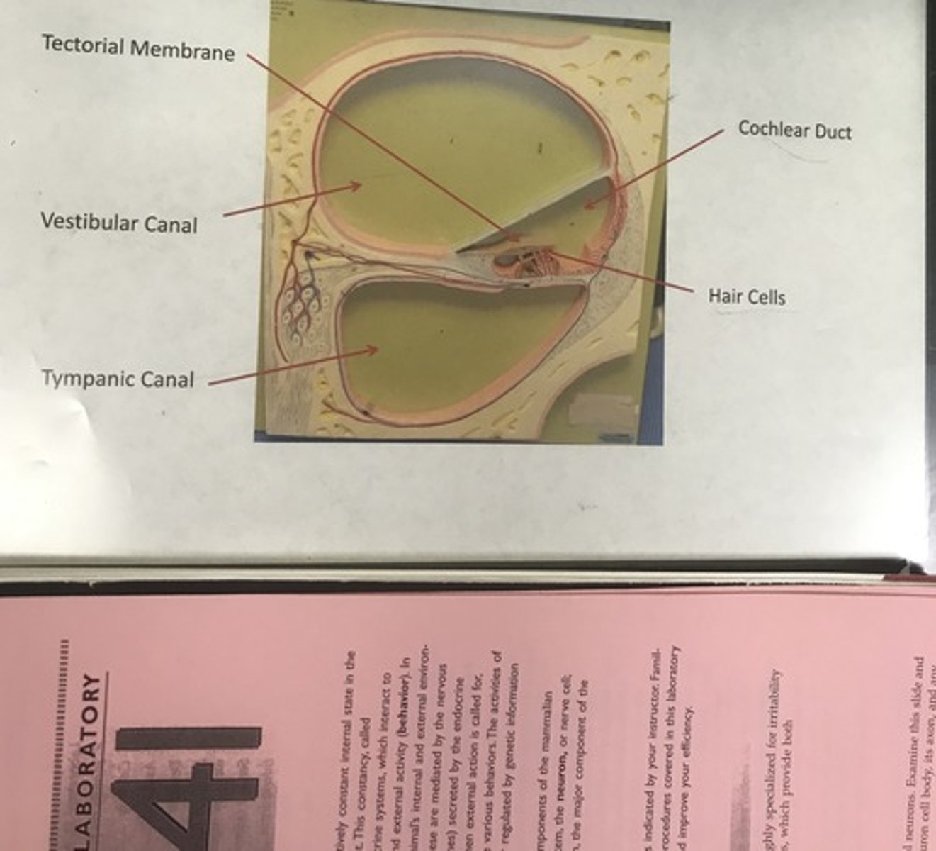
tectorial membrane
Label the structure covered in blue ink
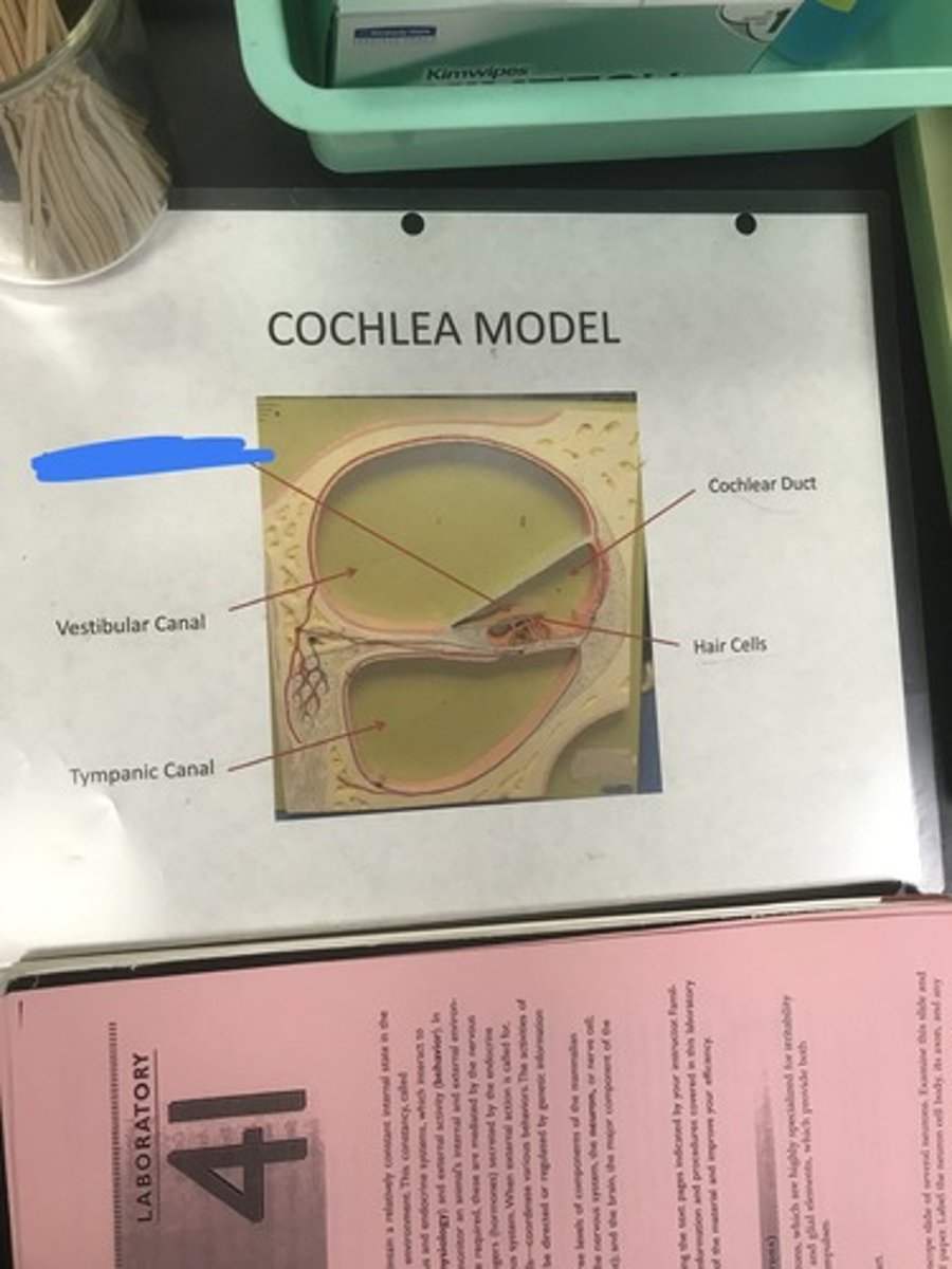
vestibular canal
- upper canal of cochlea
Label the structure covered in blue ink and define it
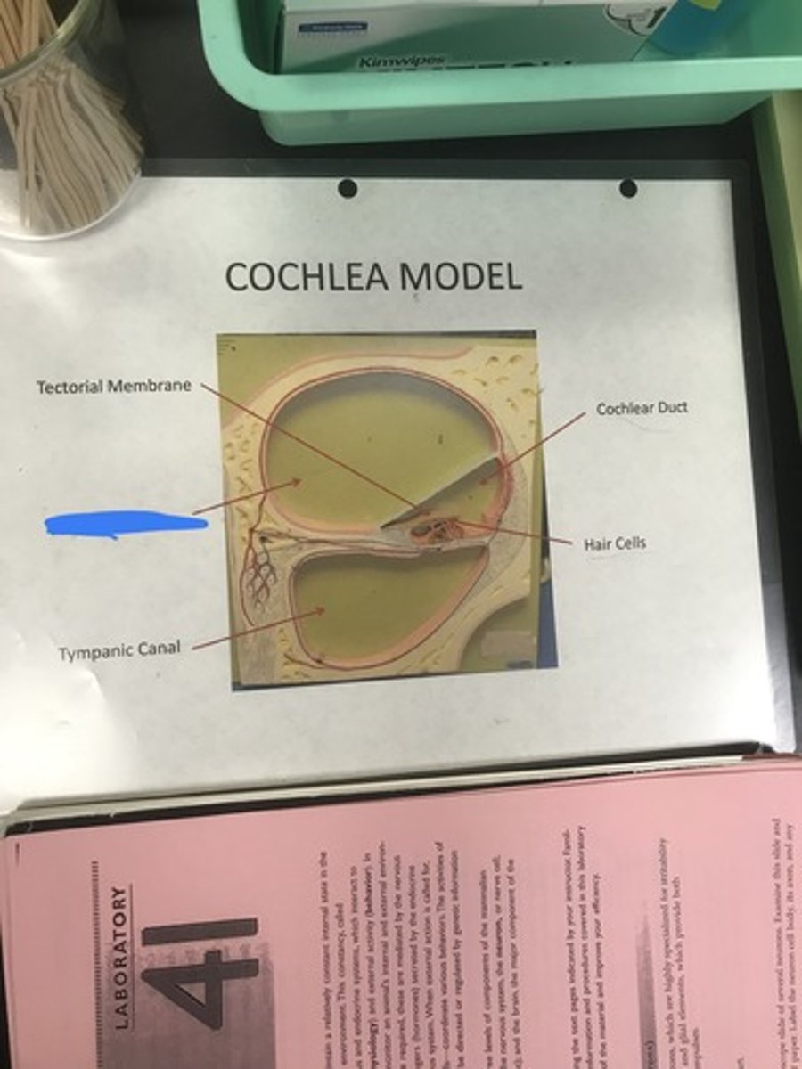
tympanic canal
- lower canal of cochlea
Label the structure covered in blue ink and give a function
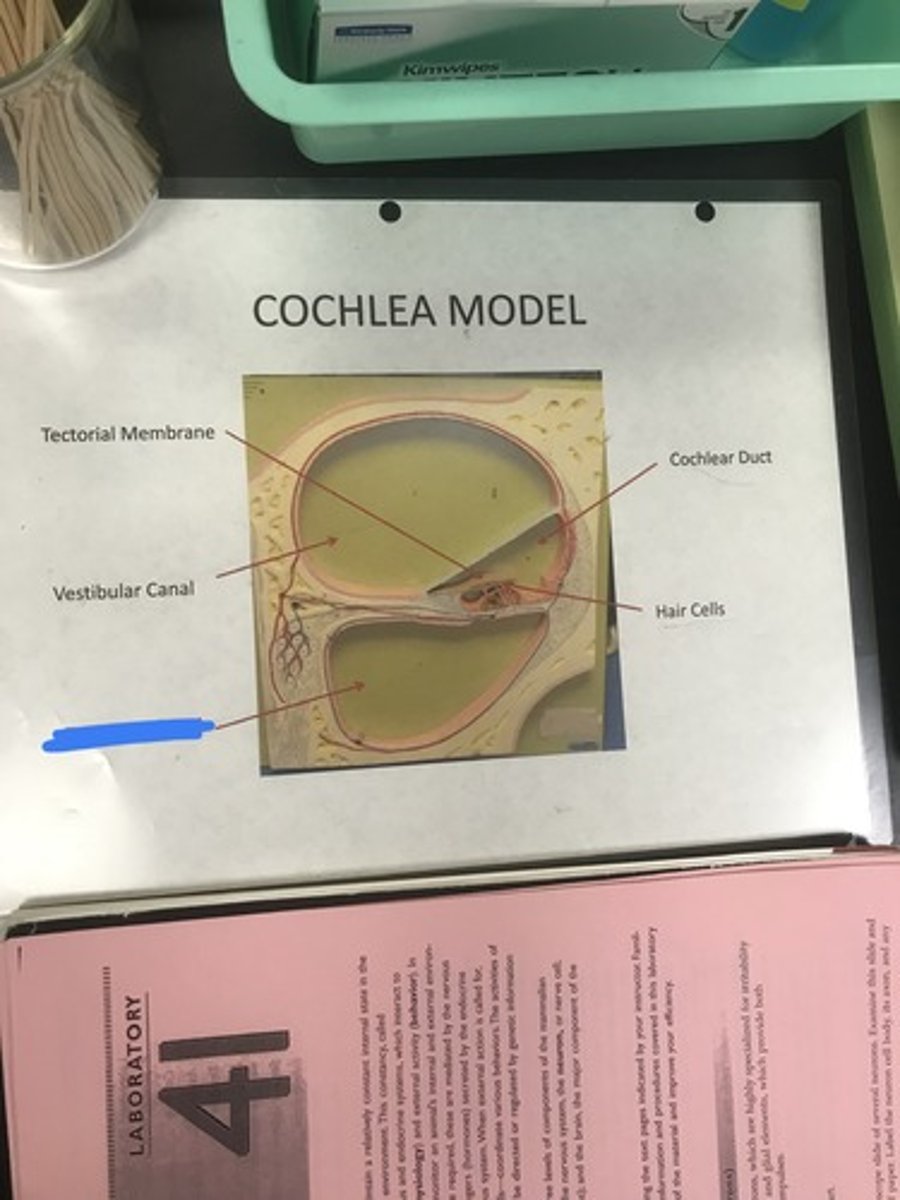
hair cells
Label the structure covered in blue ink
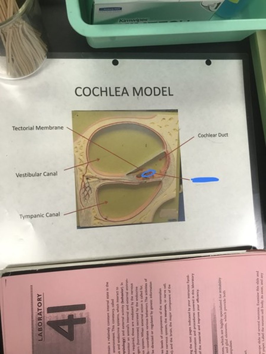
cochlear duct
Label the structure covered in blue ink
air, fluid
The outer and middle ears are filled with ________ and the inner ear is filled with __________
lower pitch
Vibration of a long hair cell will produce sound with a....
higher pitch
Vibration of a short hair cell will produce sound with a....
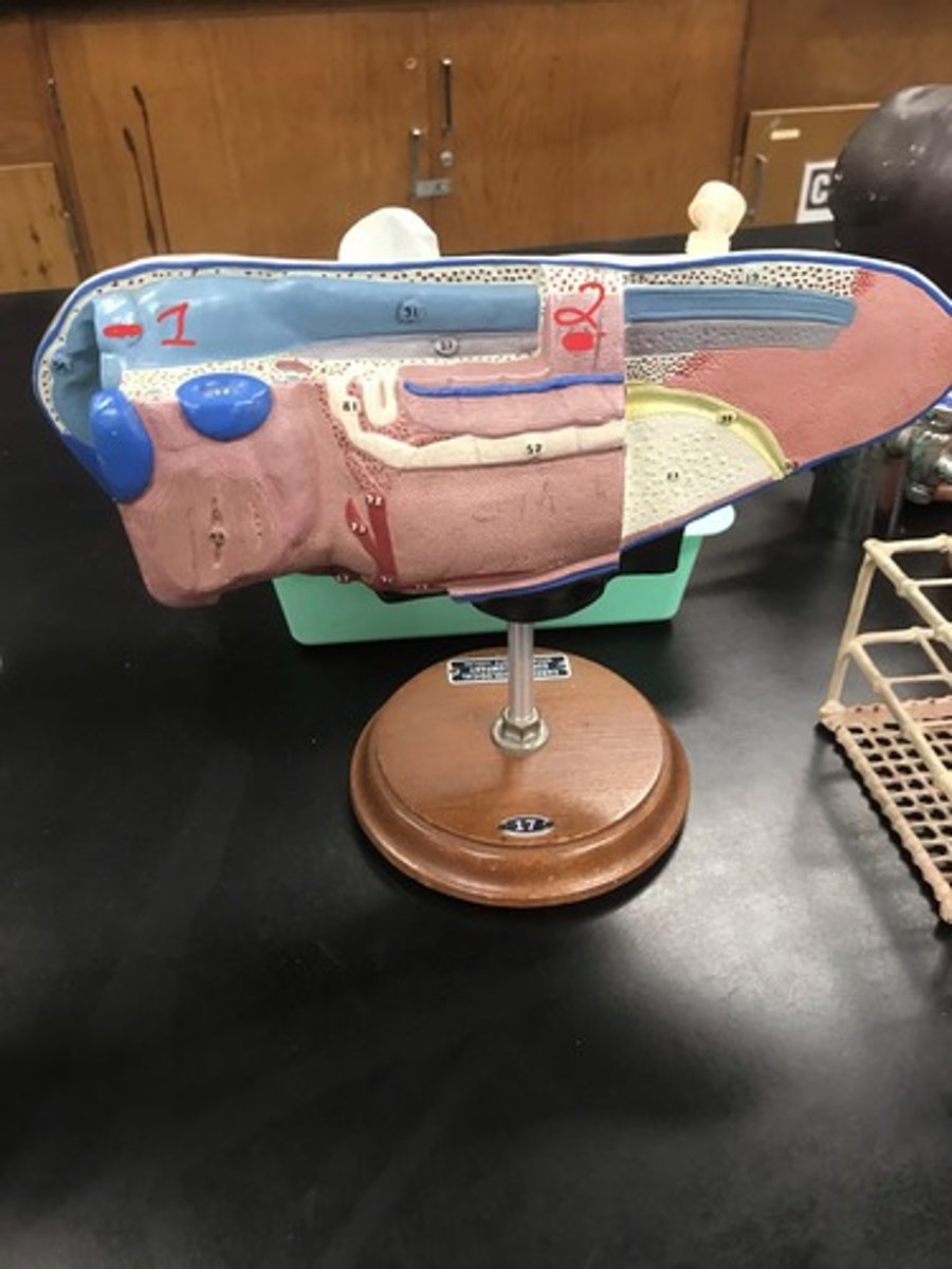
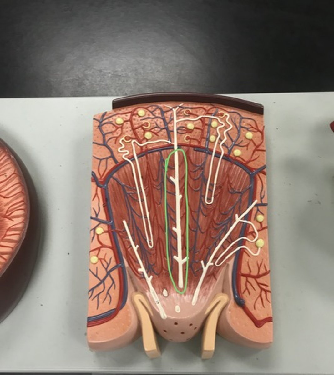
skin
Name the model
Meissner's corpuscle
- allow for discriminative touch
Label the structure covered in blue ink and give a function

Ruffini's end organ
- heat sensation
Label the structure covered in blue ink and give a function
Pacinian corpuscle
- detecting pressure/sustained touch
Label the structure covered in blue ink and give a function
End bulb of Kraus
- cold sensation
Label the structure covered in blue ink and give a function
Merkel's discs/tactile discs
- allow for discriminative touch
Label the structure covered in blue ink and give a function
nociceptor (naked dendrites in skin epidermis)
- responds to pain
Label the structure covered in blue ink and give a function
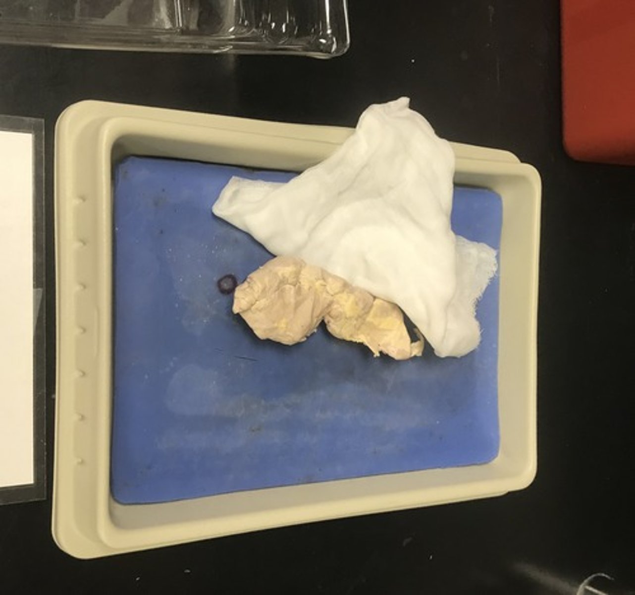
pig pancreas
Name the specimen
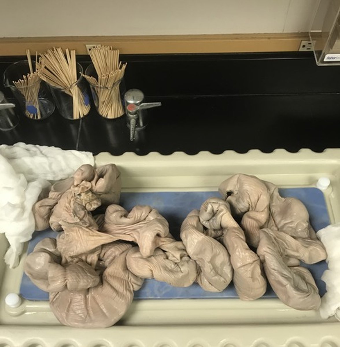
pregnant pig uterus
Name the specimen
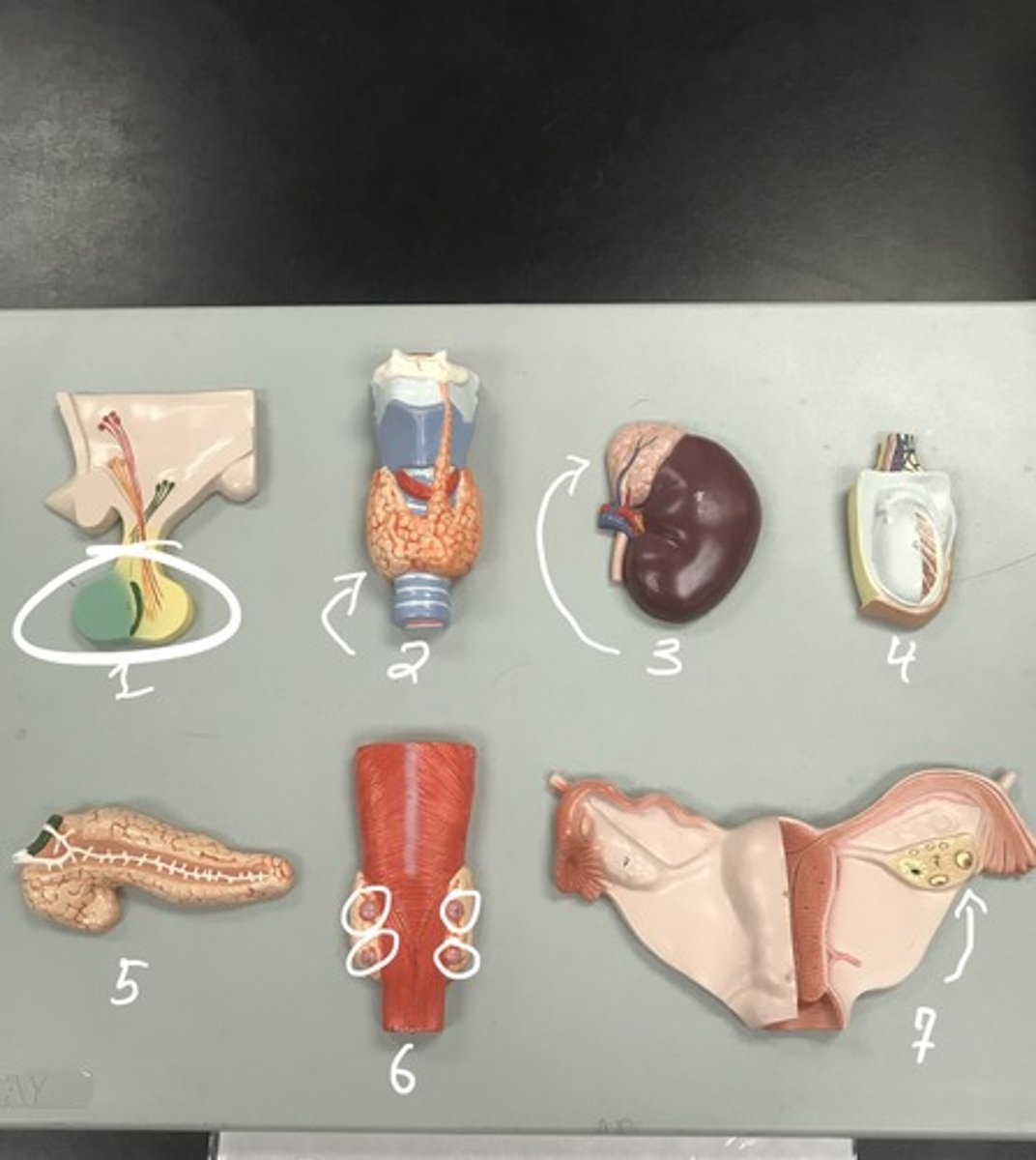
Structures:
1) hypophysis
2) thyroid gland
3) adrenal glands
4) testis
5) pancreas
6) parathyroid glands
7) ovary
Label each of the structures in numerical order
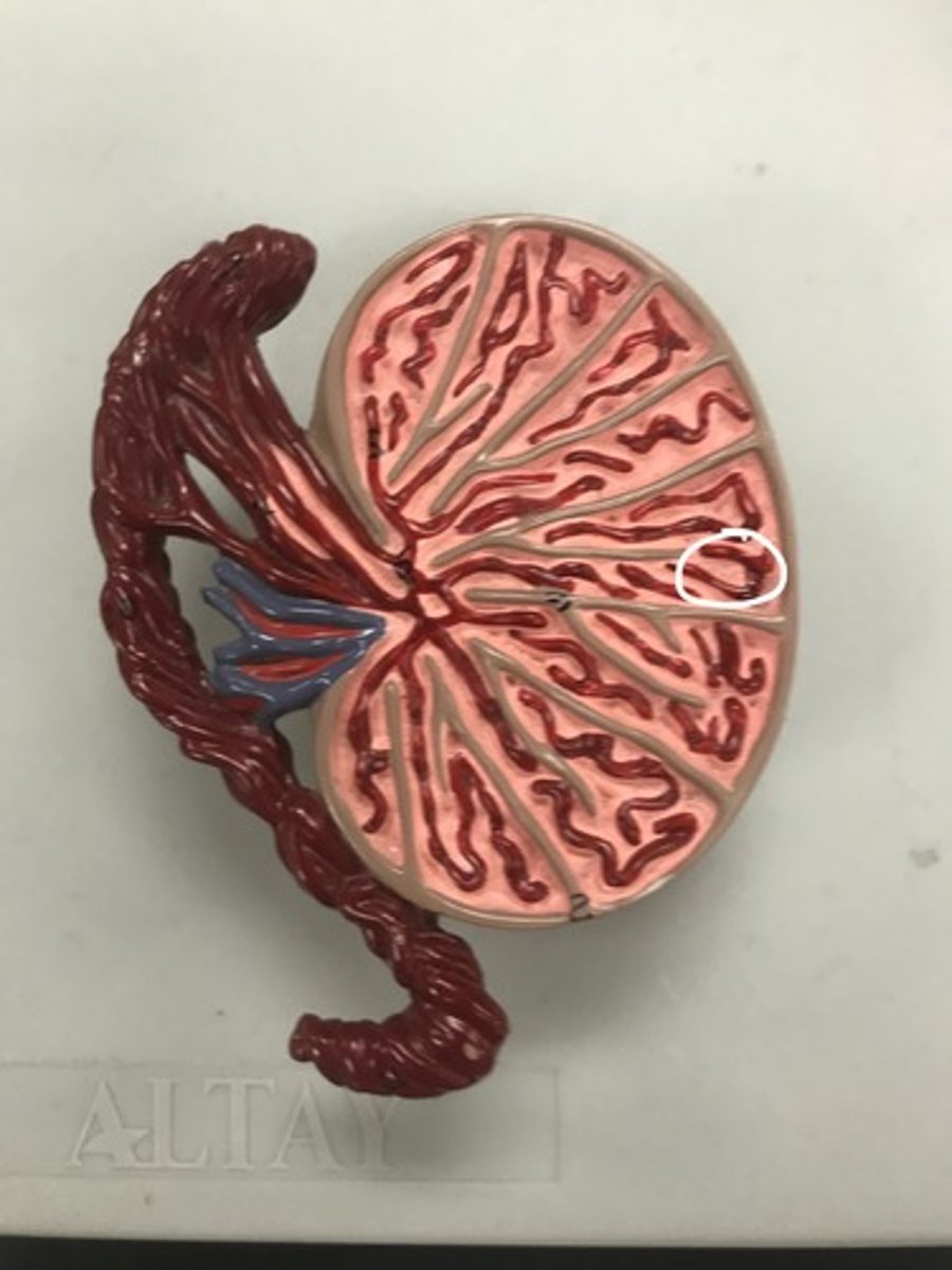
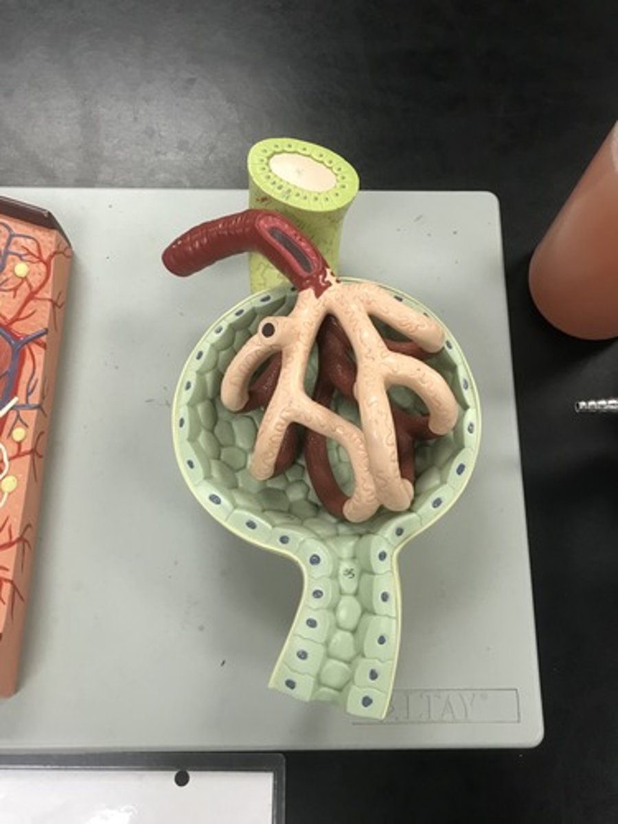
kidney nephron
Name the model
proximal convoluted tubule
Label the circled structure

Loop of Henle
Label the circled structure
descending limb
Label the circled structure

ascending limb
Label the circled structure

distal convoluted tubule
Label the circled structure

collecting duct
Label the circled structure
glomerulus in Bowman's capsule
What is this?
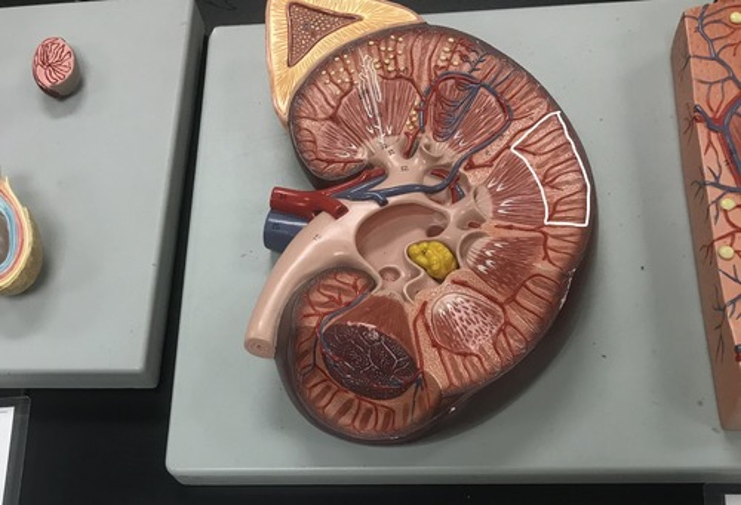
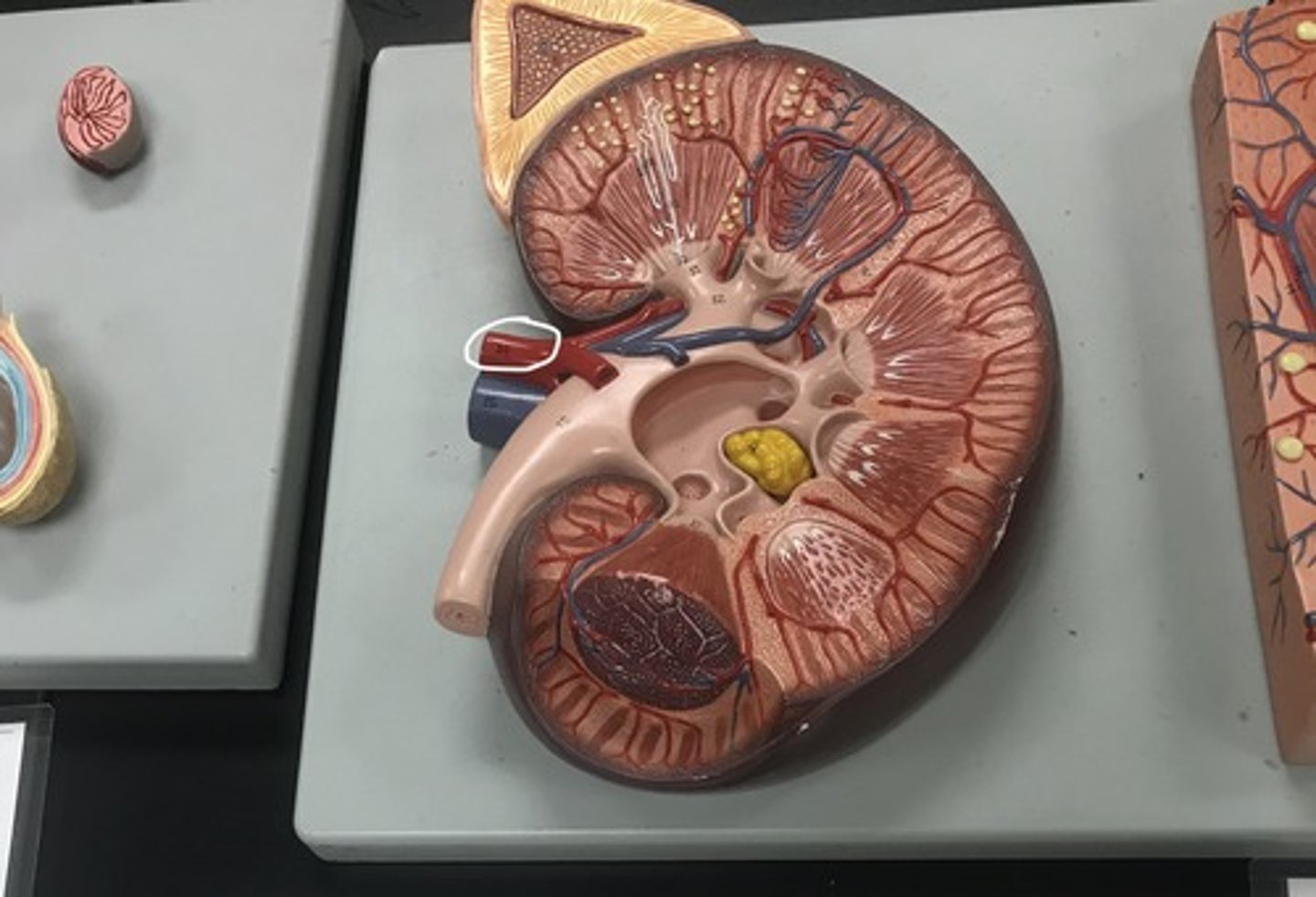
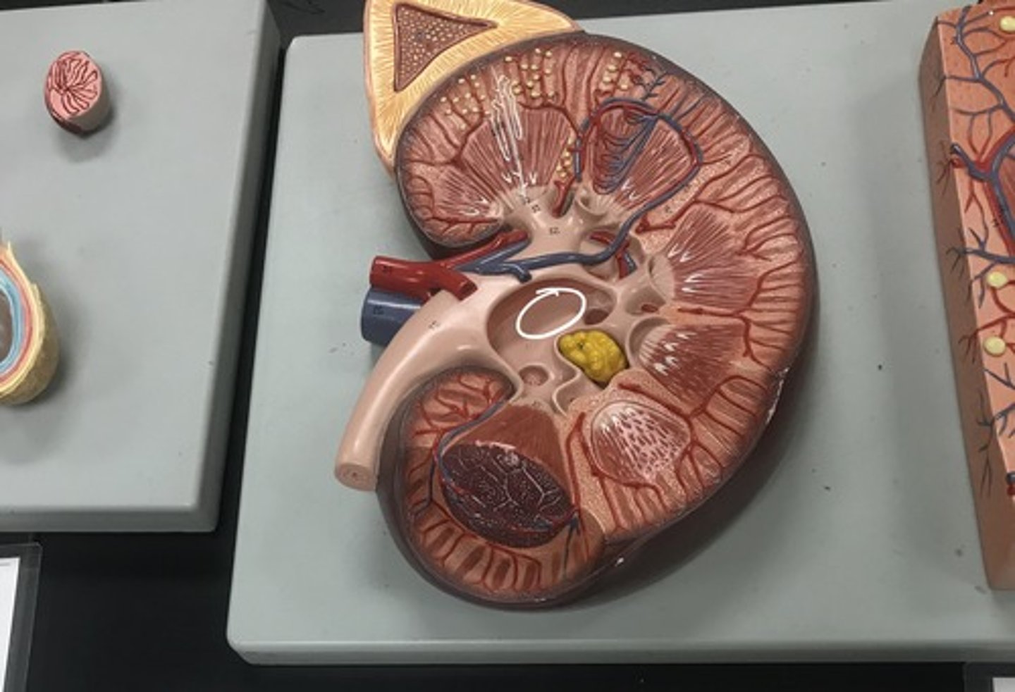
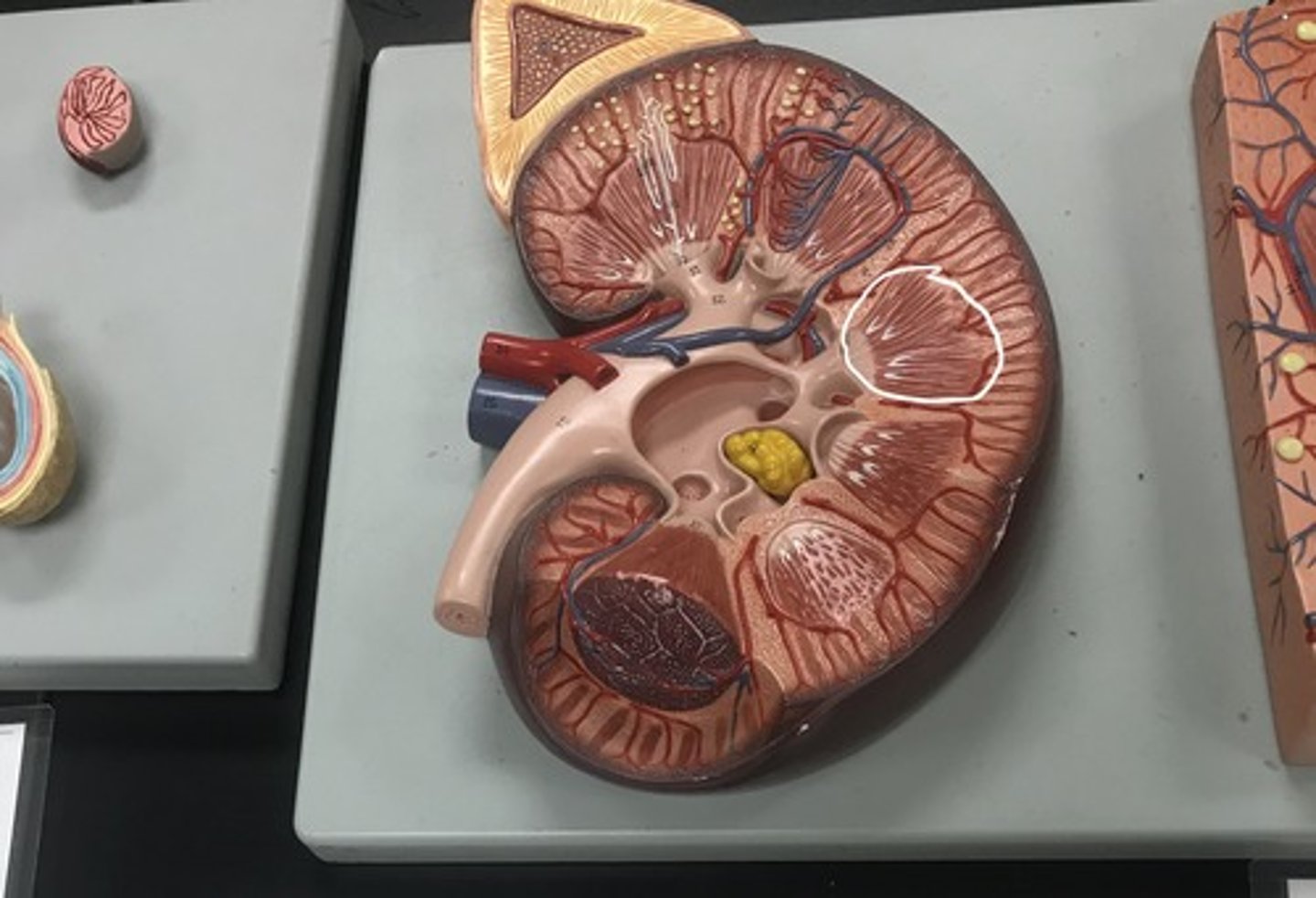
kidney
Name the model

cortex
Label the indicated structure
renal artery
Label the circled structure
renal vein
Label the circled structure

renal pelvis
Label the circled structure
medulla w/ pyramid
Label the circled structure
testis
Label the structure
contorted seminiferous tubules
Label the structure