chapter 8
1/31
Earn XP
Description and Tags
perception
Name | Mastery | Learn | Test | Matching | Spaced | Call with Kai |
|---|
No analytics yet
Send a link to your students to track their progress
32 Terms
akinetopsia
or “motion blindness,” where motion is either very difficult or impossible to perceive.
LM: stroke. couldnt percieve motion. couldnt poor a cup of T as water looked still. difficult for her to follow dialogue because she couldn’t see the motions of a speaker’s face and mouth, and people suddenly appeared or disappeared because she couldn’t see them approaching or leaving. Would see a far away car and then very suddenly it would appear near.
Functions of Motion
detecting things:
Perceiving Objects:
Perceiving Events:
Social Perception:
Taking Action: our own movement depends on our perception of movement. optic flow movement. We pay attention to other people’s movements to avoid colliding with them. Also keep track of our movements when completing a task. same as chapter 7.
detecting things:
motion is salient so attracts attnetion. needed for survival to move out of harms way. preditor prey relationship - both rely on motion.
Perceiving Objects:
our own motion relative to objects is constantly adding to the information we have about those objects. objects movement also provides us more information. Observers perceive shapes more rapidly and accurately when an object is moving. Movement also serves an organizing function, which groups smaller elements into larger units e.g. flocks of birds and movement of limbs organise with eachother to make biological movement.
Perceiving Events:
ongoing behaviour is perceived as a sequence of events (a segment of time with a clear beginning and end) e.g Example (coffee shop):Ordering coffee = one event. Taking the cup = another event. Each transition = an event boundary (Points where one event ends and another begins. Often linked to changes in movement). (Zacks et al., 2009): Participants watched everyday activities (e.g., washing dishes) and Pressed a button when they thought a new event began. Findings: Event boundaries occurred when there were changes in motion and Especially changes in speed or acceleration of movements. Motion perception helps segment behaviour into meaningful events.
Social Perception:
Movement is essential for understanding people’s actions and intentions. Example: Patient L.M. (akinetopsia) struggled socially because she couldn’t perceive motion (e.g., lip movements). Koul et al. (2019): Participants watched a hand reaching for a bottle. Task: decide if intention was to drink or pour. Findings: Judgments based on motion cues: speed (velocity), trajectory, grip type. Conclusion: People use movement characteristics to infer intentions (“why” of actions). Movement & Emotion Perception: Motion can communicate emotions and intentions even without other cues. Study: Heider & Simmel (1944). Participants watched moving geometric shapes. Findings: People created stories and personalities (e.g., “bully,” “couple”) e.g. the triangle was a bully and the circle and square was in a relationship. Conclusion: Humans naturally assign social meaning and emotions to movement. Other studies confirm motion can signal: fear, chasing, mocking, seduction, etc. Social Information from Minimal Cues. Study: Point-light walkers (Johansson). Lights placed on joints → only motion visible. Still perceived as a person moving. Another study: Two conditions: Social interaction (the characters interacted with eachother) & Non-social (independent movement). Findings: Most observers could tell if interaction was happening. People with autism spectrum disorder were less accurate. Conclusion: Movement alone provides important social interaction cues.
Taking Action:
our own movement depends on our perception of movement. optic flow movement. We pay attention to other people’s movements to avoid colliding with them. Also keep track of our movements when completing a task. same as chapter 7.
4 types of motion
real motion: something moves across our visual field.
illusory motion: the perception of the motion of stimuli that aren’t actually moving. three types:
Apparent motion: when two stimuli in slightly different locations are alternated with the correct timing, an observer perceives one stimulus moving back and forth smoothly between the two locations
Induced motion: motion of one object (usually a large one) causes a nearby stationary object (usually smaller) to appear to move. e.g. moon moves when clouds move.
Motion aftereffects: viewing a moving stimulus causes a stationary stimulus to appear to move. e.g. waterfall illusion
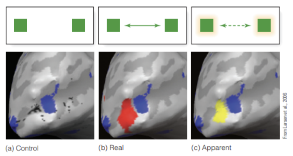
Comparing Real and Apparent Motion - Key Study: Larsen et al. (2006)
Key Study: Larsen et al. (2006)
Used fMRI to measure brain activity
Compared 3 conditions:
Control: two squares flashed at the same time → no motion perceived
Real motion: square physically moved back and forth
Apparent motion: squares flashed alternately → illusion of motion
Control condition:
Activated two separate brain areas (one for each square)
Real motion:
Activated a continuous area of visual cortex along motion path
Apparent motion:
Activation similar to real motion
Brain also activated the space between the squares, even though nothing was there
Real and apparent motion:
Use similar neural mechanisms
Are processed in similar brain areas
The brain fills in motion between positions, even without actual stimuli
Therefore, motion perception is best understood through general mechanisms that apply to both types
optic array
optic array —the structure created by the surfaces, textures, and contours of the environment—focused on
how movement of the observer causes changes in the optic
array.
what provides evidence for The Ecological Approach to Motion Perception
optic array
local disturbance in the optic array.
global optic flow
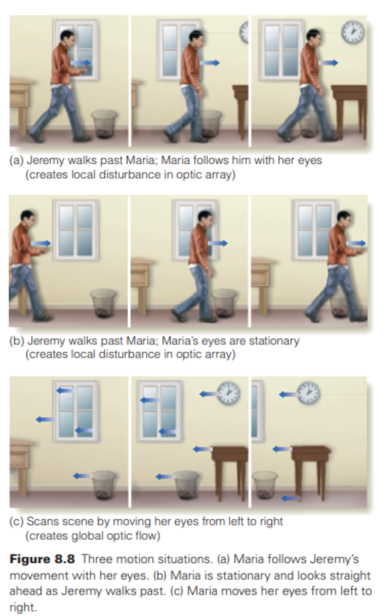
when Jeremy walks from left to right and Maria follows him with her eyes, portions of the optic array become covered as he walks by and then are uncovered as he moves on. This result is called a local disturbance in the optic array. covering and uncovering the stationary background, causes Maria to perceive Jeremy’s movement, even though his image is stationary on her retina.
The fact that everything moves at once in response to movement of the observer’s eyes or body is called global optic flow; this signals that the environment is stationary and that the observer is moving.
according to Gibson, motion is perceived when one part of the visual scene moves relative to the rest of scene, and no motion is perceived when the entire field moves, or remains stationary.
The Corollary Discharge and Motion Perception
(1) the image displacement signal, which occurs when an image moves across the retina, (2) the motor signal, which is sent from the motor area to the eye muscles to cause the eye to move, and (3) the corollary discharge signal, which is a copy of the motor signal.
Movement will be perceived if a brain structure called the comparator (actually a number of brain structures) receives just one signal—either the image displacement signal or the corollary discharge signal. movement will be perceived if the comparator receives both signals at the same time.
a shows the signals that occur when Maria is following Jeremy with her eyes. There is a CD signal, because Maria is moving her eyes. There is, however, no image displacement signal, because Jeremy’s image stays in the same place on Maria’s retina. The comparator, therefore, receives just one signal, so Maria perceives Jeremy to be moving.
b shows that if Maria keeps her eyes stationary as Jeremy walks across her field of view, there is an image movement signal, because Jeremy’s image is moving across Maria’s retina, but there is no CD signal, because Maria’s eyes are not moving. Because only one signal reaches the comparator, movement is perceived.
c shows that if Maria scans the room, there is a CD signal because her eyes are moving and an image movement signal because the scene is moving across her retinas. Because both signals reach the comparator, no movement is perceived.
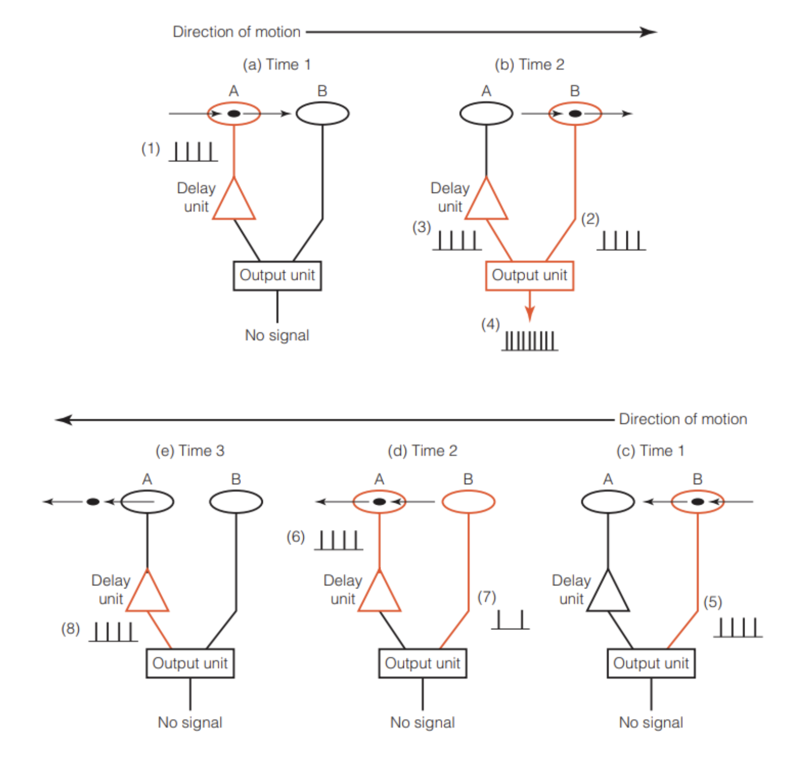
The Reichardt Detector
Overview
Explains how motion is perceived when the eye is stationary.
Proposed by Werner Reichardt (1961, 1987).
Based on a neural circuit called the Reichardt detector.
Structure of the Circuit
Contains:
Neuron A
Neuron B
Output unit
Key features:
Delay unit slows signals from neuron A.
Output unit multiplies signals from A and B to detect motion.
How It Detects Motion (Left → Right)
Neuron A activates first
Object (Jeremy) approaches from the left.
A sends a signal that is delayed by delay unit.
Neuron B activates next
Object moves right and stimulates B.
B sends a signal directly to the output unit.
Timing coincidence
Delayed signal from A + direct signal from B arrive at the same time.
Multiplication at output unit
Signals are multiplied → large response.
Result: motion is perceived.
Why Direction Matters (Right → Left)
Neuron B activates first
Signal goes directly to output.
Neuron A activates later
Signal is delayed.
Signals don’t overlap
By the time A’s delayed signal arrives, B’s signal has faded to zero. Result: no motion signal detected.
Key Property
Circuit is direction-sensitive:
Detects motion left → right
Does not detect motion right → left
These create directionally selective neurons:
Each neuron responds to a specific direction of motion.
Many such circuits work together to:
Detect movement direction across the visual field
single neuron motion and MT cortex - experiment on monkeys
Single-Neuron Responses to Motion
directionally-selective neurons were recorded from neurons in the rabbit’s retina and in the cat’s visual cortex
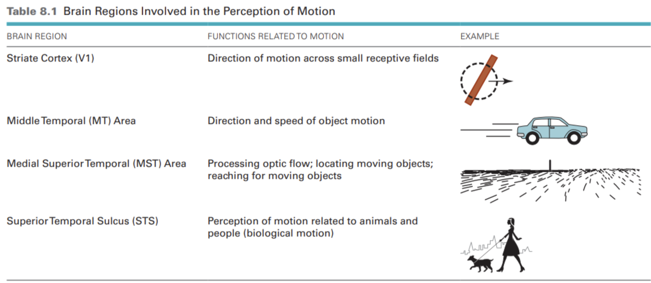
middle temporal (MT) area: contains many directionally selective neurons.
Experiments Using Moving Dot Displays:
coherence = all the dots are moving the same way. when movement is random, coherance = 0.
Newsome et al. – Motion Perception Study (MT Cortex). Purpose of Study: Investigated the relationship between:
Monkey’s ability to judge motion direction
Activity of neurons in the MT cortex
Used moving dot stimuli with varying levels of coherence
(coherence = % of dots moving in the same direction)
Key Findings
As dot coherence increased:
Monkey’s accuracy in judging direction increased
MT neurons fired more strongly
Strong correlation:
Neural activity and behaviour were closely linked
Researchers could predict one from the other
Low Coherence (0.8%)
Very few dots moving in the same direction
Results:
Monkey could not judge direction
Neuron firing ≈ baseline (no significant increase)
Higher Coherence
Increasing coherence → better performance
At 12.8% Coherence
~25 out of 200 dots move in same direction
Results:
Monkey judged direction almost perfectly
MT neuron fired above baseline every time
Conclusion
MT cortex activity directly relates to motion perception
Strong evidence that: Neural firing in MT supports perception of motion direction
effects of Lesioning the MT Cortex:
A monkey with an intact MT cortex can begin detecting the direction dots are moving when coherence is as low as 1 or 2 percent. However, after the MT is lesioned, the coherence must be 10 to 20 percent before monkeys can begin detecting the direction of motion.
effects of Deactivating / simulating the MT Cortex:
transcranial magnetic stimulation (TMS) = temporarily disrupts the normal functioning of neurons. A series of electromagnetic pulses presented to a particular
area of the brain for a few seconds interferes with brain functioning in that area for seconds or minutes. When researchers applied TMS to the MT cortex, participants had difficulty determining the direction in which a random pattern of dots was moving.
Stimulating the MT Cortex: microstimulation = lowering a small wire electrode into the cortex and passing a weak electrical charge through the tip of the electrode. causes neural firing. monkey watch dots move right. when the researchers stimulated neurons that are activated by downward motion, the monkey began responding as though the dots were moving downward and to the right.
medial superior temporal (MST) area:
involved in eye movements, so it is particularly important in localizing a moving object in space. For example, a monkey’s ability to reach for a moving object is adversely affected by both microstimulation and lesioning of the MST cortex.
aperture problem
the neuron’s receptive field is functioning like an aperture, which reveals only a small portion of the scene e.g moving left and up, not just left.
solution to the apature problem
neurons that follow the end of objects. e.g. the tip of pencil would be seen to move across and up. thus information about the end of a moving object used to determine its direction of motion. found in the striate cortex
Pooling of Neuronal Responses (MT Cortex): Pooling = combining responses from multiple neurons. Helps the brain determine the true direction of motion. Study by Pack & Born (2001). Recorded activity from MT cortex neurons in monkeys. Stimuli: moving oriented lines (e.g., a bar or pencil).
Early Response (~70 ms): Neuron response based on orientation of the object, not true motion.
Example:
Vertical bar moving:
Horizontally right
Diagonally up-right
→ Same neuronal response
Later Response (~140 ms)
Neurons begin responding to the actual direction of motion.
Indicates additional processing has occurred.
Explanation
MT neurons:
Receive input from many neurons in the striate cortex (V1).
Combine (pool) these signals.
This allows the system to:
Resolve ambiguity
Accurately detect true motion direction
Key Idea
Motion perception is a two-stage process:
Initial response → based on orientation
Later response → reflects true motion direction via pooling
shortest path constraint
apparent movement tends to occur along the shortest path between two stimuli.
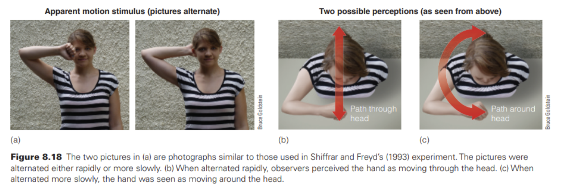
apparent motion in a hand going through a head image
(1) They show that the visual system needs time to process information in order to perceive the movement of complex meaningful stimuli. (2) They suggest that there may be something special about the meaning of the stimulus—in this case, the human body—that influences the way movement is perceived.
When objects such as boards are used as stimuli, the likelihood of perceiving movement along the longer path does not increase at lower rates of alternation, as it does for pictures of humans
• ⁃ found that both movement through the head and movement around the head activated areas in the parietal cortex associated with movement. However, when the observers saw movement as occurring around the head, the motor cortex was activated as well. Thus, the motor cortex is activated when the perceived movements are humanly possible but isn’t activated when the perceived movements are not possible. This connection between the brain area associated with perceiving movement and the motor area reflects the close connection between perception and taking action
Biological Motion Studied by Point-Light Walkers
created by placing small lights on people’s joints and then filming the patterns created by these lights when people move.
shows that motion of the body creates perceptual organization by causing the movements of the individual dots to become organized into “a person moving.” When the person is stationary, nno significant pattern is identified by observers, but as soon as the dots start moving in a human-like walk, the motion of the lights is immediately perceived as being caused by a walking person. This self-produced motion of a person or other living organism is called biological motion.
found that a small area in the superior temporal sulcus (STS) was more active when viewing biological motion than viewing scrambled motion in all eight of their observers
• ⁃ both the FFA and the portions of the PFC that contain mirror neurons are activated more by biological motion than by scrambled motion.
brain regions involved in motion perception
perception of motion is decreased by lesioning the MT cortex and is influenced by stimulating neurons in the MT cortex
disrupting operation of the MT cortex decreases a monkey’s ability to perceive the direction of moving dots
• ⁃ using transcranial magnetic stimulation (TMS) to disrupt the operation of the STS in humans decreases the ability to perceive biological motion
Grossman (2005) – Biological Motion Perception
Aim: Investigate how the brain perceives biological motion (movement of living beings).
Method: Participants viewed: Point-light biological motion (e.g., walking, kicking, throwing), Scrambled motion (same dots, random movement)
Task: Decide whether motion was: Biological or Scrambled
Difficulty increased by: Adding noise dots
Noise adjusted so performance ≈ 71% accuracy
Key Brain Area: Focus on STS (superior temporal sulcus): Activated during biological motion perception
Key Findings: transcranial magnetic stimulation Applied to STS = Significant decrease in ability to detect biological motion. TMS Applied to Other Areas: MT cortex = no effect on biological motion perception. Prefrontal cortex = also impairs biological motion perception.
Conclusion: STS is essential for perceiving biological motion
what is the STS
superior temporal sulcus
what is the MT
middle temporal area
what is MST
medial superior temporal area
implied motion:
still picture depicts an action involving motion
study: Aim: Test whether people mentally anticipate motion in static images.
Method: Participants shown:
Initial image (person midair)
After a pause, a second image:
Same image
Time-forward image (person closer to ground)
Time-backward image (person higher up)
Task: Decide as quickly as possible if second image is: Same orDifferent
Key Findings: Participants took longer to respond to Time-forward images than they did Time-backward images
Explanation: Viewers mentally predict future motion (e.g., falling downward)
This causes Confusion between:
What they actually saw and What they expected to happen next
Conclusion: The brain automatically extrapolates motion forward in time
Static images can trigger implied motion perception
representational momentum:
motion depicted in a picture tends to continue in the observer’s mind.
found that the area of the brain that responds to actual motion also responds to pictures of motion, and that implied-motion pictures caused a greater response than no-implied-motion pictures.
motion after effect: Winawer showed participants images with implied motion (left or right) and then asked them to judge the direction of random moving dots. After viewing rightward implied motion, participants were more likely to see the dots moving left, and vice versa. This opposite effect is the same as real motion adaptation, suggesting that implied motion reduces activity in direction-specific neurons and is processed similarly to real motion.