Case 3: Daniel Gatto
1/62
There's no tags or description
Looks like no tags are added yet.
Name | Mastery | Learn | Test | Matching | Spaced | Call with Kai |
|---|
No analytics yet
Send a link to your students to track their progress
63 Terms
Joints
Connection between bones
Joint Types
Synovial
Cartilaginous
Fibrous
Synovial Joints
Freely movable (diarthrosis)
Connected by intraarticular space with articular cavity
Contain synovial fluid = Reduce friction
Synovial Joints: Bone Articulation
Articular Head: Convex bone end moving in socket joint
Socket Joint: Concave bone end containing articular head
Synovial Joints: Joint Capsule
Surround joint
Fibrous Membrane: Dense connective tissue
Synovial Membrane: Synovial cells (synoviocytes)
Type A: Macrophage-like cells remove debris
Type B: Fibroblast-like cells produce glycoproteins → Synovial fluid
Synovial Joint Types
Ball and socket
Acetabulofemoral
Glenohumeral
Condyloid
Saddle
Hinge
Pivot
Modified hinge
Plane
Patellofemoral
Cartilaginous Joints
Slightly movable (amphiarthrosis)
Connected by cartilage
Ex:
Tibiofibular
Carpus
Tarsus
Fibrous Joints
Fixed (synarthrosis)
Connected by connective tissue (usually collagen)
Ex:
Skull sutures
Between ulna + radius
Between tibia + fibula
Joint Menisci
Fibrocartilaginous discs in joints
Absorb force + prevent abnormal movement
In:
Knee
Wrist
Acromioclavicular
Sternoclavicular
Temporomandibular
Joint Cruciate Ligaments
Ligaments crossing in X shape
In knee only
Joint Collateral Ligaments
Ligaments on side of joint
In many joints
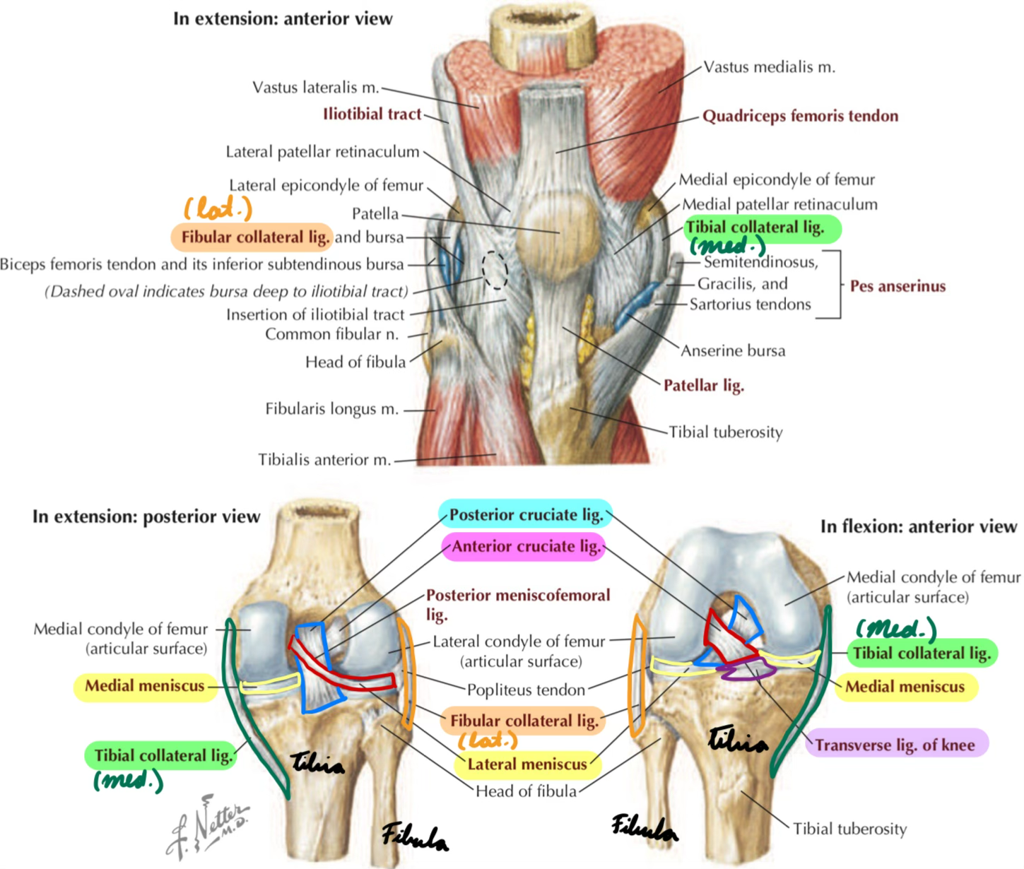
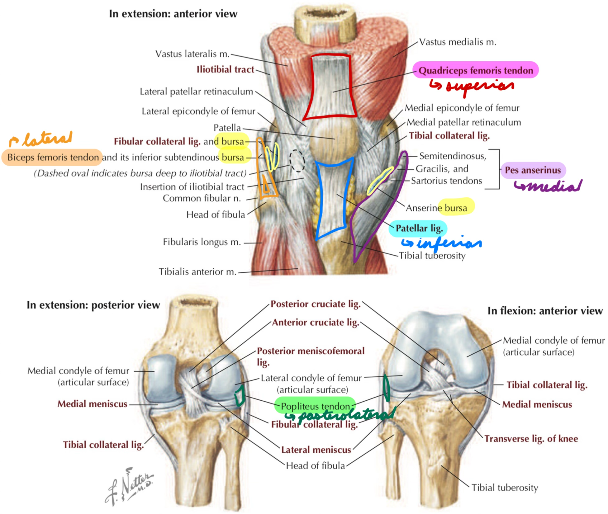
Knee: Description
Synovial hinge joint
2 articulations
Tibiofemoral: Between tibia + femur
Patellofemoral: Between femur + patella
Knee: Movements
Flexion/extension
External/internal rotation
Knee: Bones
Femur: Superior
Tibia: Inferior
Fibula: Not part of knee joint
Patella: Kneecap
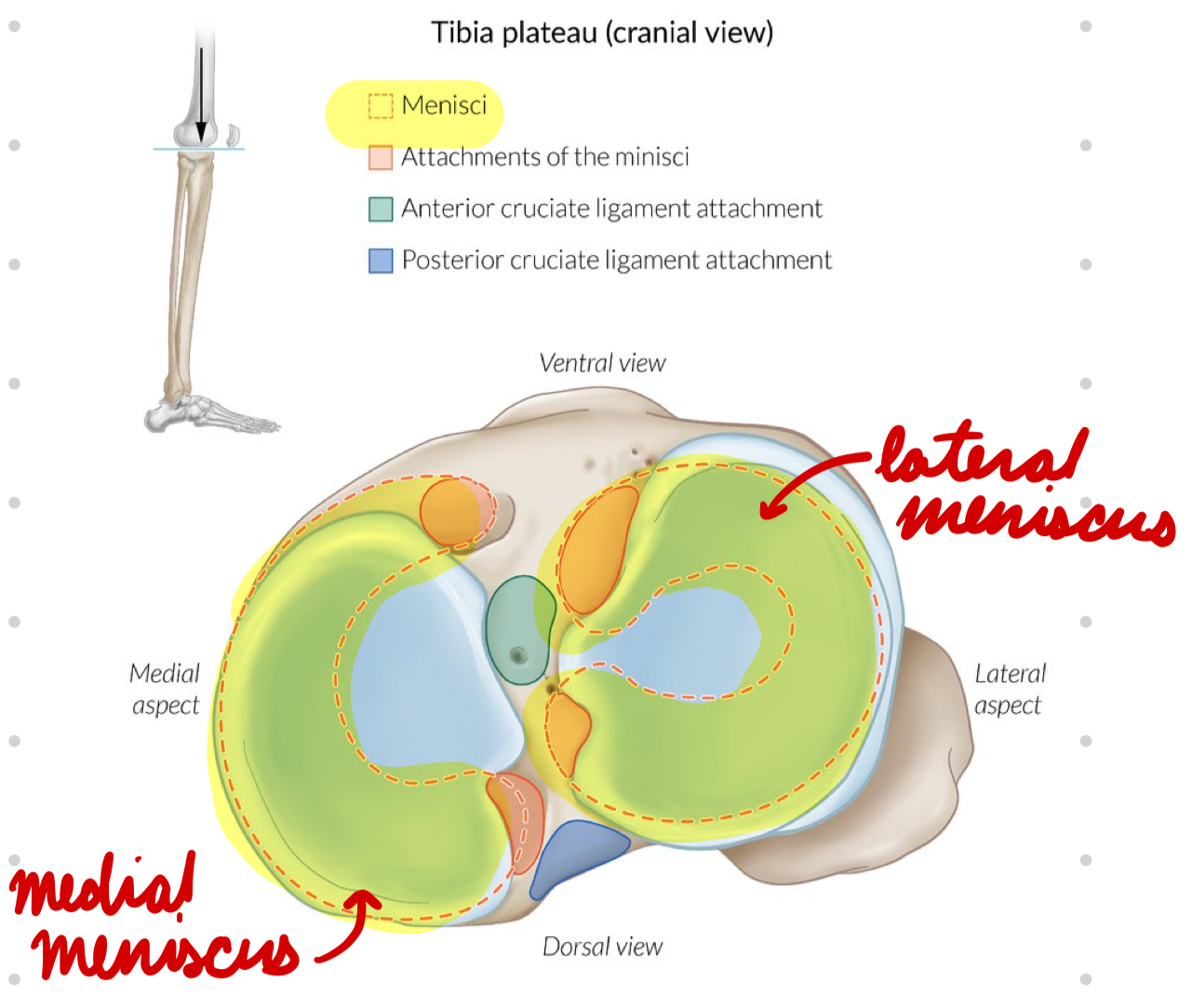
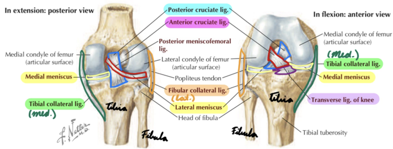
Knee: Menisci
Medial: On medial tibia
C-shaped
Attached to MCL
Lateral: On lateral tibia
Circular
Knee: Ligaments
Cruciate:
Anterior (ACL)
Posterior (PCL)
Collateral:
Medial (MCL)
Lateral (LCL)
Popliteofibular (popliteal)
Transverse
Anterolateral
Knee Cruciate Ligament: ACL
Anterior tibia → Lateral femur
Prevent anterior tibia/femur displacement
Prevent hyperextension

Knee Cruciate Ligament: PCL
Posterior tibia → Medial femur
Thicker than ACL
Prevent posterior tibia/femur displacement
Prevent hyperflexion
Knee Collateral Ligament: MCL
Medial femur → Medial tibia
Prevent medial tibia/femur displacement
Limit abduction

Knee Collateral Ligament: LCL
Lateral femur → Fibula head
Prevent lateral tibia/femur displacement
Limit extension + adduction

Knee Popliteofibular Ligament
Popliteus tendon → Fibula head
Stabilize posterolateral knee
Knee Transverse Ligament
Between menisci
Stabilize + decrease pressure on menisci

Knee Anterolateral Ligament
Lateral femur → Tibia
Stabilize tibia during internal rotation
Knee: Tendons
Quadriceps femoris tendon: Superior
Connect 4 quad muscles to patella
Patellar tendon/ligament: Inferior
Connect patella to tibia
Popliteus tendon: Posterolateral
Biceps femoris tendon: Lateral
Pes anserinus tendon: Medial

Knee: Bursae
Decrease friction between muscles, tendons, and bones
Elbow: Description
3 synovial hinge joints
Humeroulnar: Between humerus + ulna
Humeroradial: Between humerus + radius
Proximal radioulnar: Between radius + ulna
Elbow: Movements
Flexion/extension
Pronation/supination (radioulnar joint)
Elbow: Bones
Humerus: Superior
Ulna + Radius: Inferior
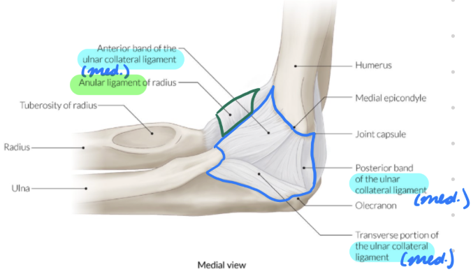
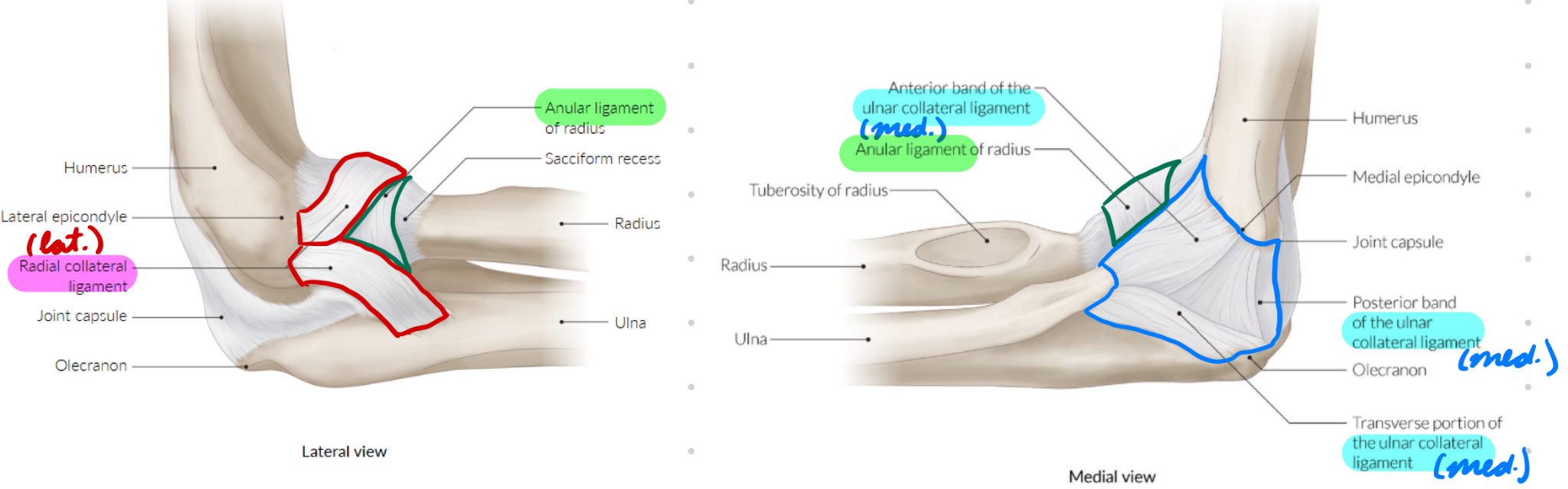
Elbow: Ligaments
Collateral:
Ulnar
Radial
Annular
Elbow: Ulnar Collateral Ligament
Medial humerus → Ulna + annular ligament
(Medial ligament)
Elbow: Radial Collateral Ligament
Lateral humerus → Ulna + olecranon
(Lateral ligament)
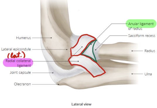
Elbow: Annular Ligament
Around radius head (radioulnar joint)
Anchor radius head to ulna radial notch
Elbow: Tendons
Anterior:
Biceps tendon
Brachialis tendon
Posterior: Triceps tendon
Medial: Common flexor tendon
Lateral: Common extensor tendon
Wrist: Description
4 joint types
Radiocarpal: Synovial ellipsoid
Between radius + proximal carpal bones (not pisiform)
Midcarpal: Synovial
Between proximal + distal carpal bones
Intercarpal: Amphiarthroses
Between carpal bones
Carpometacarpal (CMC)
2nd-5th: Amphiarthroses
Between carpal + metacarpal bones
Thumb: Synovial saddle
Between trapezium + 1st metacarpal bone
Wrist: Movements
Flexion/extension
Abduction/adduction
Thumb CMC: Opposition + circumduction
Wrist: Bones
8 carpals
2 rows: Lateral to medial (Some Lovers Try Positions That They Can’t Handle)
Proximal:
Scaphoid (deep to anatomical snuff box)
Lunate
Triquetrum
Pisiform
Distal:
Trapezium
Trapezoid
Capitate
Hamate
Wrist: Ligaments
Collateral:
Ulnar
Radial
Retinaculum:
Flexor (Transverse Carpal Ligament)
Extensor (Dorsal Carpal Ligament)
Radiocarpal:
Palmar
Dorsal
Wrist: Ulnar Collateral Ligament
Ulna → Triquetrum
Medial wrist stability
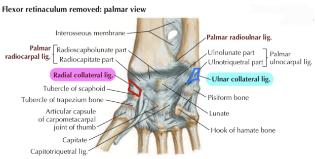
Wrist: Radial Collateral Ligament
Radius → Scaphoid
Lateral wrist stability

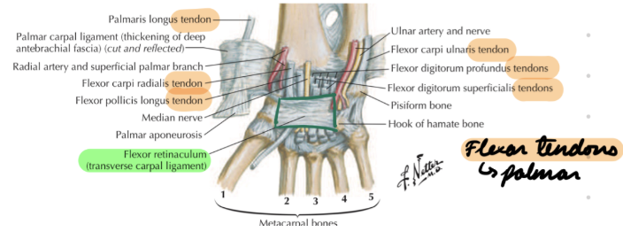
Wrist: Flexor Retinaculum
Pisiform + hamate (medial carpals) → Scaphoid + trapezium (lateral carpals)
Anchor flexor tendons (palmar)
Roof of carpal tunnel
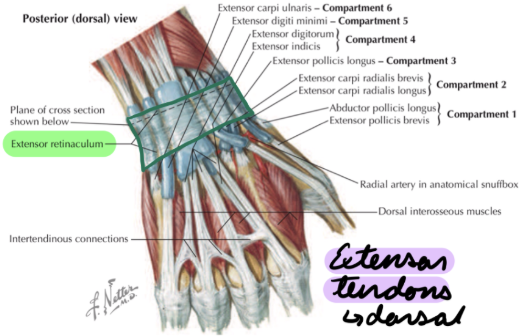
Wrist: Extensor Retinaculum
Pisiform + triquetrum → Radius
Anchor extensor tendons (dorsal)
Wrist: Radiocarpal Ligaments
Stabilize wrist + constrain carpal bone motion
Palmar: Radius → Lunate + capitate
Dorsal: Distal radius → Dorsal carpal bones
Wrist: Tendons
Flexor Tendons: Palmar
Extensor Tendons: Dorsal
Wrist: Channels
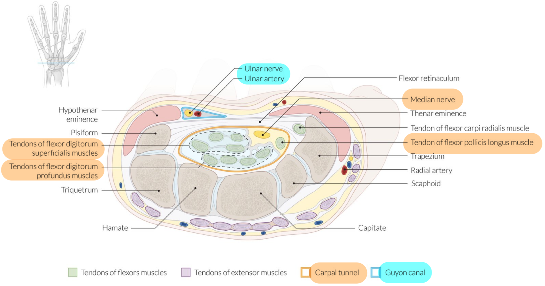
Carpal tunnel
Ulnar/guyon canal
Wrist: Carpal Tunnel
Passage for:
Median n.
Tendons
Flexor pollicis longus
Flexor digitorum profundus
Flexor digitorum superficialis
Borders:
Medial: Pisiform + hamate
Lateral: Scaphoid + trapezium
Superior: Flexor retinaculum
Inferior: Carpal groove (between carpal bones)

Wrist: Guyon Canal
Passage for ulnar nerve + artery
Borders:
Medial: Pisiform
Lateral: Hamate
Superior: Palmar carpal ligament
Inferior: Flexor retinaculum + hypothenar muscles
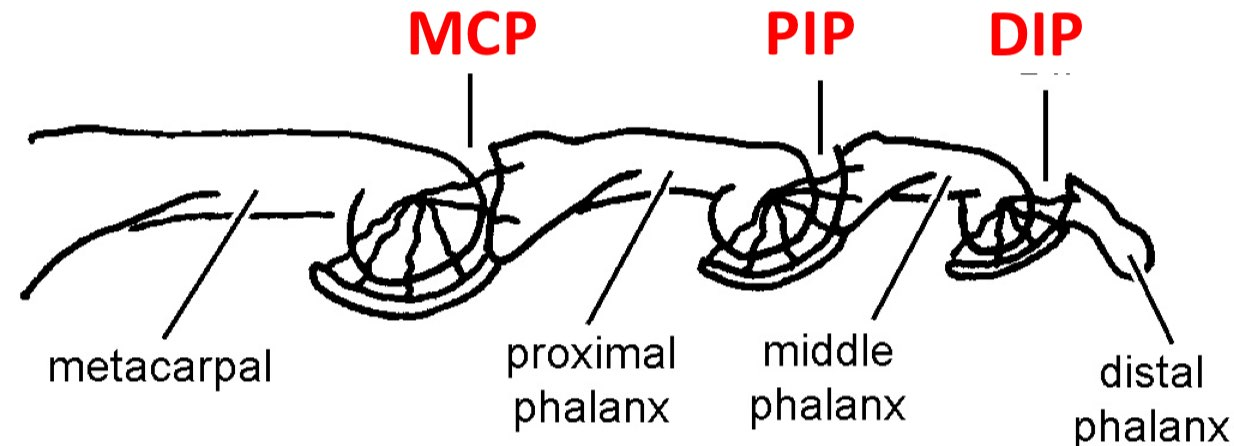
Finger Joints: Description
3 synovial joints
Metacarpophalangeal (MCP): Between metacarpals + proximal phalanges
Interphalangeal (IP):
Proximal (PIP): Between proximal + middle phalanges
Distal (DIP): Between middle + distal phalanges
Finger Joint: Movements
Flexion/extension
MCP: Abduction/adduction
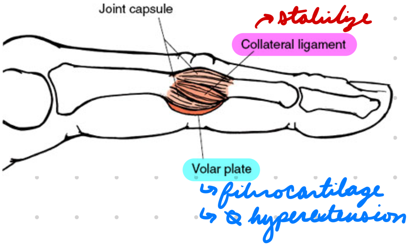
Finger Joint: Ligaments
Collateral
Volar plate
Finger Joint: Collateral Ligaments
Surround + stabilize joints

Finger Joint: Volar Plate
Fibrocartilaginous structure on palm side of each joint
Prevent hyperextension
Knee Joint Lesions: Description
Meniscus Tear: Rupture/tear to meniscus
Cruciate Tear: ACL/PCL injury
Collateral Tear: MCL/LCL injury
Patellofemoral Pain Syndrome (PFPS): Pain behind/around patella aggravated by weight-bearing activities (squatting, running, climbing stairs)
Meniscus Tear: Pathogenesis
Traumatic: Excess external force (twisting/pivoting planted foot or hard landing) = Damage meniscus
Degenerative: Overuse/chronic stress = Damage meniscus
Cruciate Tear: Pathogenesis
ACL: Knee extension + valgus force (foot out, knee in)
Usually non-contact injury
PCL: Knee hyperflexion + excess force on foot = Drive tibia posterior
Usually contact injury
Collateral Tear: Pathogenesis
MCL: Force on outer knee = Valgus force + external rotation
LCL: Force on inner knee = Varus force (foot in, knee out) + internal rotation
PFPS: Pathogenesis
Muscle/tendon weakness = Abnormal patella tracking over femur condyles = Overload patellofemoral joint = Pain
Knee Joint Lesions: Investigations
Trauma vs no trauma
Physical exam
Imaging
Knee Joint Lesion Investigations: Physical Exam
Meniscus Tear:
Thessaly test
McMurray’s test
Apley’s test
Cruciate Tear:
ACL
Lachman test
Anterior drawer test
Pivot shift test
PCL
Posterior drawer test
Posterior sag test
Quadriceps active test
Collateral Tear:
MCL: Valgus stress test
LCL: Varus stress test
PFPS:
Clarke test
Patellar apprehension/displacement test
Patellar tilt test
Knee Joint Lesion Investigations: Imaging
XR:
Ottawa Criteria: Needed if 1 of…
≥ 55 years
Patella/fibula head tenderness
Flexion < 90º
Cannot bear weight
US:
Effusions
Assess bursa
CT:
Fractures
Trauma
MRI
Knee Joint Lesions: Management
Conservative
Surgery
Knee Joint Lesions: Conservative Management
Initial management
Rest
Ice
Elevate
NSAIDs
Knee brace
Physical therapy + strength training
Knee Joint Lesions Management: Surgery
Meniscus Tear:
Indication: Complex traumatic tear
Contraindication: Degenerative tear
Method:
Repair/reconstruct meniscus
Partial menisectomy
Cruciate/Collateral Tear:
Indication:
Recurrent tears
Dysfunction (lax knee)
Methods:
Reconstruction: Graft from tissue
Repair: Suture torn ligament to bone
PFPS: Management
POLICE principles
P: Protection (rest)
OL: Optimal loading
Physical therapy + strength training
Low-impact activities
I: Ice
C: Compression
E: Elevation