Autonomic Nervous System & Skeletal Muscle Physiology: Key Concepts and Structures
1/169
There's no tags or description
Looks like no tags are added yet.
Name | Mastery | Learn | Test | Matching | Spaced | Call with Kai |
|---|
No analytics yet
Send a link to your students to track their progress
170 Terms
Ganglion
A collection of neuronal cell bodies in the peripheral nervous system.
Preganglionic neuron
The first of two ANS lower motor neurons; cell body is located in brainstem or spinal cord & axon projects to autonomic ganglion.
(Post)Ganglionic neuron
The second of two ANS lower motor neurons; cell body is located in an autonomic ganglion & axon projects to effector/target.
Thoracolumbar division
Another name for the sympathetic division of the ANS; refers to location of preganglionic cell bodies in T1 - L2 lateral horns of the spinal cord.
Craniosacral division
Another name for the parasympathetic division of the ANS; refers to location of preganglionic cell bodies in the brainstem or S2-S4 lateral horns of the spinal cord.
Neuronal convergence
Occurs when numerous preganglionic axons synapse on and influence the activity of a single ganglionic neuron.
Neuronal divergence
Occurs when a single preganglionic axon branches to synapse on and influence the activity of numerous ganglionic neurons.
Dual innervation
Effectors of the ANS are innervated by postganglionic axons of both the sympathetic and parasympathetic divisions of the ANS.
Antagonistic control
The effects of the sympathetic and parasympathetic innervation of a single target oppose one another.
Somatic Nervous System
Voluntary control of skeletal muscles.
Autonomic Nervous System
Involuntary control of cardiac muscle, smooth muscle, and glands.
Hypothalamus
Region of the CNS that controls the autonomic nervous system.
Lower motor neurons
In the somatic nervous system, one lower motor neuron extends from the anterior horn of the spinal cord to the effector.
Ganglionic neuron
In the autonomic nervous system, a ganglionic neuron extends to the effector.
Excitation only
Response of the effector in the somatic nervous system.
Excitation or inhibition
Response of the effector in the autonomic nervous system.
Sympathetic Division
Maintains homeostasis during exercise, emergency, or stress. 'Fight or flight'.
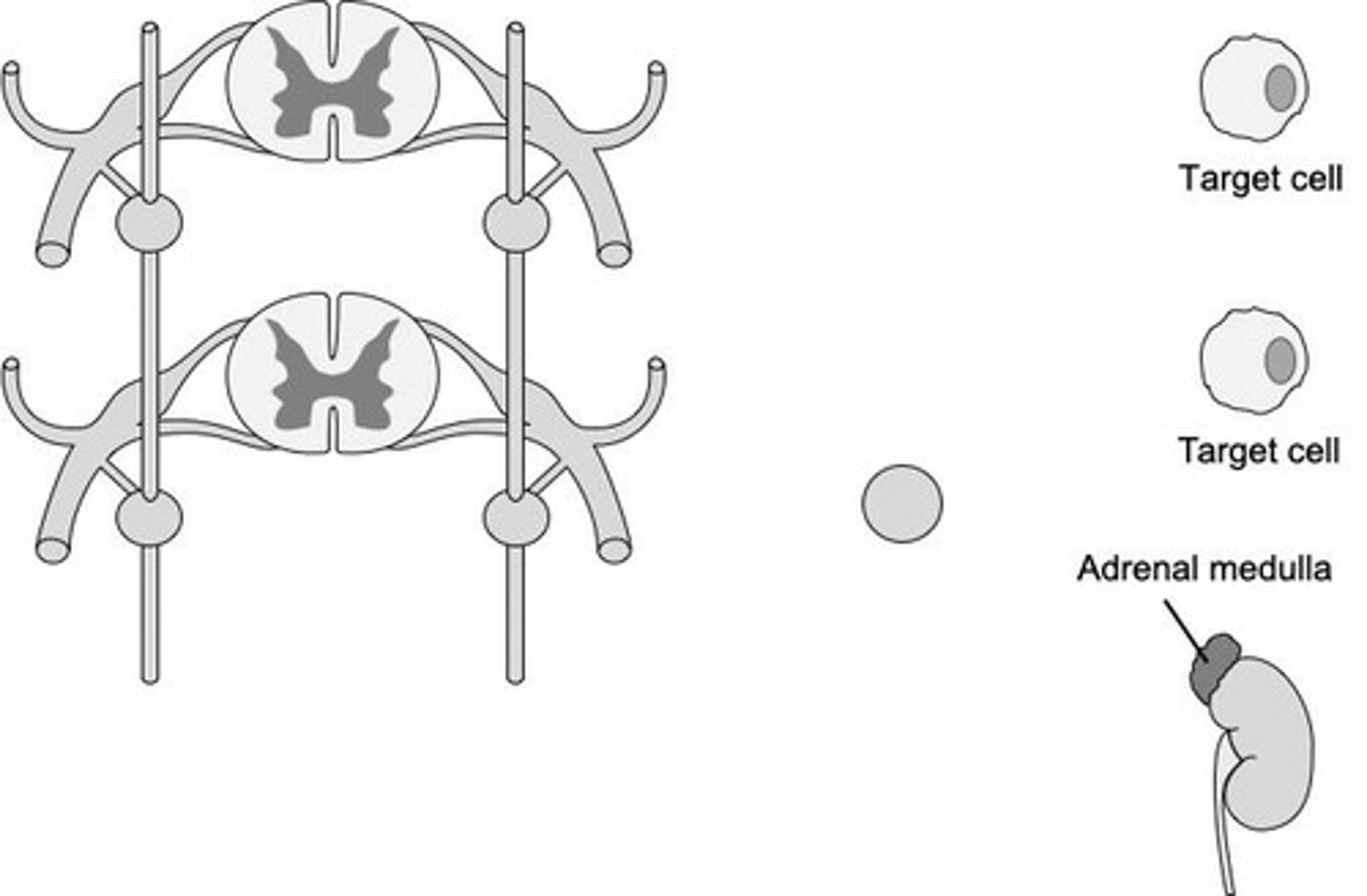
Parasympathetic Division
Maintains homeostasis during rest. 'Rest and digest'.
Preganglionic Soma Location (Sympathetic)
Cell bodies located in lateral horns of T1-L2 spinal cord.
Preganglionic Soma Location (Parasympathetic)
Cell bodies located in cranial nerve nuclei in the brainstem and lateral horns of S2-S4 spinal cord.
Ganglia Location (Sympathetic)
Ganglia located close to the spinal cord - sympathetic trunk or prevertebral ganglia.
Ganglia Location (Parasympathetic)
Ganglia located close to or within effectors.
Preganglionic Axon Length (Sympathetic)
Long.
Preganglionic Axon Length (Parasympathetic)
Short.
Postganglionic Axon Length (Sympathetic)
Short.
Postganglionic Axon Length (Parasympathetic)
Long.
Degree of Response (Sympathetic)
Mass activation: stimulate many body systems simultaneously.
Degree of Response (Parasympathetic)
Local response: stimulate one or only a few body systems at one time.
Cranial Nerve III
Oculomotor.
Cranial Nerve VII
Facial.
Cranial Nerve IX
Glossopharyngeal.
Cranial Nerve X
Vagus.
Muscarinic Receptors
Receptors that are all effectors of parasympathetic targets.
Alpha Adrenergic Receptors
Receptors that are effectors of sympathetic targets.
Beta Adrenergic Receptors
Receptors that can be either excitatory or inhibitory, depending on subtype.
Nicotinic Receptors
Receptors located at the cell body of all ganglionic neurons in the ANS.
Acetylcholine
Neurotransmitter bound by muscarinic and nicotinic receptors.
Norepinephrine
Neurotransmitter bound by adrenergic receptors.
Ionotropic Receptors
Receptors that are typically excitatory.
Metabotropic Receptors
Receptors that can be either excitatory or inhibitory, depending on subtype.
Excitability
Ability to respond to stimulus, causing a change in membrane potential.
Conductivity
Ability to propagate an action potential along the plasma membrane.
Contractility
Ability to shorten.
Extensibility
Ability to lengthen/stretch.
Elasticity
Ability to return to original shape after contracting or elongating.
Skeletal Muscle
An organ consisting of muscle fibers, connective tissue, blood vessels, and nerves.
Fascicle
A bundle of muscle fibers.
Skeletal Muscle Fiber
Cells of skeletal muscle tissue; aka myofibers.
Tendon
Thick, cordlike structure that attaches muscle to bone/ligament/fascia.
Aponeurosis
Flat sheet of dense regular connective tissue that attaches muscle to bone/ligament/fascia.
Sarcoplasm
Cytoplasm of a skeletal muscle fiber.
Sarcolemma
Plasma membrane of a skeletal muscle fiber.
T-tubule
Extensions of the sarcolemma that extend down into the muscle fiber.
Myofibril
Long, cylindrical structures filling the sarcoplasm; composed of bundles of contractile proteins.
Sarcoplasmic Reticulum
Intracellular membrane complex that fits around each myofibril like a sleeve.
Terminal Cisternae
Sacs at the ends of individual sections of sarcoplasmic reticulum that act as reservoirs for calcium.
Triad
A central T-tubule and two terminal cisternae that sit on either side of it.
Myofilaments
Contractile proteins bundled within myofibrils; includes thick and thin filaments.
Myosin
Major protein of the thick filament.
Actin
Contractile protein of the thin filament.
Tropomyosin
A rope-like protein in the thin filament that covers the myosin binding site on G-actin.
Troponin
A ball-like protein in the thin filament that contains a binding site for calcium.
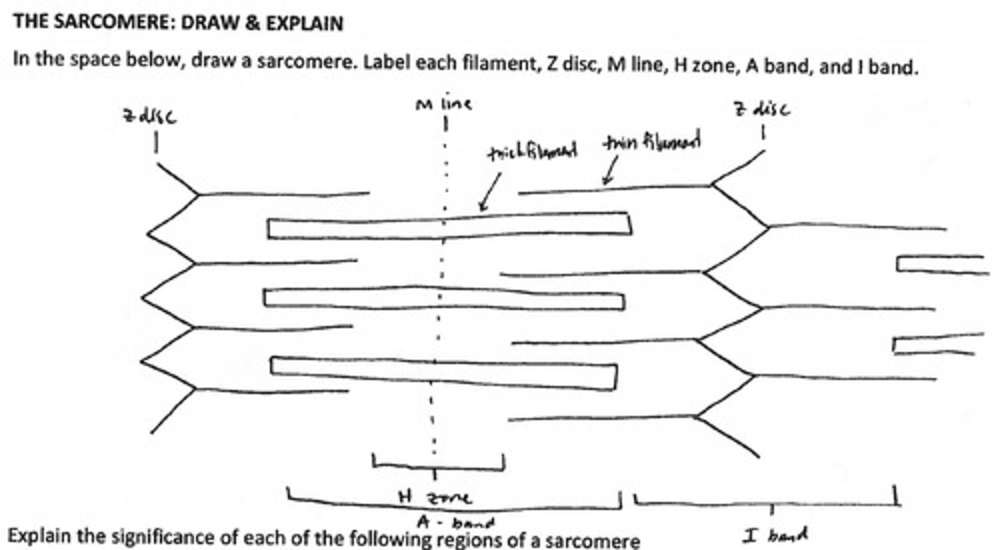
Sarcomere
A repeating cylindrical unit composed of overlapping thick and thin filaments.
Motor Unit
A motor neuron and all the muscle fibers it innervates.
Neuromuscular Junction
The specific location where the axon terminal of a motor neuron innervates a skeletal muscle fiber.
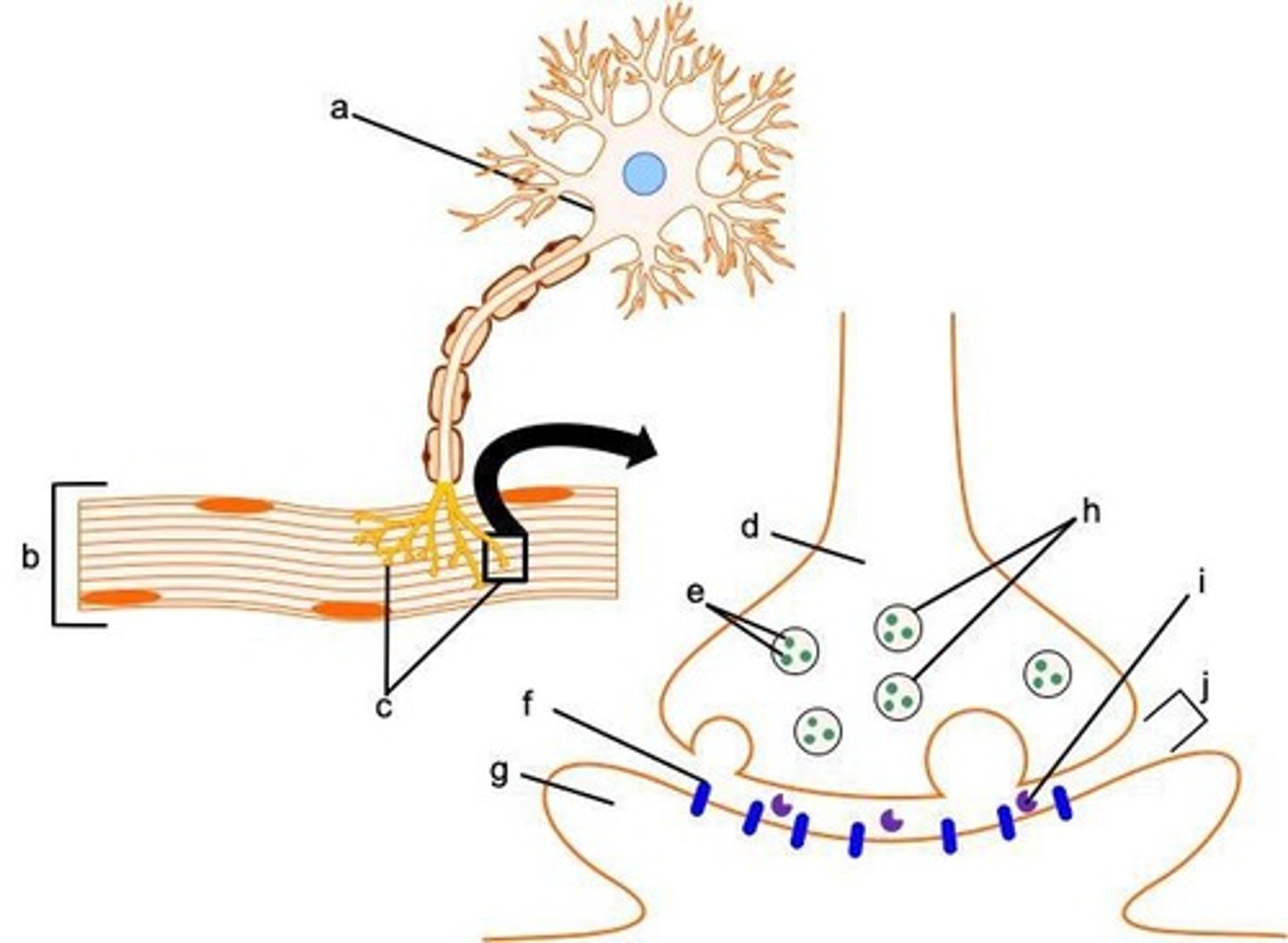
Synaptic Knob
The axon terminal containing synaptic vesicles filled with acetylcholine.
Motor End Plate
Specialized region of the sarcolemma innervated by the motor neuron.
Acetylcholinesterase
An enzyme that breaks down acetylcholine.
Muscle fiber
An individual skeletal muscle fiber.
Myofilament
Consists of actin, troponin, and tropomyosin.
Endomysium
Connective tissue wrapping around a muscle fiber.
Perimysium
A bundle of skeletal fibers.
Epimysium
A skeletal muscle.
Thick cord
A structure formed by connective tissue.
Multinucleated
Skeletal muscle fibers are multinucleated because multiple embryonic muscle cells, called myoblasts, fuse to form a single muscle fiber.
Functional significance of multinucleated
Multiple nuclei help to coordinate necessary cellular processes due to high energy demands.
Z disc
Ends of each sarcomere; anchor for thin filaments.
I bands
Region containing only thin filaments.
A bands
Central region of sarcomere containing entire thick filament and partially overlapped thin filament.
H zone
Center of the A-band; containing only thick filaments.
M line
Very center of the H-zone; anchor for thick filaments.
Striations
Produced by the partial overlapping of thick and thin filaments in a sarcomere.
Thin filament
Consists of actin.
Thick filament
Consists of myosin.
Voltage-gated calcium channels
Open when an action potential arrives at the terminal and allows calcium to enter the synaptic knob and initiate ACh release.
Calcium pumps
Restore the concentration gradient for calcium, which is needed to initiate the release of ACh.
Acetylcholine Receptors
Chemically gated cation channels expressed on the motor end plate that establish the end plate potential.
Resting membrane potential
The electrical charge difference across the sarcolemma at rest, with a value of -90 mV.
Sodium (Na+)
More concentrated in the interstitial fluid when a skeletal muscle fiber is at rest.
Potassium (K+)
More concentrated in the cytosol when a skeletal muscle fiber is at rest.
Excitation
The first phase of muscle contraction associated with the neuromuscular junction.
Excitation - Contraction Coupling
The second phase of muscle contraction associated with the sarcomere.
Crossbridge Cycling
The third phase of muscle contraction associated with the sarcolemma and T-tubules.
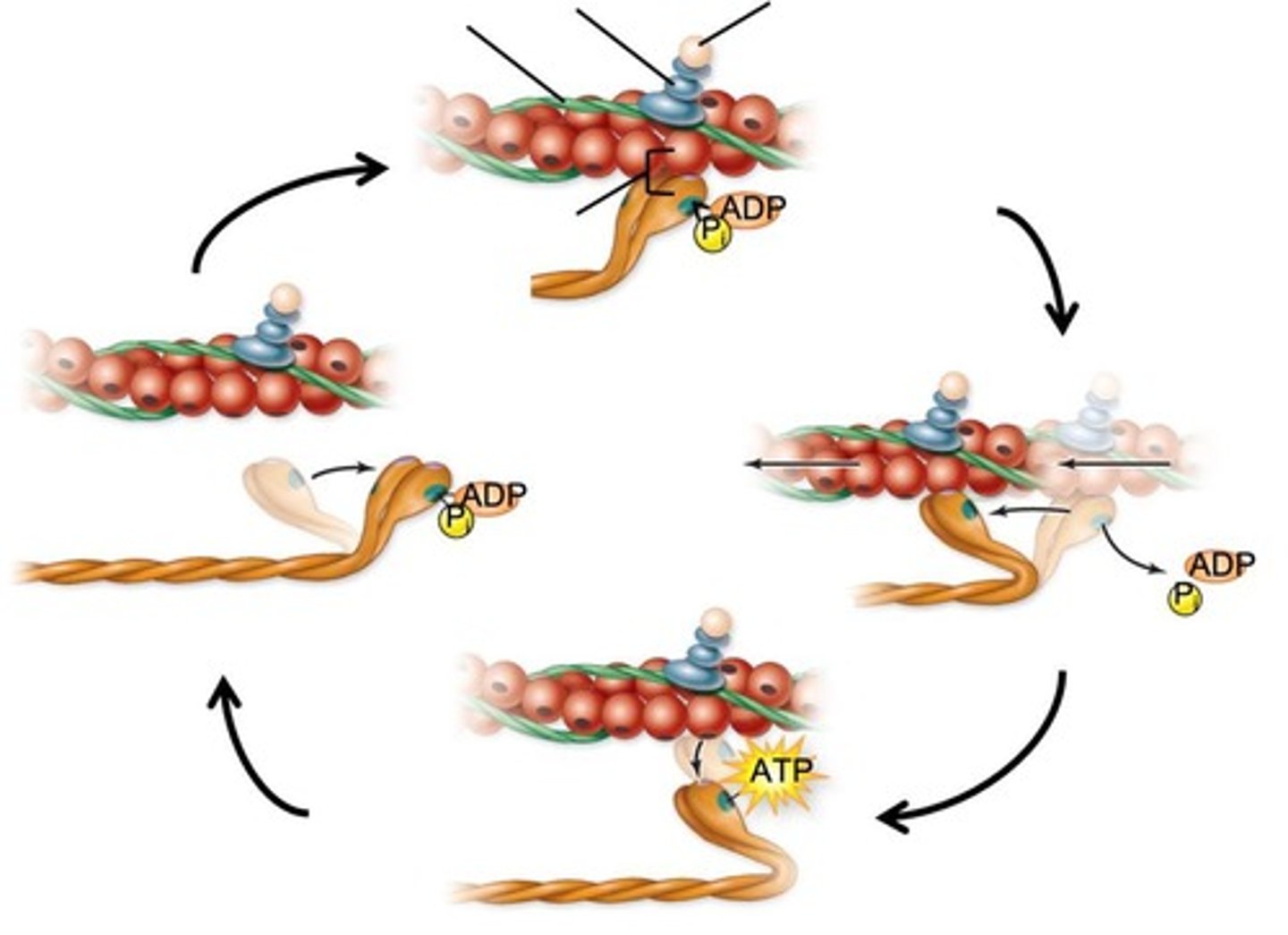
Calcium ion channels
Open in the sarcoplasmic reticulum when T-tubule depolarization occurs.
End-plate potential
Stimulated by the entry of Na+ which depolarizes the sarcolemma locally.
Sliding filament mechanism
Thin filaments are pulled past thick filaments toward the center of the sarcomere.
Crossbridge
Formed when the myosin head binds actin after calcium binds to troponin.
Power Stroke
Occurs when ADP and Pi are released as the myosin head swivels, pulling the thin filament toward the center of the sarcomere.
ATP hydrolysis
Resets the myosin head into a 'cocked' position.
Skeletal muscle relaxation
Involves the dissociation of calcium from troponin and the breakdown of ACh by acetylcholinesterase.