B1.1
1/46
There's no tags or description
Looks like no tags are added yet.
Name | Mastery | Learn | Test | Matching | Spaced | Call with Kai |
|---|
No analytics yet
Send a link to your students to track their progress
47 Terms
What is a eukaryotic cell?
A eukaryotic cell is a complex cell that can be a unicellular or (usually) part of a multicellular organism
What do eukaryotic cells contain?
It contains a true nucleus
It contains membrane-bound organelles (e.g. mitochondria)
How big are eukaryotic cells?
They are relatively large compared to prokaryotic cells (10-100 micrometres
Give examples of eukaryotic cells
Examples include plant cells, animal cells, fungi (e.g. yeast), and protista
What is a prokaryotic cell?
A prokaryotic cell is a relatively simple, unicellular organism
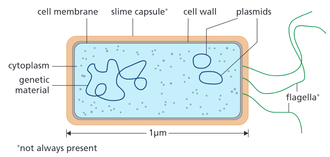
What do prokaryotic cells contain?
It lacks a true nucleus, instead the genetic material is stored in a singular strand, and as small rings of DNA, called plasmids
It lacks membrane-bound organelles, so it respires anaerobically only, using the cytoplasm
Plasmids - small rings of DNA, which code for specific features (e.g. antibiotic resistance)
Slime capsule (not always present) - protects the cell from drying out and immune system attacks, and allows them to adhere to surfaces
Flagellum (not always present) - long strand of protein that lashes about, used for movement
How big are prokaryotic cells?
They are relatively small compared to eukaryotic cells (0.2-2 micrometres)
Give examples of prokaryotic cells
Examples include bacteria and archaea
Define a cell
Cells are the basic units of all forms of life
(Usually) how large are animal cells
Animal cells are around 10-30 micrometres
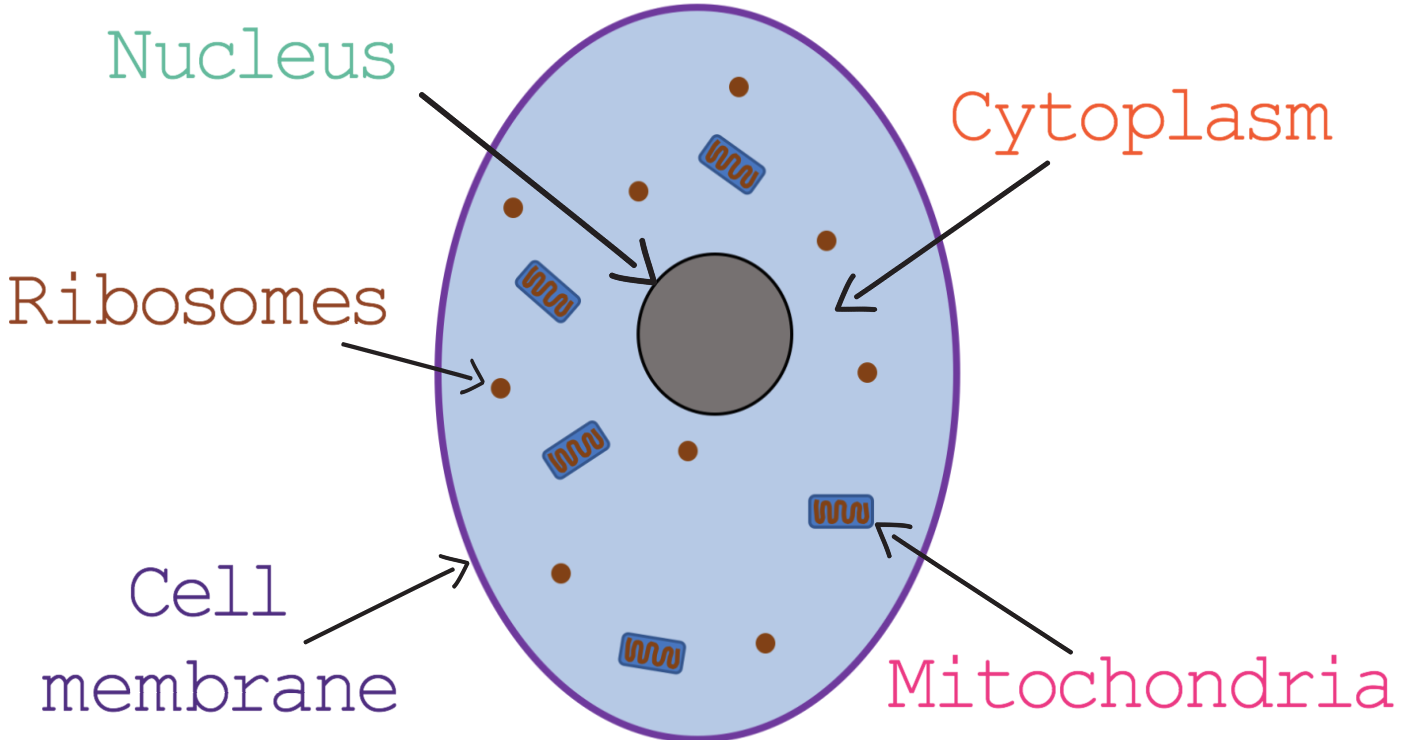
What organelles do animal cells usually contain?
Nucleus - contains genetic material of the cell, which controls the cell's activities
Mitochondria - is the site of aerobic respiration, and releases the energy used by the cell
Cell membrane - Controls the movement of substances in and out of the cell (because it has a structure that is permeable to some substances but not to others)
Cytoplasm - a gelatinous liquid that contains the organelles and also enzymes, and is the site of chemical reactions in the cell
Ribosomes - is the site of protein synthesis

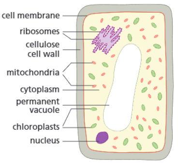
(Usually) how big are plant cells?
Plant cells are around 10-100 micrometres
What organelles do plant cells contain?
In addition to the 5 main organelles that animal cells have, plants specifically have:
Cell wall (cellulose) - strengthens the cell and provides support
Chloroplasts (larger than mitochondria) - contain the pigment chlorophyll, which absorbs light necessary for photosynthesis (chloroplasts are the site of photosynthesis)
Permanent vacuole - contains cell sap and keeps the cell rigid
What does cell specialisation mean?
Cells can become specialised (have certain adaptations), which allow them to carry out their function(s) efficiently.
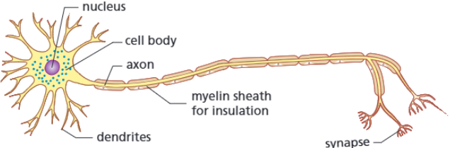
Function and adaptations for nerve cell
Function:
Carries electrical impulses around the body of an animal. (They provide a rapid communication system between the different parts of the body).
Adaptations:
Long axon
Myelin sheath
Synapses contain transmitter chemicals
How adaptations help:
Axons carry the electrical impulses around the body
The myelin sheath insulates the cell, increasing the speed and efficiency of carrying the electrical impulses
The transmitter chemicals help carry the electrical impulses across nerve cells
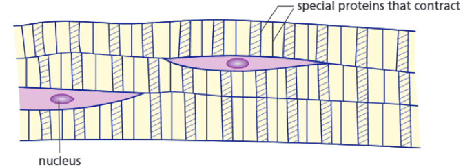
Function and adaptations for muscle cell (striated)
Function:
Contracts and relaxes which enables movement.
Skeletal muscle (which is all striated) contract and relax to move bones
Smooth muscle contracts to move food through the digestive system
Cardiac muscle contracts and relaxes to pump blood throughout the body
Adaptations:
Contains actin and myosin
Lots of mitochondria
Stores glycogen (a type of stored glucose)
How adaptations help:
Actin and myosin are proteins that enable muscle contraction (by converting ATP into mechanical force)
The mitochondria transfer energy needed for the chemical reactions that occur as the cell contracts/relaxes
Glycogen can be broken down and used in respiration by the mitochondria
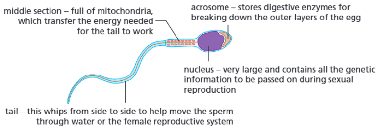
Function and adaptations for sperm cell
Function:
Travels to and fertilises the egg during sexual reproduction, and contains the genetic information of the male parent.
Adaptations:
Long tail
Lots of mitochondria (in midsection)
Acrosome
How adaptations help:
The tail allows the cell to move (long distances) to the egg
Mitochondria transfer the energy needed for the tail to work
Acrosome contains digestive enzymes for breaking down the outer layers of the egg
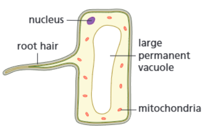
Function and adaptations for root hair cell
Function:
Take water and mineral ions from the soil (by active transport) so that they can be carried by the xylem tissue to the rest of the plant.
Adaptations:
Large projection
Many mitochondria
Permanent vacuole
How adaptations help:
The projection increases the surface area available for the water and mineral ions to move into the cell
The mitochondria transfer energy needed for the active transport of the mineral ions into the cell
The permanent vacuole speeds up the movement of water by osmosis
Function and adaptations for photosynthetic cell
Function:
Carry out photosynthesis (the process of converting carbon dioxide and water into glucose and oxygen).
Adaptations:
Contains chloroplasts
Permanent vacuole
Tightly-packed (and regular shape)
How adaptations help:
The chlorophyll in the chloroplasts absorbs the light energy necessary for photosynthesis
The permanent vacuole helps to keep the cell rigid, and helps keep the leaf spread out
The tightly-packed arrangement (in the leaves and outer layers of the stem) allows them to absorb enough light

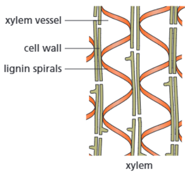
Function and adaptations for xylem cell
Function:
The xylem tissue carry water and mineral ions from the roots to the (highest) leaves and shoots, and supports the plant.
Adaptations:
Cell walls contain spirals and rings of lignin
The xylem cells are alive at first, but after the production of lignin (in the spirals of the cell walls) they die and form long hollow tubes
How adaptations help:
Lignin is a polymer that provides strength and support to the cell, and allows it to withstand the water pressure
The water and mineral ions can move through the long hollow tubes easily
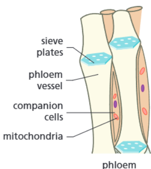
Function and adaptations for phloem cell
Function:
The phloem tissue carry the sugars and amino acids (food produced by photosynthesis) around the plant.
Adaptations:
Cell walls between the cells have broken down to form sieve plates (perforated ends that connect sieve elements)
There are companion cells, which contain mitochondria
How adaptations help:
The sieve plates allow water that carries dissolved food to move freely up or down the tubes to where it is needed
The companion cells (which support and help keep the phloem cells alive) contain mitochondria, which transfer the energy needed to move the dissolved food
Define cell differentiation and state why it is important
Cell differentiation is the process in which a cell changes to become specialised, which occurs as an organism develops. It is important as it allows cells to develop certain features that allow it to do its functions within the organism efficiently
When do animal cells differentiate
Most types of animal cell differentiate at an early stage - before birth/the egg hatching (mature animal cells can divide to create more of the same cell, for repair and replacement)
When do plant cells differentiate
Many types of plant cells retain the ability to differentiate throughout life (most mature plant cells can differentiate, which is necessary for continuous growth and the repair of damaged tissues).
(EXTRA) What is the difference between the spongy and palisade mesophyll
The palisade mesophyll is made of photosynthetic cells, whereas the spongy mesophyll allows gas exchange within the leaf to occur, and adaptations include the fact they are loosely arranged and have large air pockets, and that they have are covered with a thin layer of water, which enables the gases to dissolve before diffusing into the cells.
Define magnification
The number of times greater an image appears compared to the real object/specimen (the size at which an image can be seen).
Define resolution
A measure of the degree of detail that can be seen in an image (the ability to distinguish between two separate points).
How do light microscopes work
They are more basic than electron microscopes and use light, which is shone through the specimen and passes through an objective lens and then the eyepiece, which produces a magnified image.
How do electron microscopes work
They are more complex than light microscopes and use a beam of electrons to produce an image on a screen.
State some advantages and disadvantages of light microscopes
Advantages:
Cheap, light, and easy to use
Can produce coloured images
Can keep the cells alive
Disadvantages:
Maximum useful magnification is 2000x
Limited resolution (200mm)
They can only produce 2D images
State some advantages and disadvantages of electron microscopes
Advantages:
Maximum useful magnification is around 2000000x
Great resolution (less than 1nm)
Can produce 3D images
Disadvantages:
Expensive, importable, difficult to use
Only produces B&W images
Cannot view cells when they are alive (because they are placed in a vacuum)
State the formula for magnification, real size, and image size:
magnification = size of image / size of real object (remember I = AM)
Convert between units
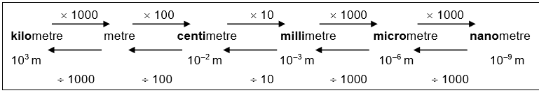
What are orders of magnitude
Sizes can be compared using orders of magnitude. Orders of magnitude make approximate comparisons between numbers of objects.
How are orders of magnitude represented as?
They are shown using powers of 10:
10x bigger = one order of magnitude bigger = 101
100x bigger = two orders of magnitude bigger = 102
How do you calculate orders of magnitude
Check both values have the same units
Divide the larger number by the smaller number
If the answer is <10 - same order of magnitude
If the answer is around 10 - 1 order of magnitude bigger
If the answer is around 100 - 2 orders of magnitude bigger
How do you use a light microscope
Clip the slide onto the stage of the microscope
Adjust the mirror/light source so that light passes through the specimen
Turn the coarse focus knob to bring the stage down as far as possible
Position the lowest-power objective lens over the slide
(Look into the eyepiece and) use the coarse focus knob the raise the stage until the specimen comes into focus
Use the fine focus knob to get a clearer and sharper image
To observe the image with a higher magnification, change the objective lens to a higher power and refocus
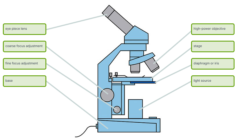
What are the parts of a light microscope?
Labels | Description |
Eye piece lens | The lens you look through – normally 10x magnification |
Coarse focus adjustment | Moves the lens up or down and adjusts focus |
Fine focus adjustment | Moves the lens in order to make very small adjustments to gain better focus |
Base | The bottom of the microscope used for stability |
High-power objective | For increased magnification – usually 10x, 40x and 100x magnification |
Stage | Where the slide is held/placed |
Diaphragm or iris | Varies intensity of the light projected upwards onto the slide |
Light source | Sends light onto the specimen/slide |
(Stage clips) | Holds the slide in place |
What is the aim of RP1 (microscopy)
Use a light microscope to investigate a selection of plant and animal cells.
What equipment do you need for RP1?
Light microscope
Microscope slide
Cover slip
Onion/cheek
Forceps
Iodine solution/methylene blue
(Any other prepared slides)
How do you prepare a slide containing onion cells?
Peel off one layer of cells from the epidermal tissue of an onion
Using a pipette add a drop of water to a clean slide, then lay the specimen on top of it (using forceps), ensuring it is flat
Add a drop of iodine solution, using a pipette, and cover with a cover slip (do so by holding the coverslip in the left hand, so that one side touches the slide, and then at a 45 degree angle, resting the opposite side on a mounted needle held in the right hand, lower it gently until flat, to reduce air bubbles being trapped). Touch a piece of paper towel to any liquid that spreads from under the cover slip.
How do you prepare a slide for cheek cells?
Take a cotton bud from a newly-opened pack, and move it over the inside of the cheek on one side of the mouth, and along the outer lower side of the gum.
Smear the cotton bud over a small area of a clean microscope slide.
Immediately place the used cotton bud into a small volume of disinfectant in a suitable container.
Add methylene blue stain from a dropper pipette onto the smear and cover with a cover slip (do so by holding the coverslip in the left hand, and then at a 45 degree angle resting the opposite side on a mounted needle held in the right hand, lower it gently until flat, to reduce air bubbles being trapped). The smear and stain will spread out under the cover slip.
If the stain is unevenly distributed or too dark, place a drop of water at one slide of the cover slip and hold a small piece of blotting/filter paper on the opposite side of the coverslip. The stain can then be drawn through, making the cells easier to observe.
State safety precautions for RP1
Wear safety goggles (and maybe gloves) when handling the iodine solution
Handle slides and microscopes with care
When cutting, use a chopping board
Why do you start with the lowest objective lens
You use the lowest objective lens first because it provides the widest field of view, allowing the specimen to be located easier, and because it minimises the risk of crashing the lens into the slide (because it is the longest)
What solutions are used to stain the organelles in the onion and cheek cells
Iodine solution is used to stain the organelles in the onion cells
Methylene blue is used to stain the organelles in the cheek cell
How do you calculate field of view (FOV)
To calculate the FOV (the lit circle seen under a microscope):
Find the lowest power dFOV (diameter of field of view) - do this by measuring the diameter of the FOV using a ruler for the lowest objective lens
Calculate all FOVs from this for all magnifications, using the equation M1 x dFOV1 = M2 x dFOV2
Place the slide into position and increase the magnification until individual cells can be viewed
State the criteria for a biological drawing
The drawing should be big (at least 1/3 of the page)
Draw using a sharp pencil, but label using a pen
Do not sketch, instead draw using clear lines
Only include large organelles (nucleus, chloroplast, large vacuole), and don't use shading or dots
Labels should be in capital letters
Label lines should be in pencil, drawn using a ruler, without arrowheads, and they shouldn't cross
Include the title (underlined), magnification, and scale bar (to draw a scale bar draw a horizontal I, and write down the actual size underneath)