Practical 3 BSCI201
1/146
There's no tags or description
Looks like no tags are added yet.
Name | Mastery | Learn | Test | Matching | Spaced | Call with Kai |
|---|
No analytics yet
Send a link to your students to track their progress
147 Terms
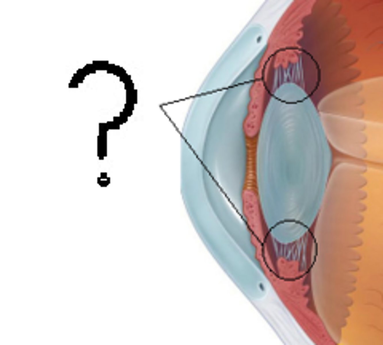
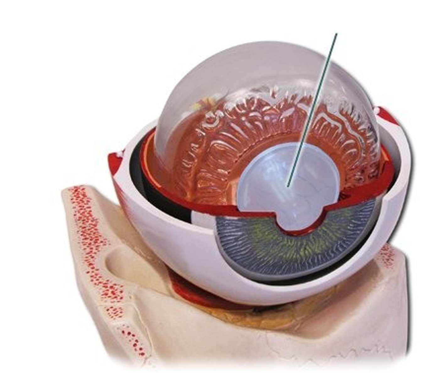
Zonules — Fibers that extend from the ciliary process and hold the lens upright.
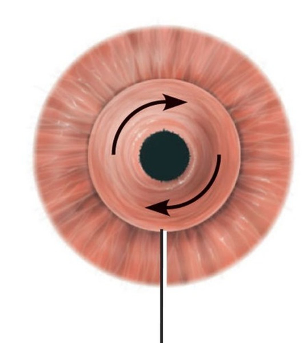
Sphincter pupillae — Muscles that constrict the pupil for close vision and bright light.
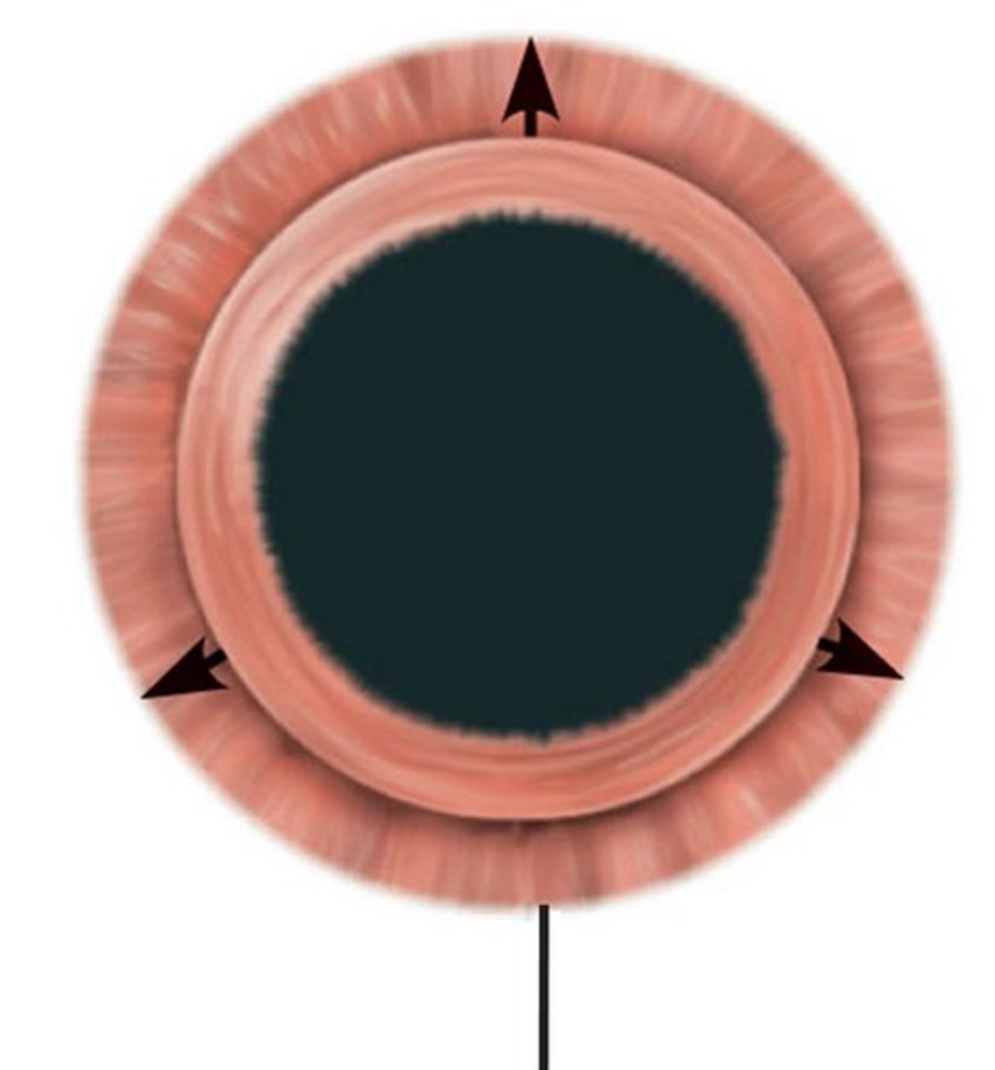
Dilator pupillae — Muscles that dilate the pupil for distance and dim light.
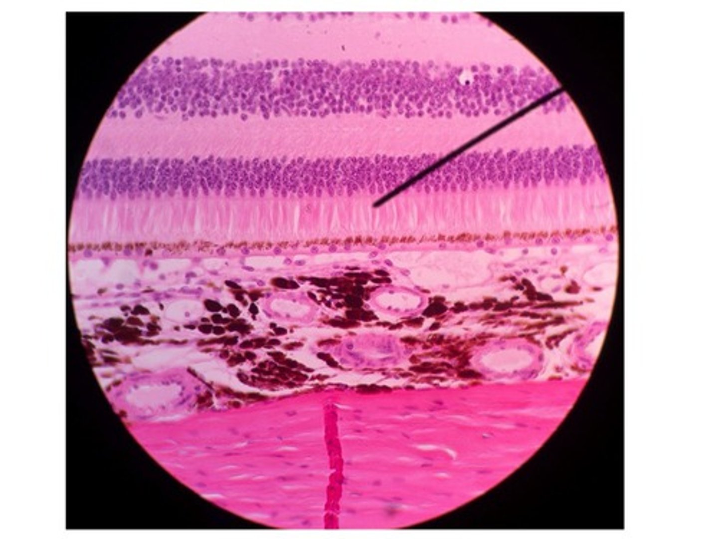
Pigmented layer — Outer layer of retina containing melanocytes; absorbs light.
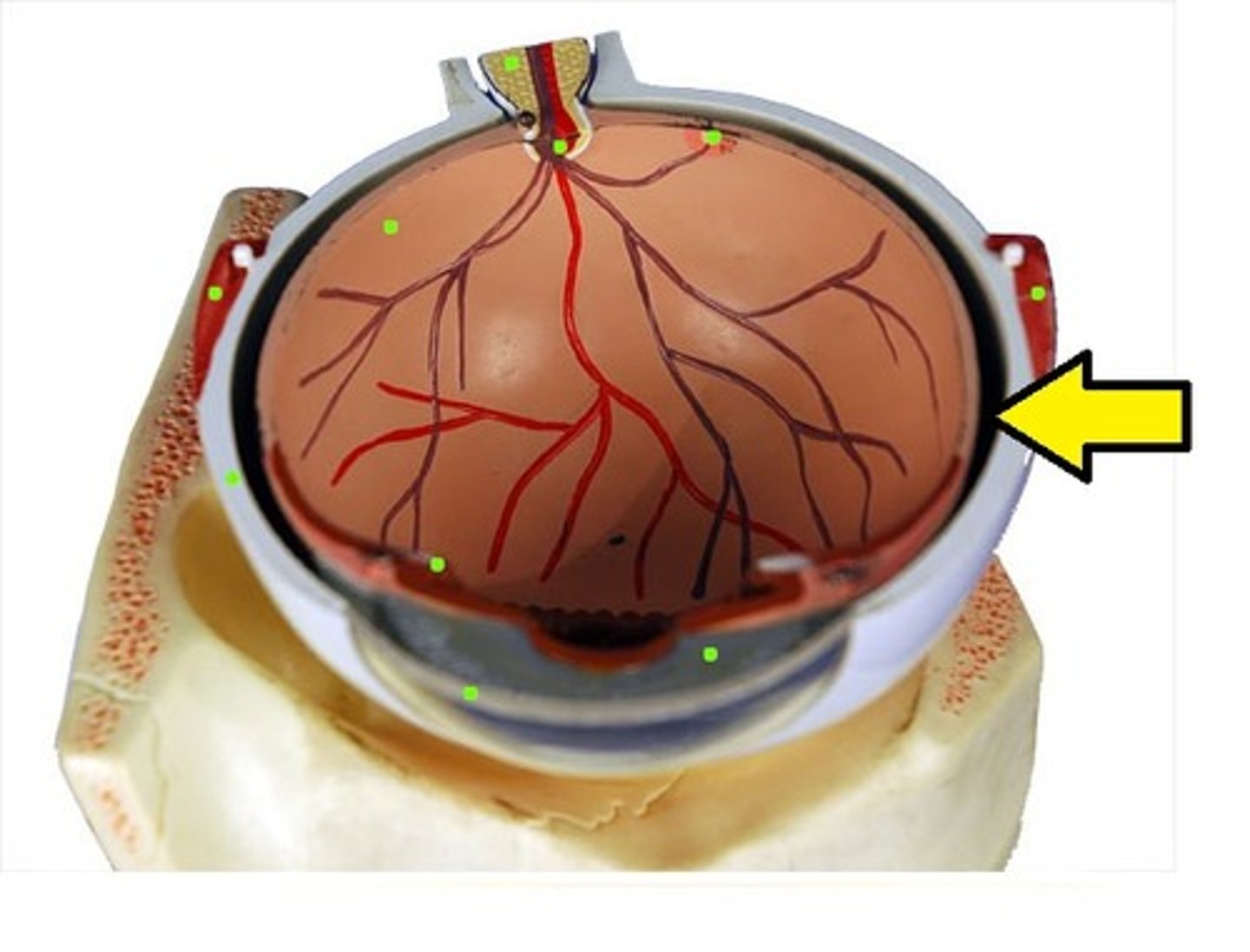
Neural layer — Converts light energy into nerve impulses sent to the brain.
Photoreceptors — Cells that change membrane potential in response to light.
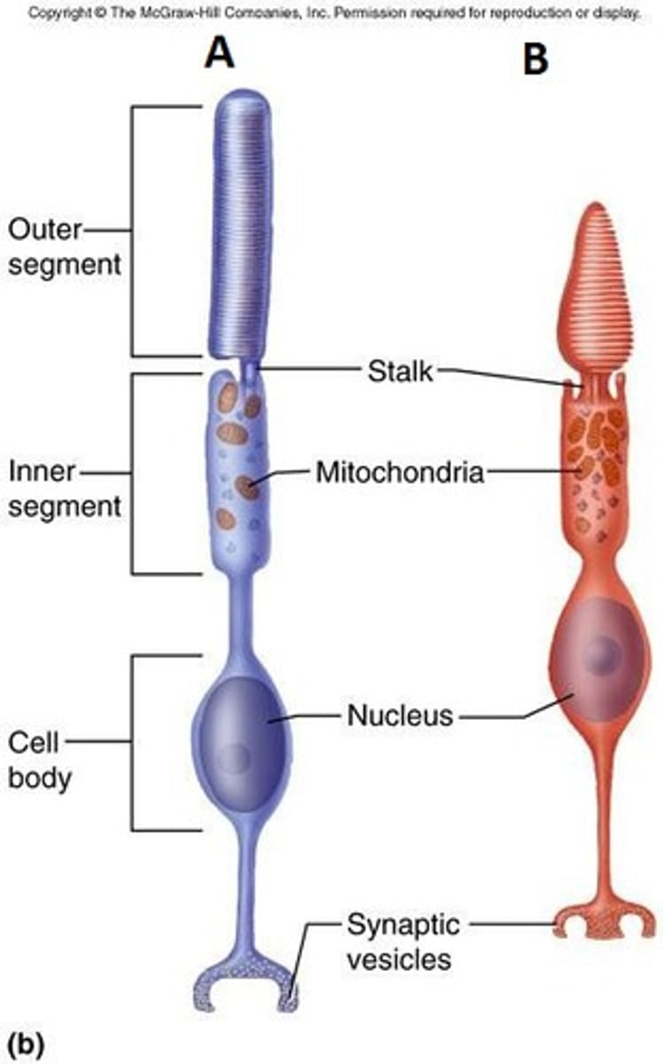
Rods — Photoreceptors specialized for dim light; numerous in the periphery.
Cones — Photoreceptors for bright light and color; most numerous in macula lutea and fovea centralis.
Fovea centralis — Region of retina with only cones; area of highest visual acuity.
Bipolar cells — Neurons stimulated by photoreceptors; relay signals to ganglion cells.
Ganglion cells — Neurons whose axons form the optic nerve.
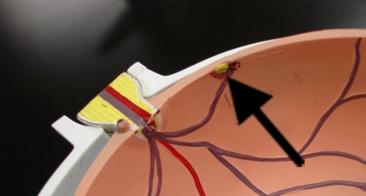
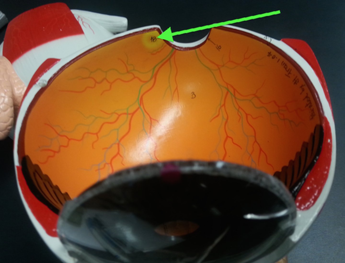
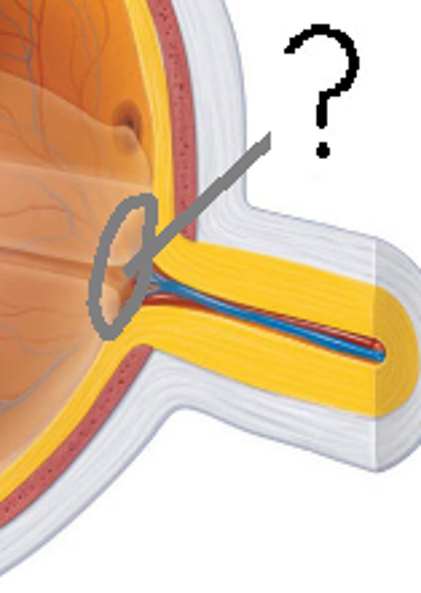
Optic disc (blind spot) — Region where optic nerve exits eye; contains no photoreceptors.
Anterior segment — Region between cornea and lens; contains aqueous humor.
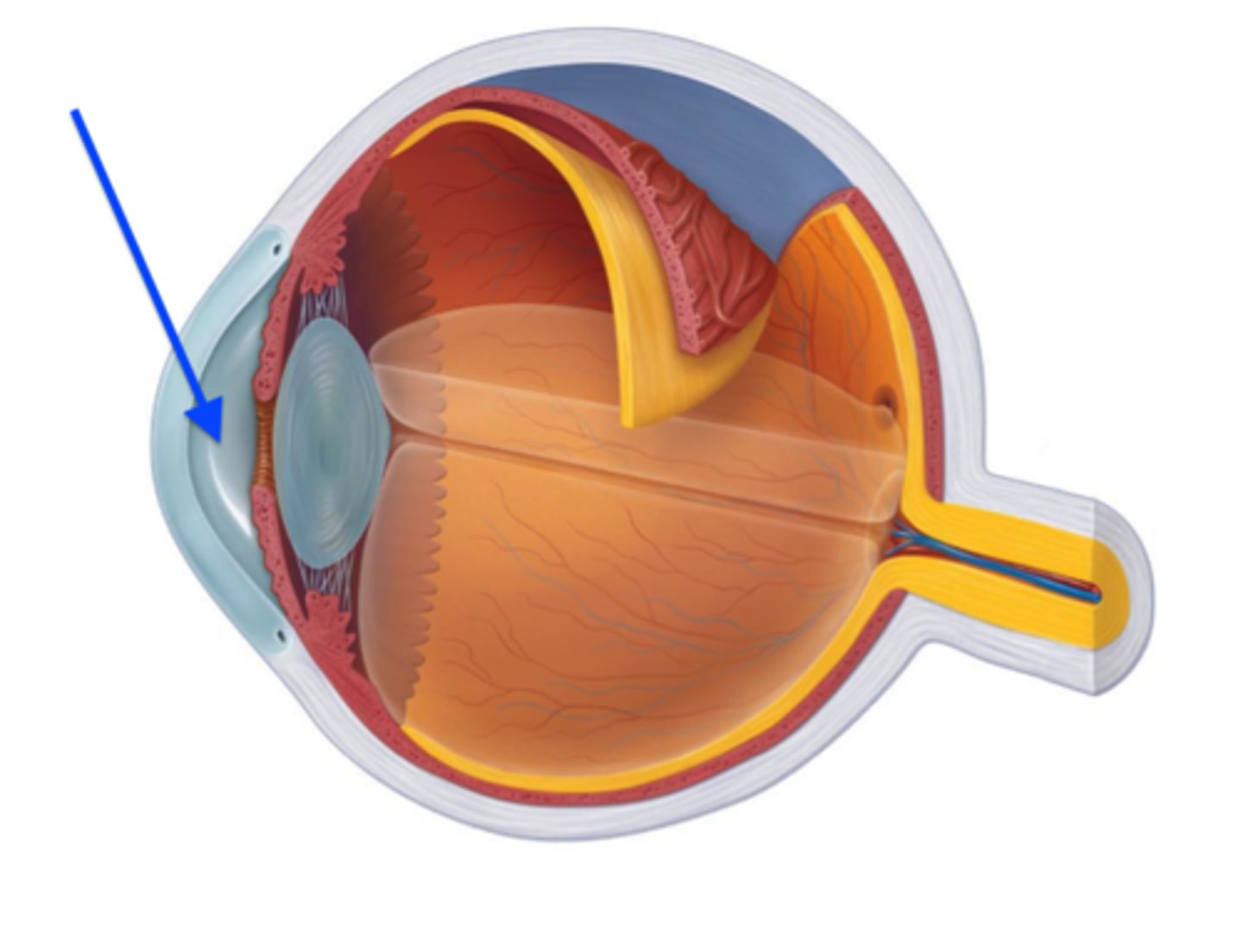
Aqueous humor — Watery fluid in anterior segment that refracts light.
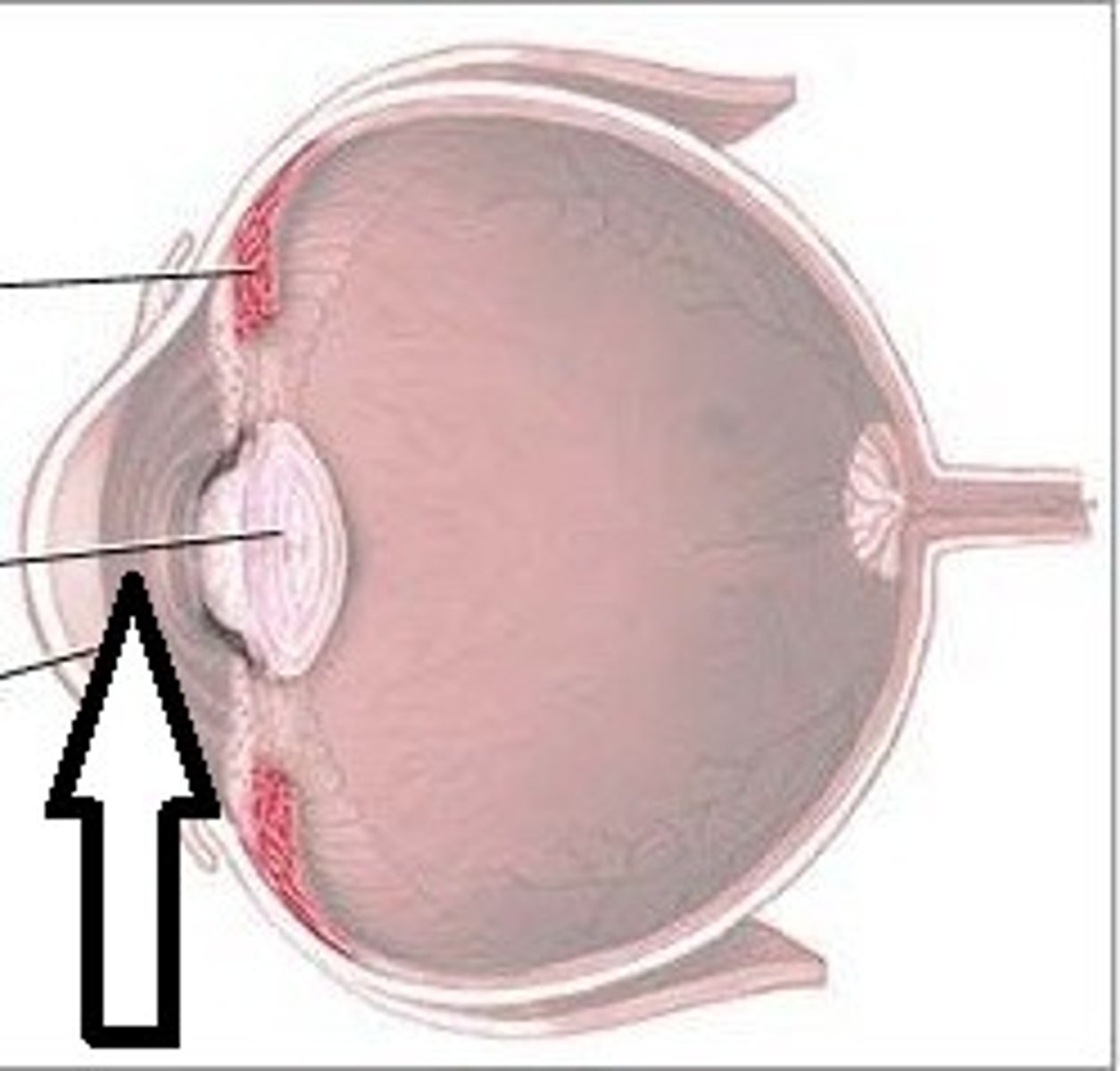
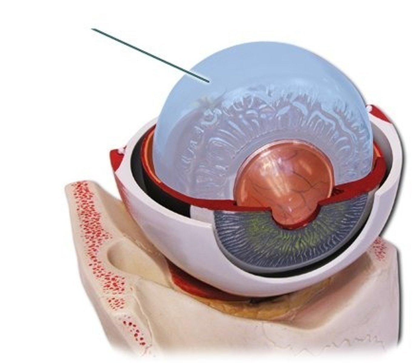
Lens — Biconvex transparent structure that refracts light.
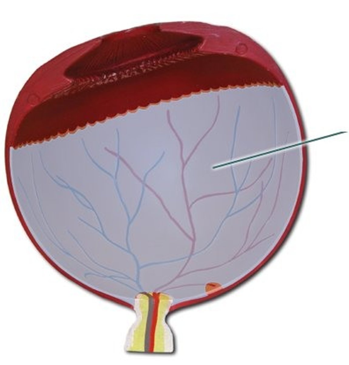
Posterior segment — Region posterior to the lens; contains vitreous humor.
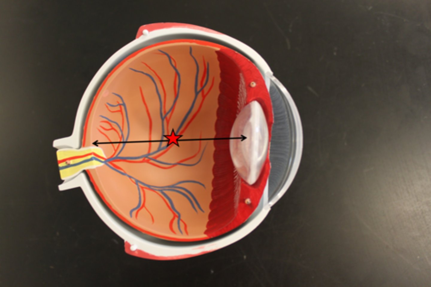
Vitreous humor — Gel-like fluid in posterior segment; maintains shape and refracts light.
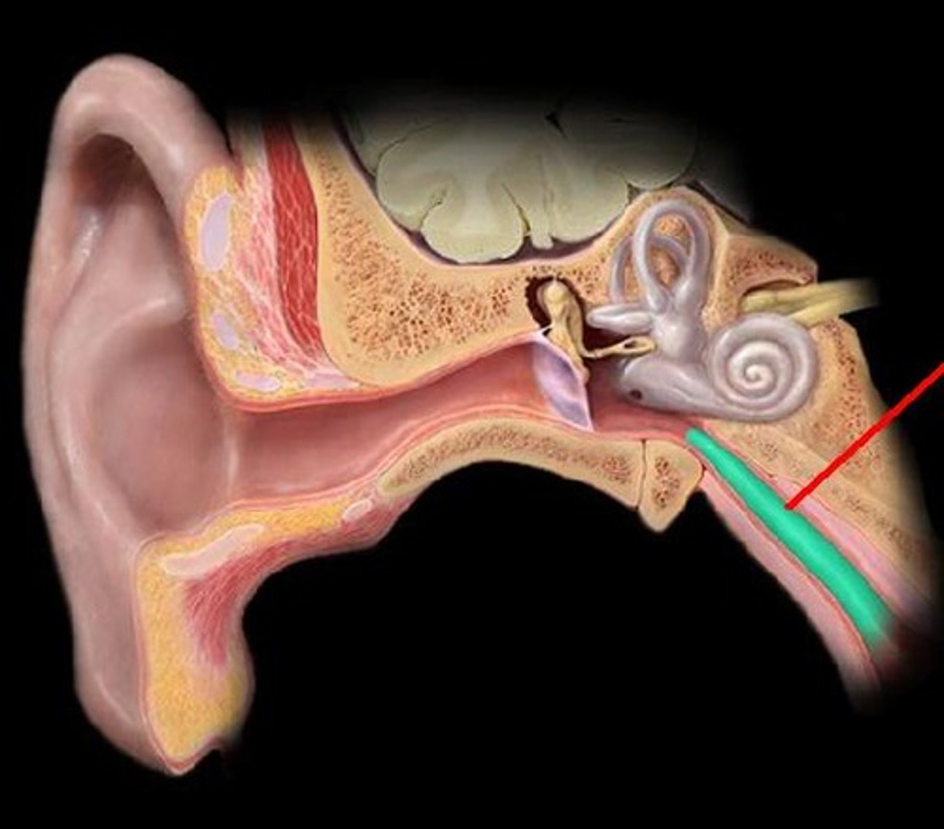
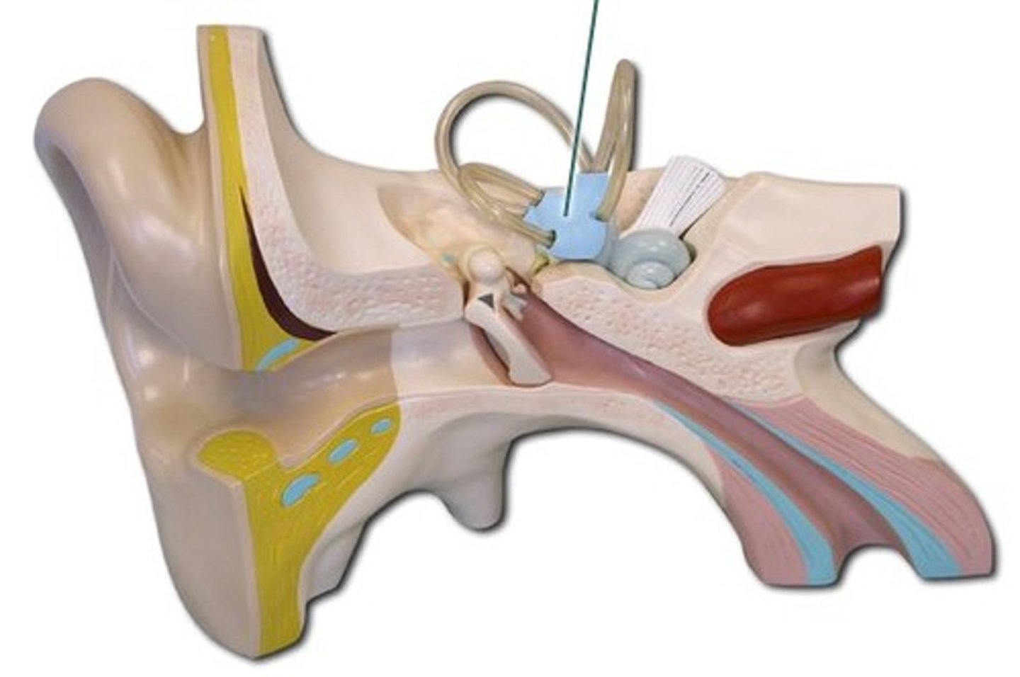
Pinna — External ear structure made of elastic cartilage.
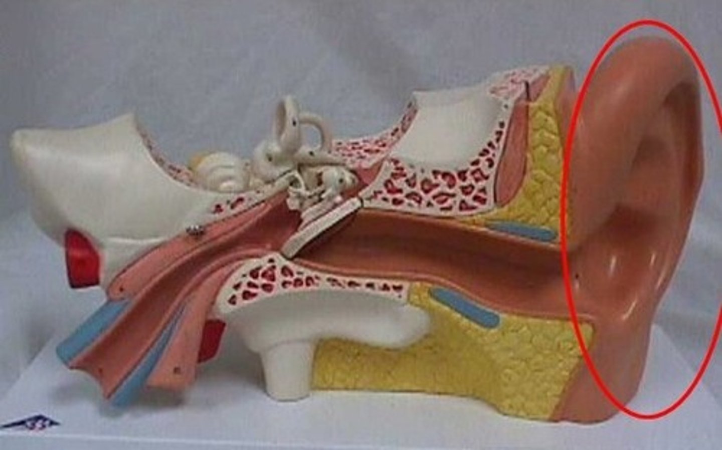
External acoustic meatus — Ear canal lined with ceruminous glands.
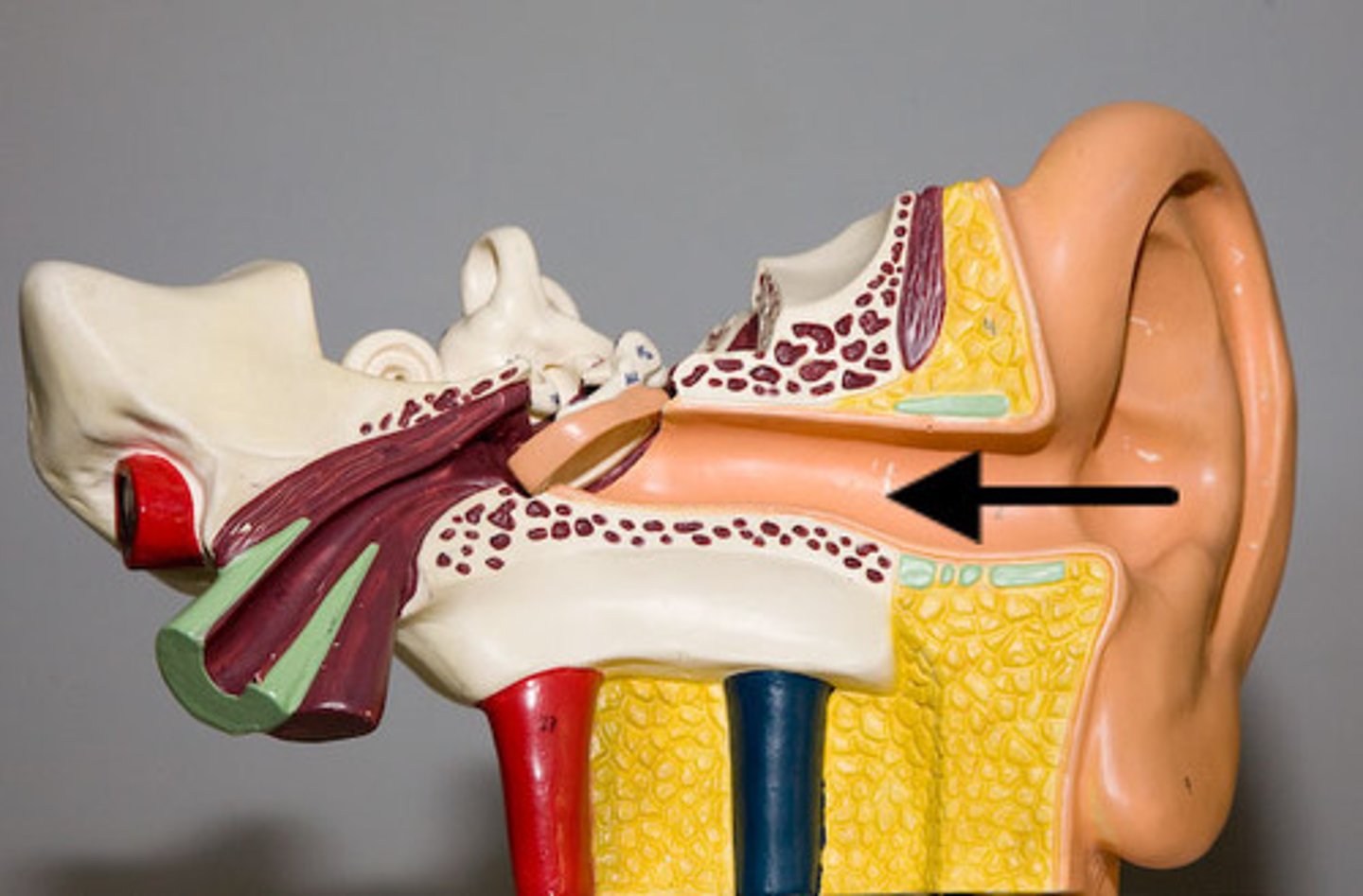
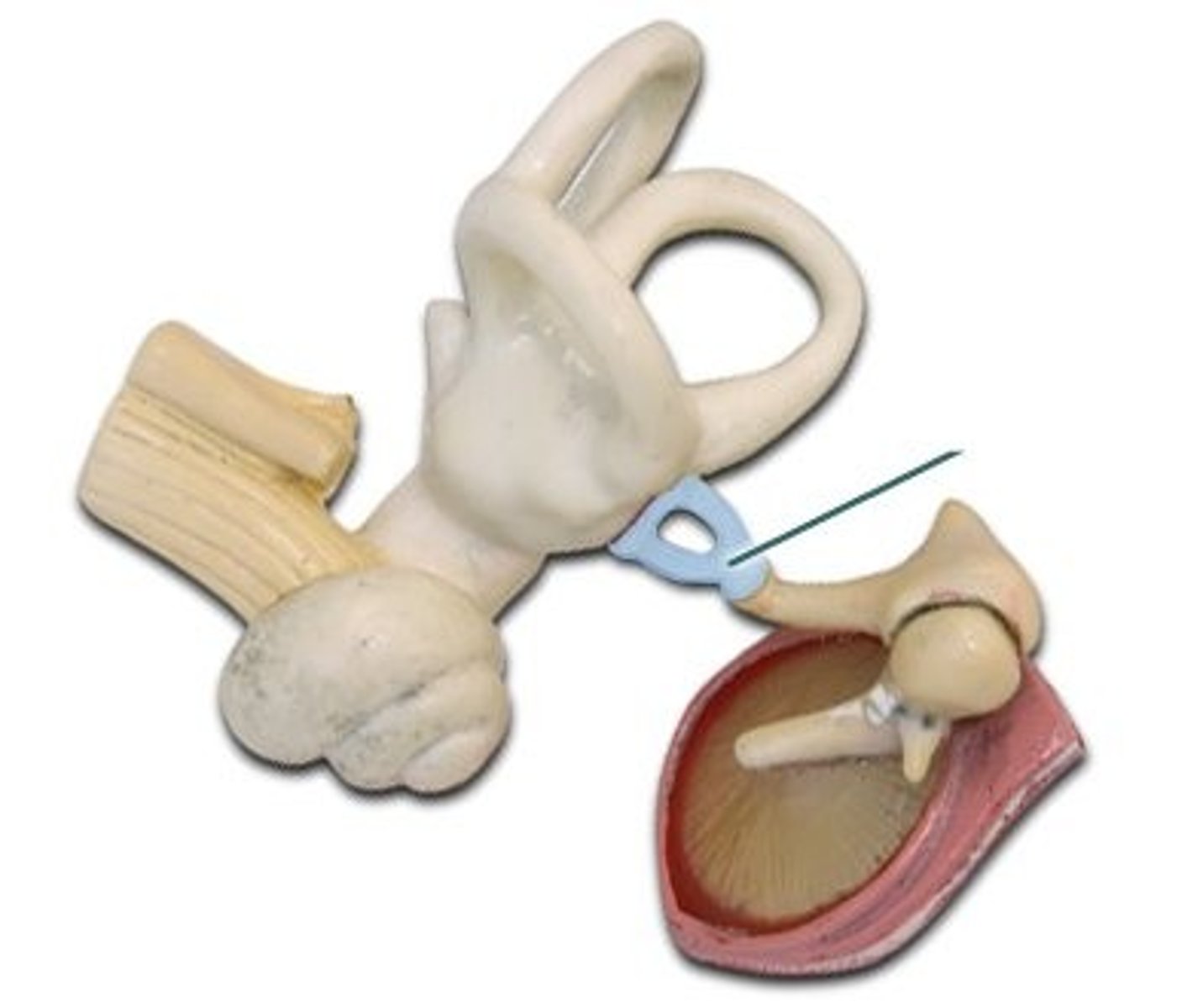
Tympanic membrane — Structure separating external and middle ear; vibrates with sound.
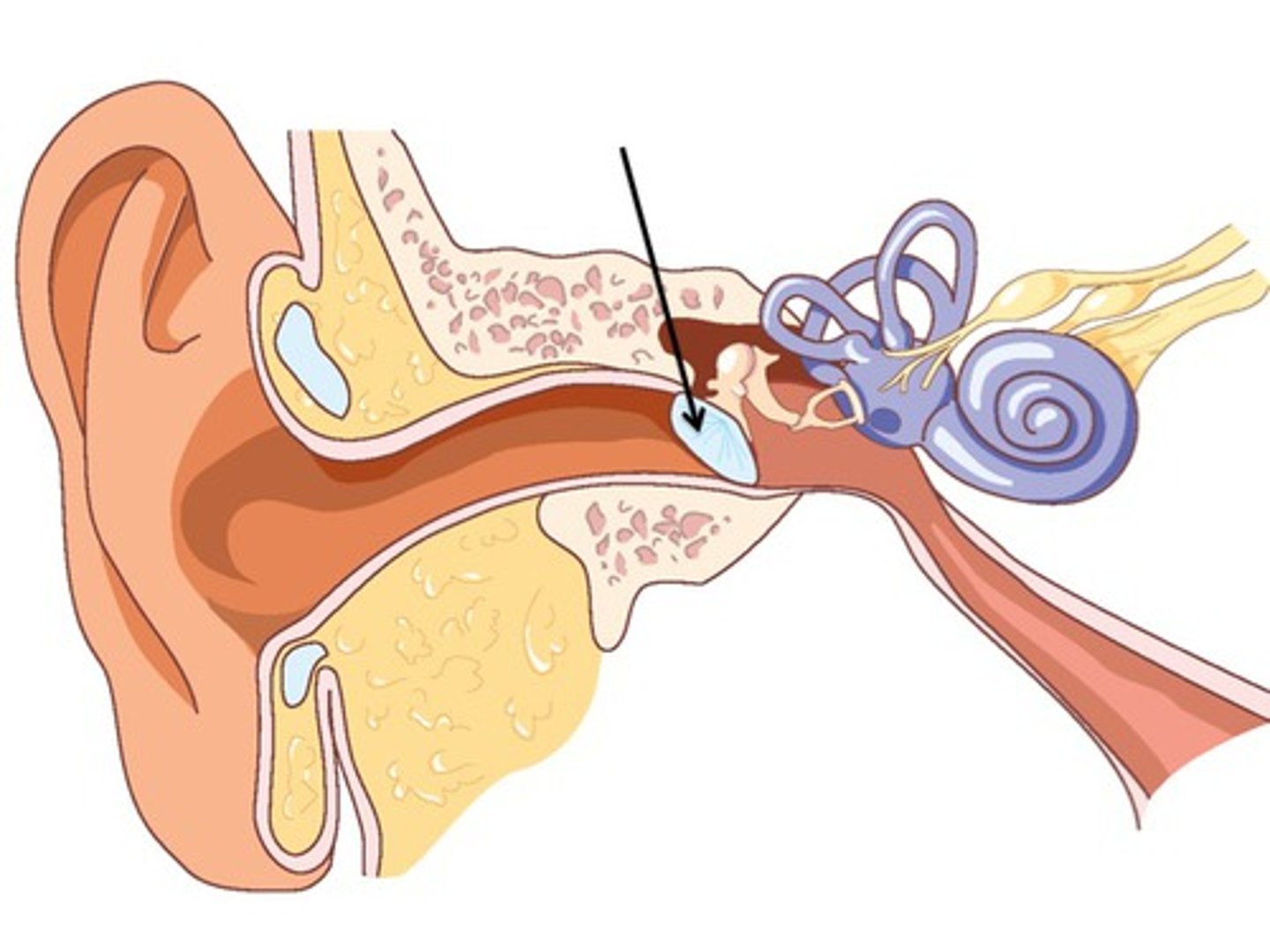
Ossicles — Three small bones of the middle ear.
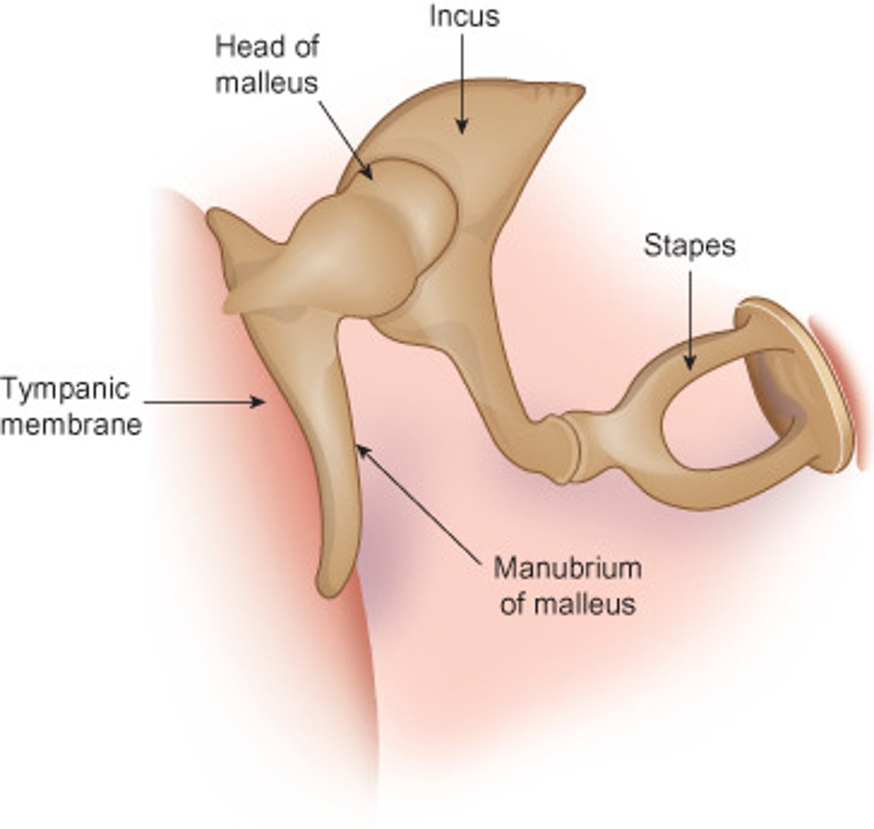
Malleus — Ossicle attached to tympanic membrane and incus.
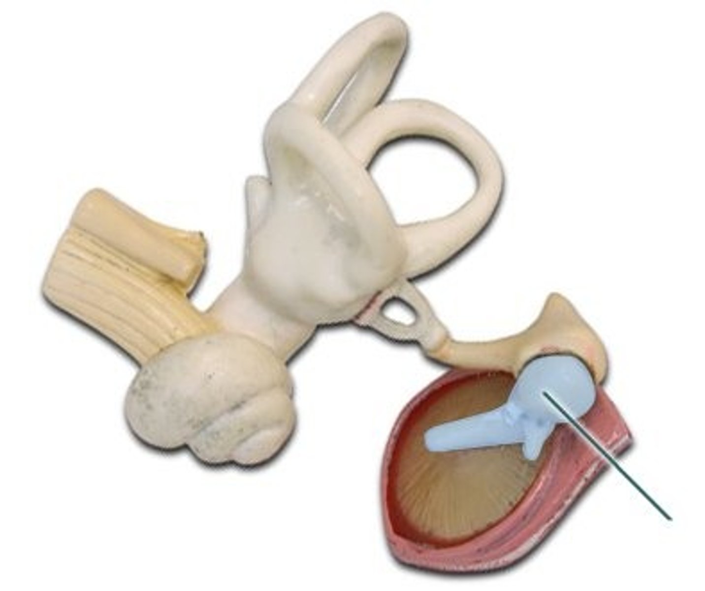
Incus — Ossicle articulating with malleus and stapes.
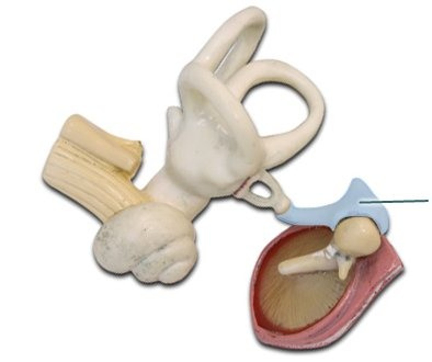
Stapes — Ossicle attaching to oval window; transmits vibrations to inner ear.
Pharyngotympanic tube — Connects middle ear to throat; equalizes air pressure.
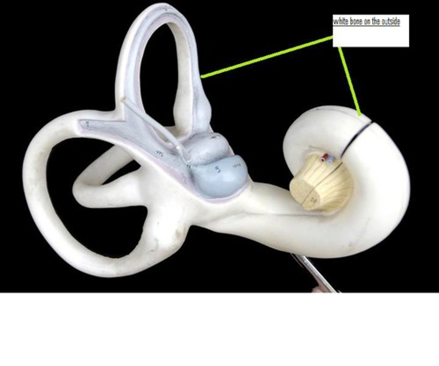
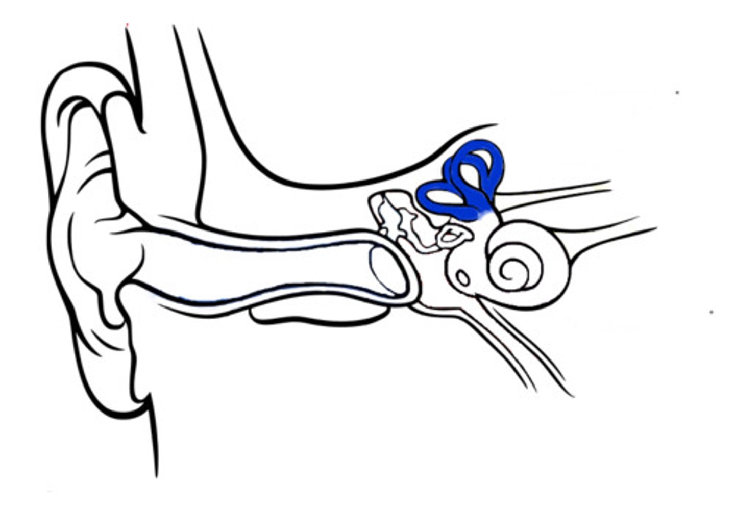
Bony labyrinth — Cavities in temporal bone filled with perilymph; includes cochlea, vestibule, semicircular canals.
Perilymph — Fluid filling the bony labyrinth.
Membranous labyrinth — Soft inner passages suspended in perilymph; filled with endolymph.
What is the membranous labyrinth
Endolymph — Fluid within membranous labyrinth.
Fluid within membranous labyrinth.
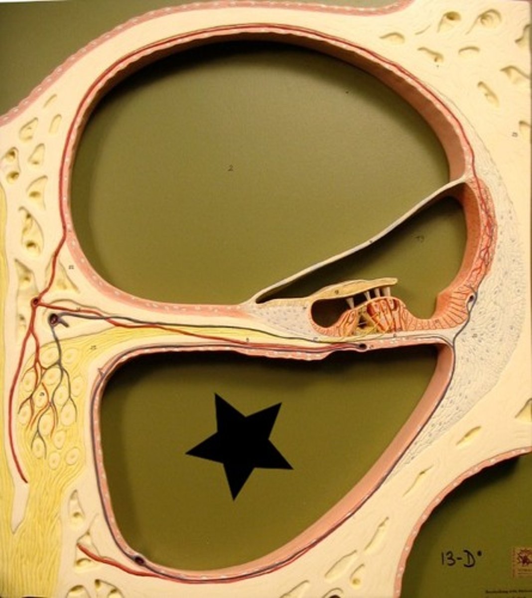
Cochlear duct — Endolymph-filled duct inside cochlea.
Endolymph-filled duct inside cochlea.
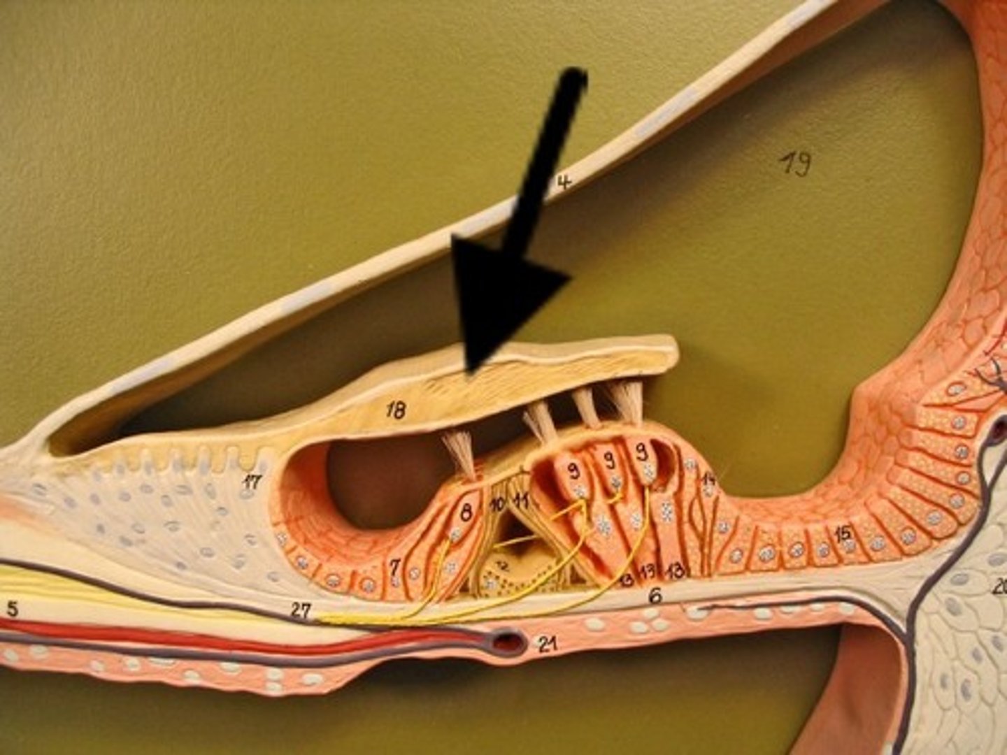
Organ of Corti — Organ of hearing located within cochlear duct.

Auditory receptors (hair cells) — Receptor cells resting on basilar membrane with stereocilia in tectorial membrane.
Receptor cells resting on basilar membrane with stereocilia in tectorial membrane.
Basilar membrane — Membrane supporting hair cells; movement generates impulses.
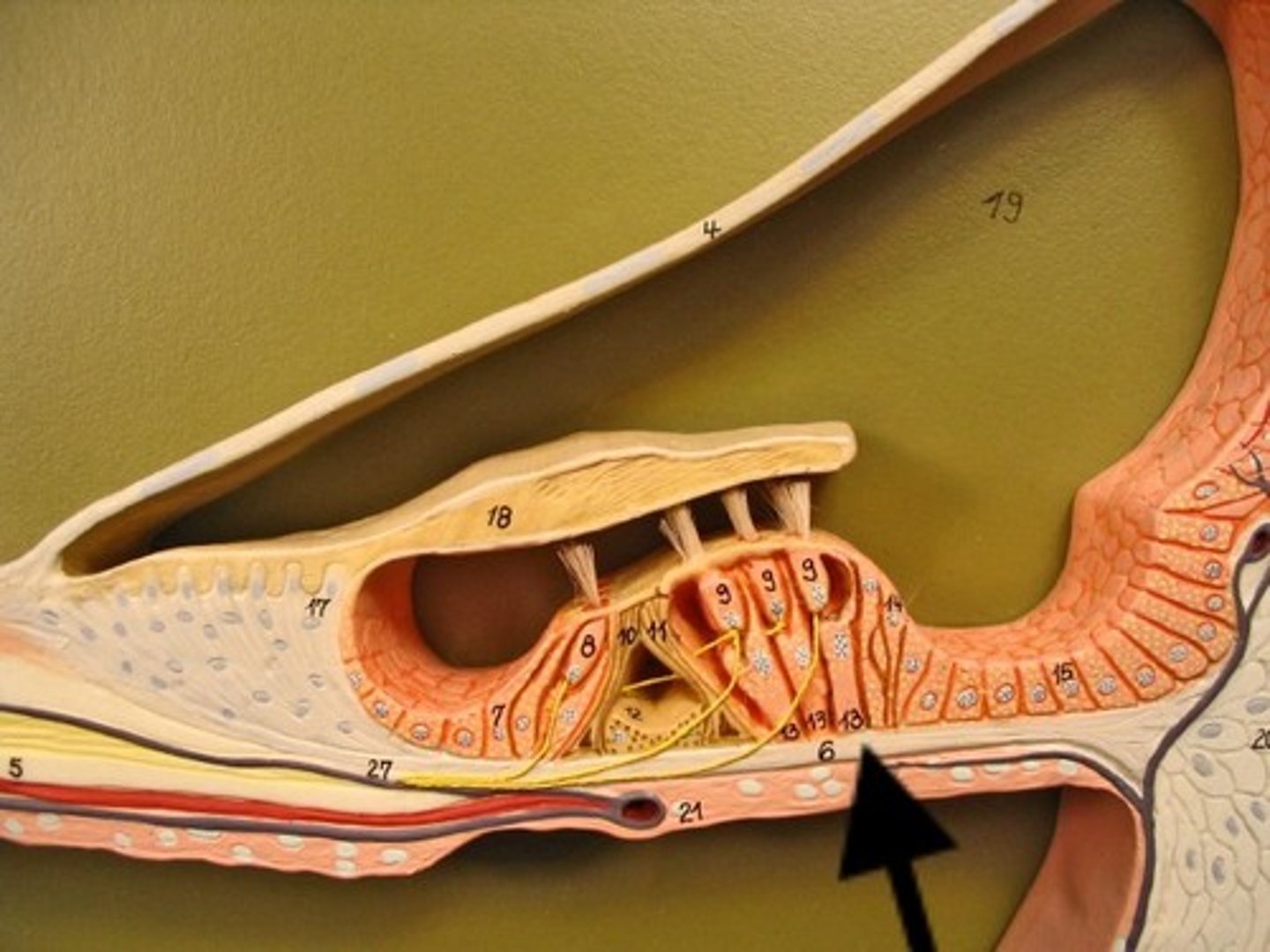
Tectorial membrane — Membrane contacting stereocilia of hair cells.
Semicircular ducts — Endolymph-filled ducts that detect rotational movement.
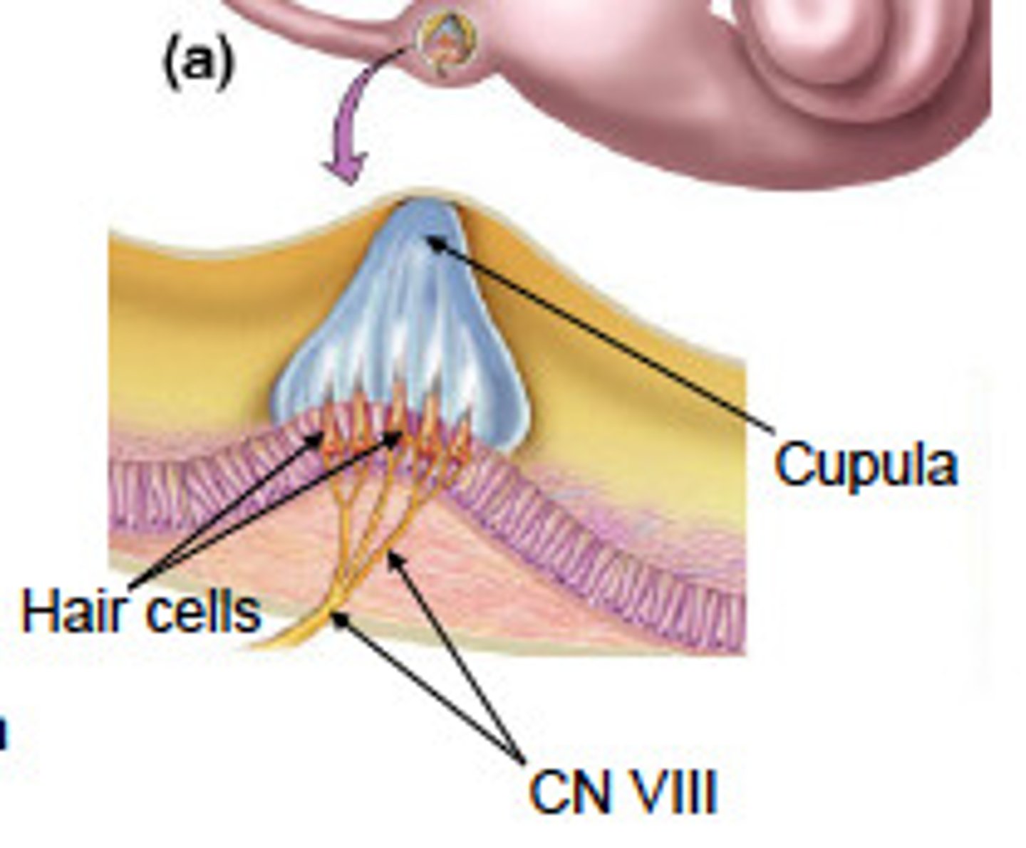
Cristae ampullaris — Receptors for rotational equilibrium located in semicircular duct ampullae.
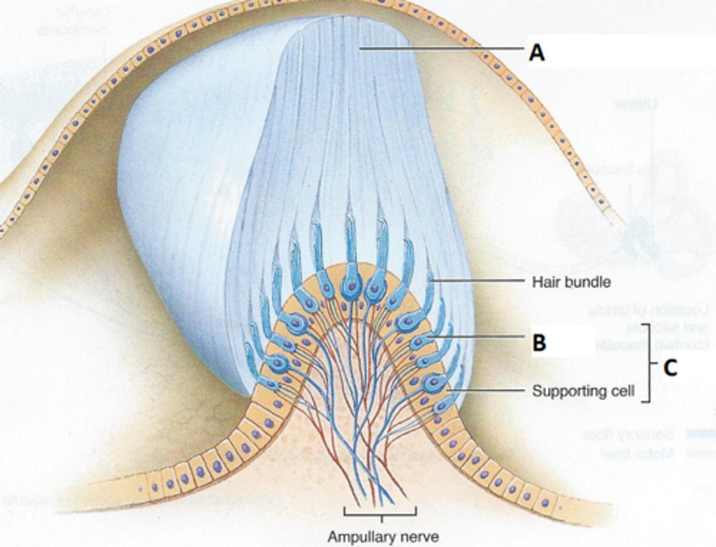
Cupula — Gel-like structure moved by endolymph to stimulate equilibrium receptors.
Saccule and Utricle — Receptors detecting linear acceleration and head position.
Chemoreceptors — Receptors for olfaction and gustation responding to chemicals in solution.
chemoreceptors
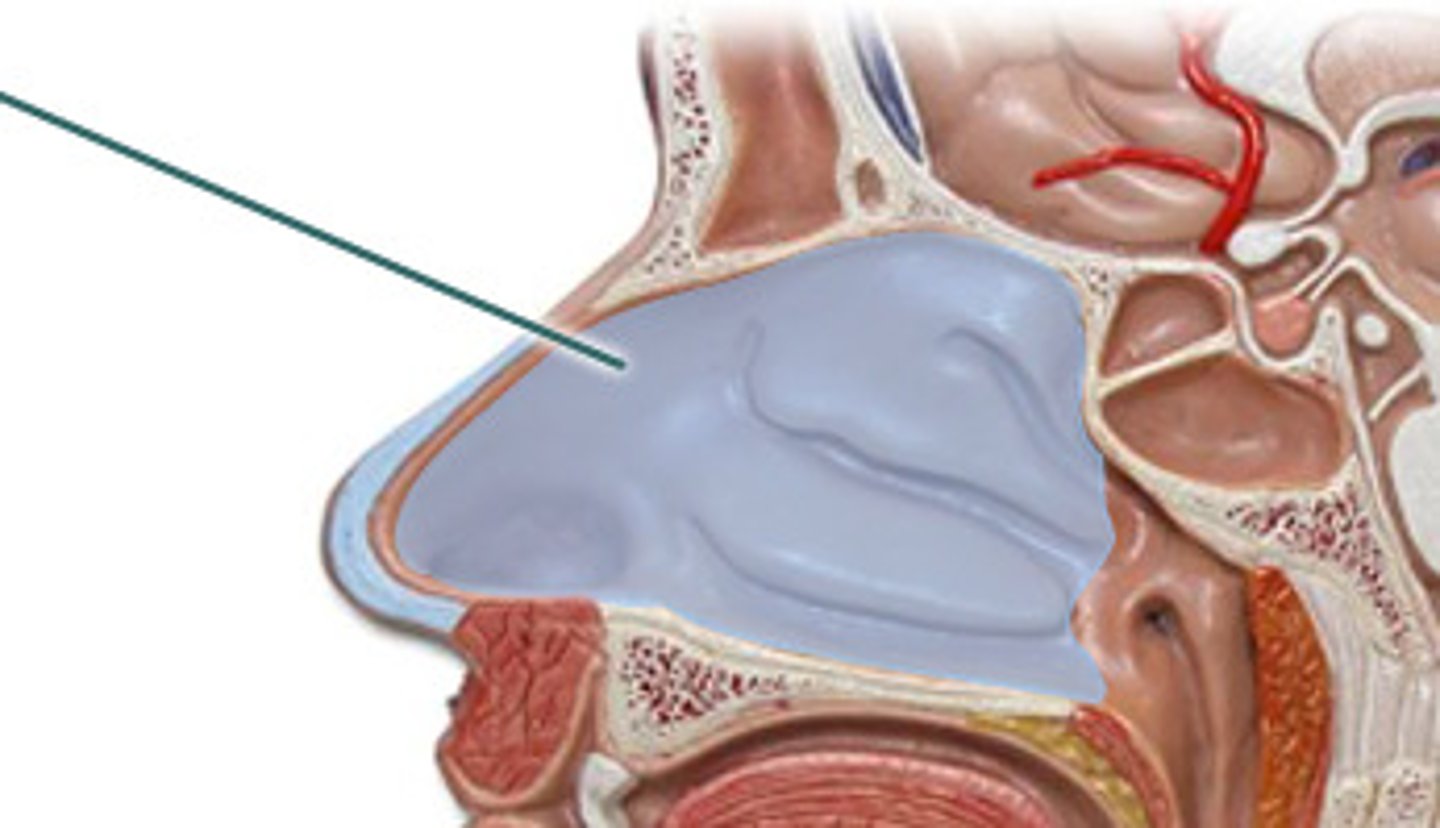
Olfactory epithelium — Organ of smell located in nasal cavity roof.
Olfactory sensory neurons — Bipolar neurons; axons form olfactory nerve; replaced every 30-60 days.
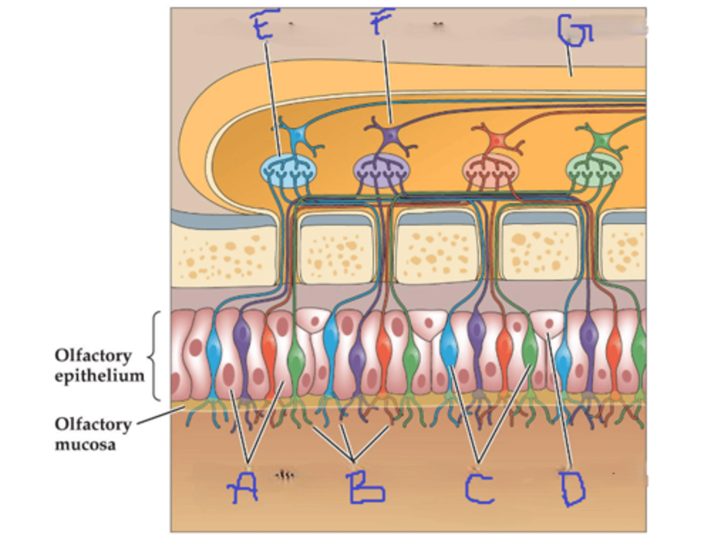
Supporting cells (olfactory) — Cells that cushion and surround olfactory neurons.
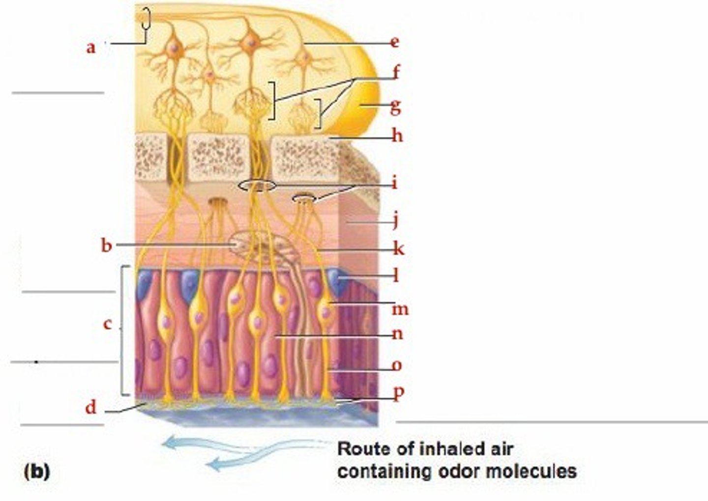
Olfactory stem cells — Stem cells replacing olfactory sensory neurons.

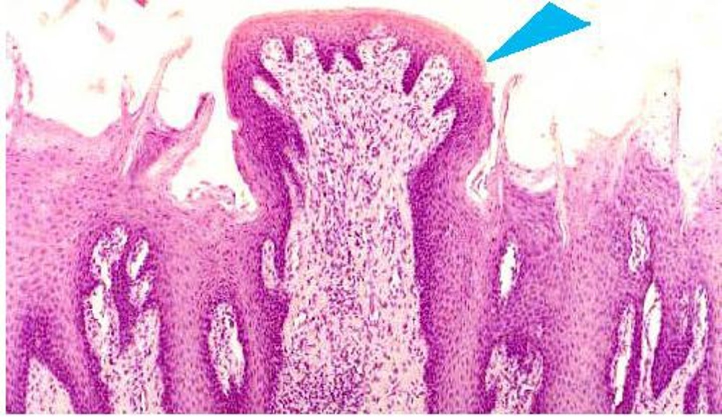
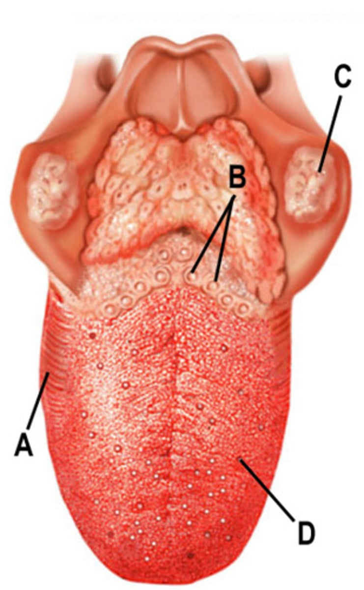
Taste buds — Receptor organs for taste found in papillae.
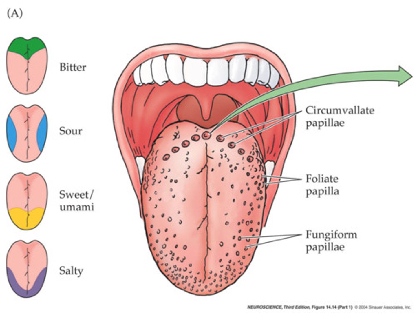
Papillae — Tongue structures where taste buds are located.
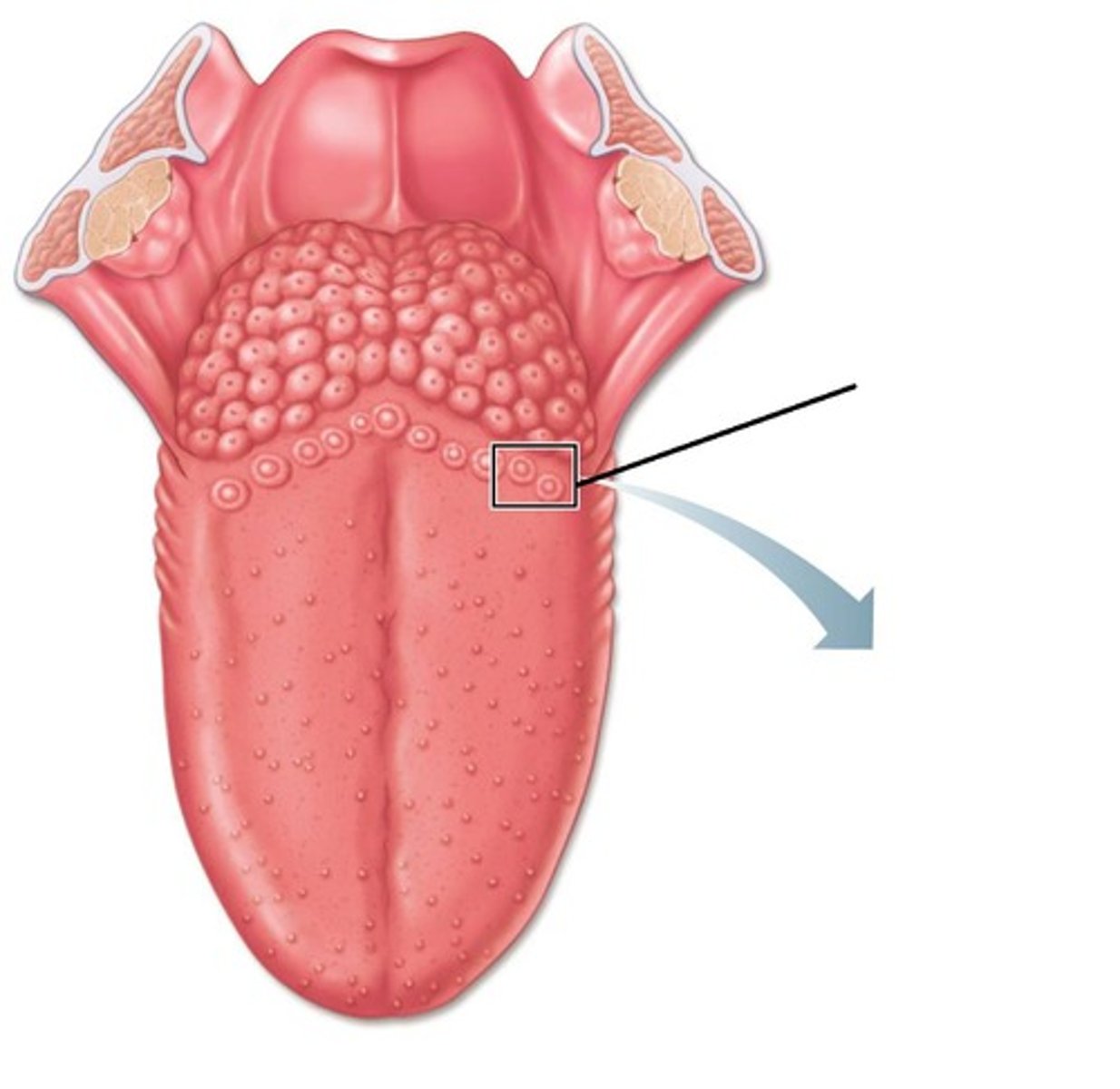
Fungiform papillae — Most numerous papillae; taste buds on superior surface.
Foliate papillae — Papillae on tongue sides; taste buds in side walls.
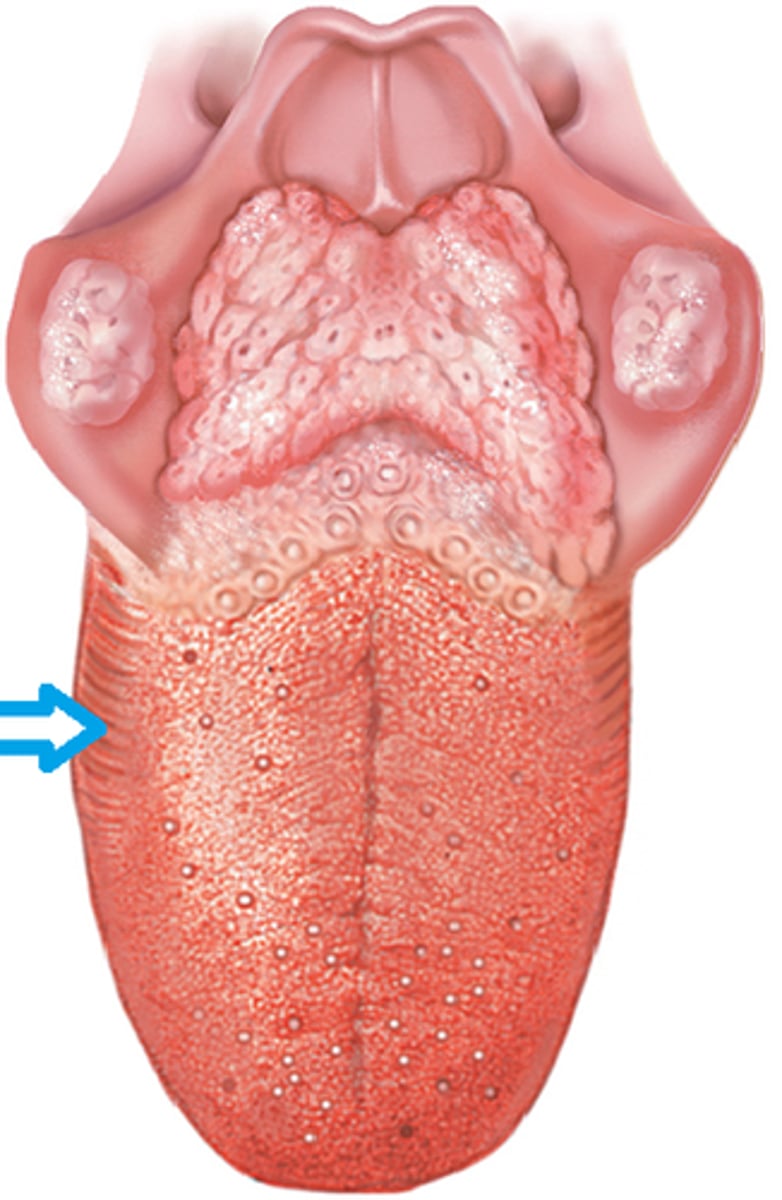
Vallate papillae — Large V-shaped papillae; taste buds in side walls.
Gustatory epithelial cells — Taste receptor cells within taste buds.
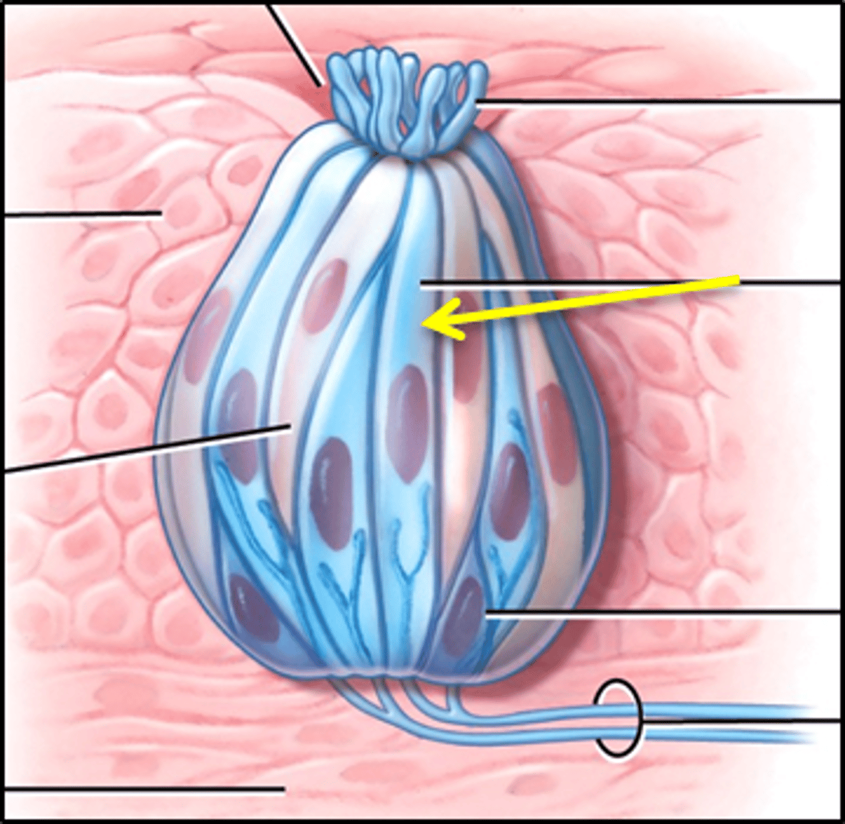
Microvilli (gustatory hairs) — Projections that enter taste pore and contact saliva.
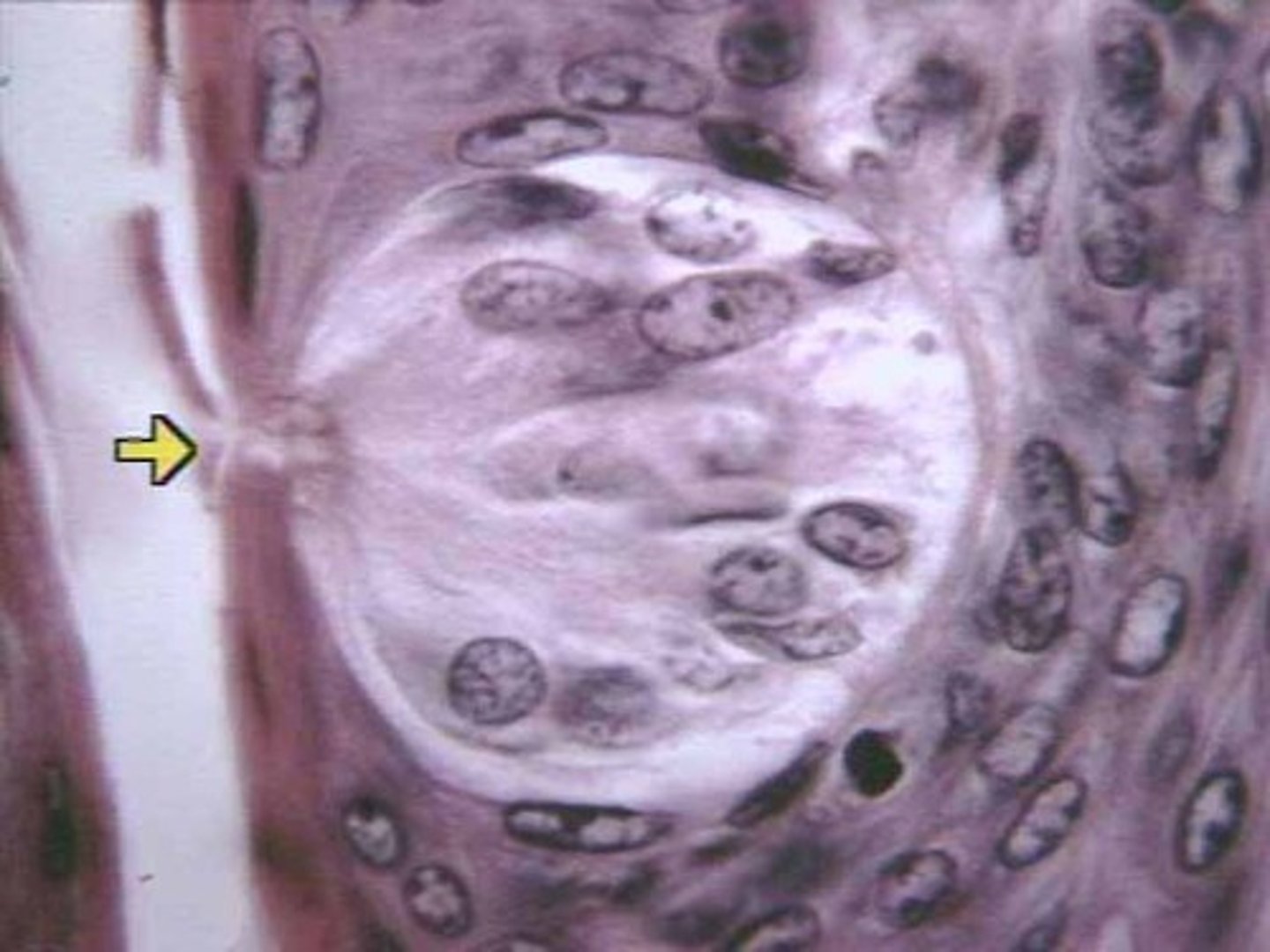
Basal epithelial cells — Stem cells replacing gustatory cells every 7 days.
Sweet — Taste produced by sugars, alcohol, saccharin, some amino acids.
sweet
Sour — Taste produced by hydrogen ions (H+).
sour
Salt — Taste produced by metal ions (Na+, K+).
salt
Bitter — Taste from alkaloids such as nicotine or quinine.
bitter
Umami — Taste from glutamate or aspartate; "savory."
umami
Nervous System — Monitors and processes sensory information and body functions.
Nervous system
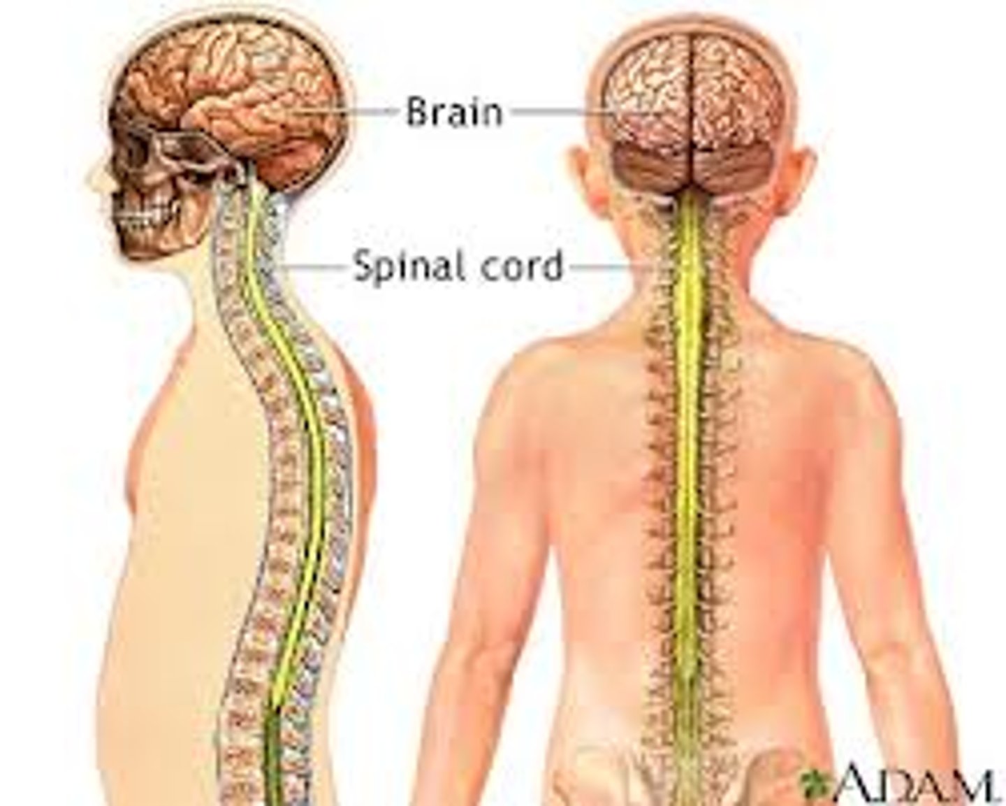
Central Nervous System (CNS) — Brain and spinal cord.
Peripheral Nervous System (PNS) — Cranial nerves, spinal nerves, ganglia, sensory receptors.
PNS
Neurons — Conducting cells of nervous tissue.
Neuroglia (glial cells) — Supporting cells that protect, brace, and myelinate neurons.
Glial Cells
Excitability — Ability of neurons to respond to stimuli and generate impulses.
Excitability
Conductivity — Ability to transmit impulses to other neurons, muscles, glands.
Conductivity
Amitotic — Property of neurons meaning they cannot divide.
Amitotic
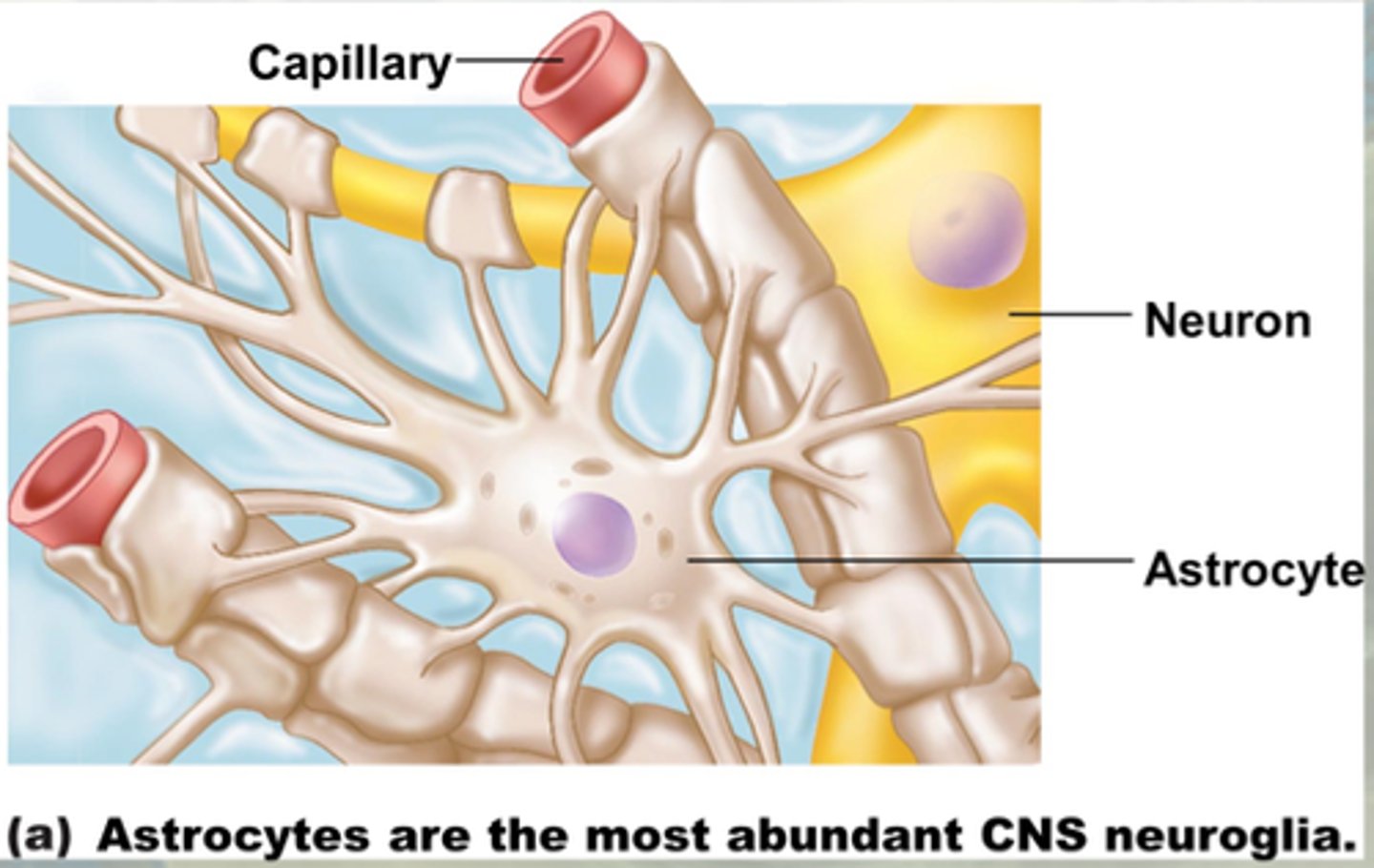
Astrocytes — CNS cells that regulate chemical environment and assist capillaries.
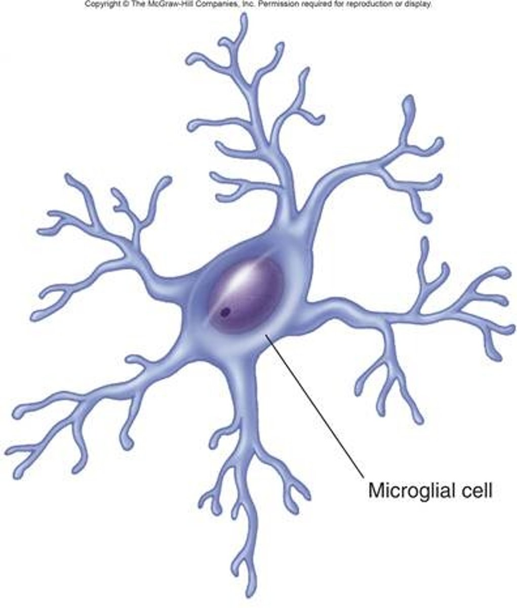
Microglia — CNS phagocytes removing debris and pathogens.
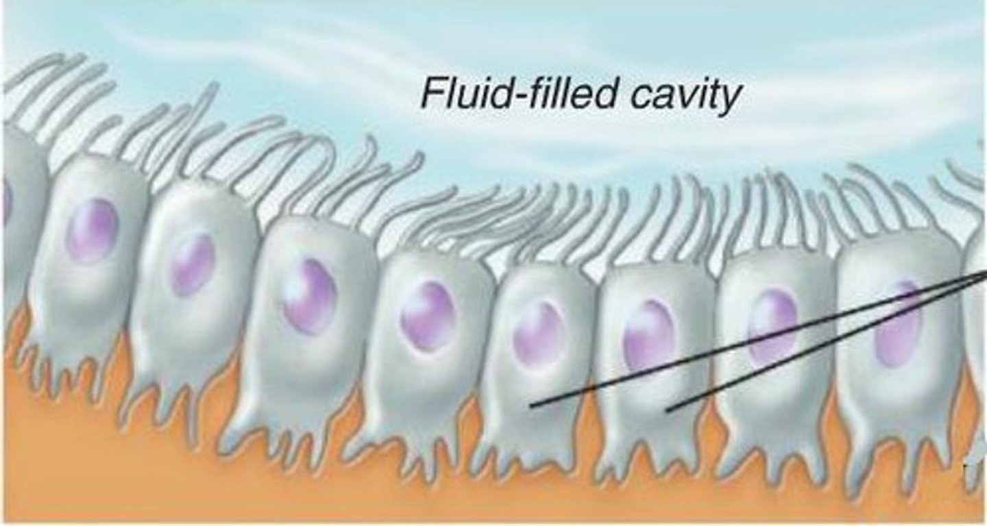
Ependymal cells — CNS cells lining CSF cavities; circulate CSF.
Oligodendrocytes — CNS cells producing myelin.
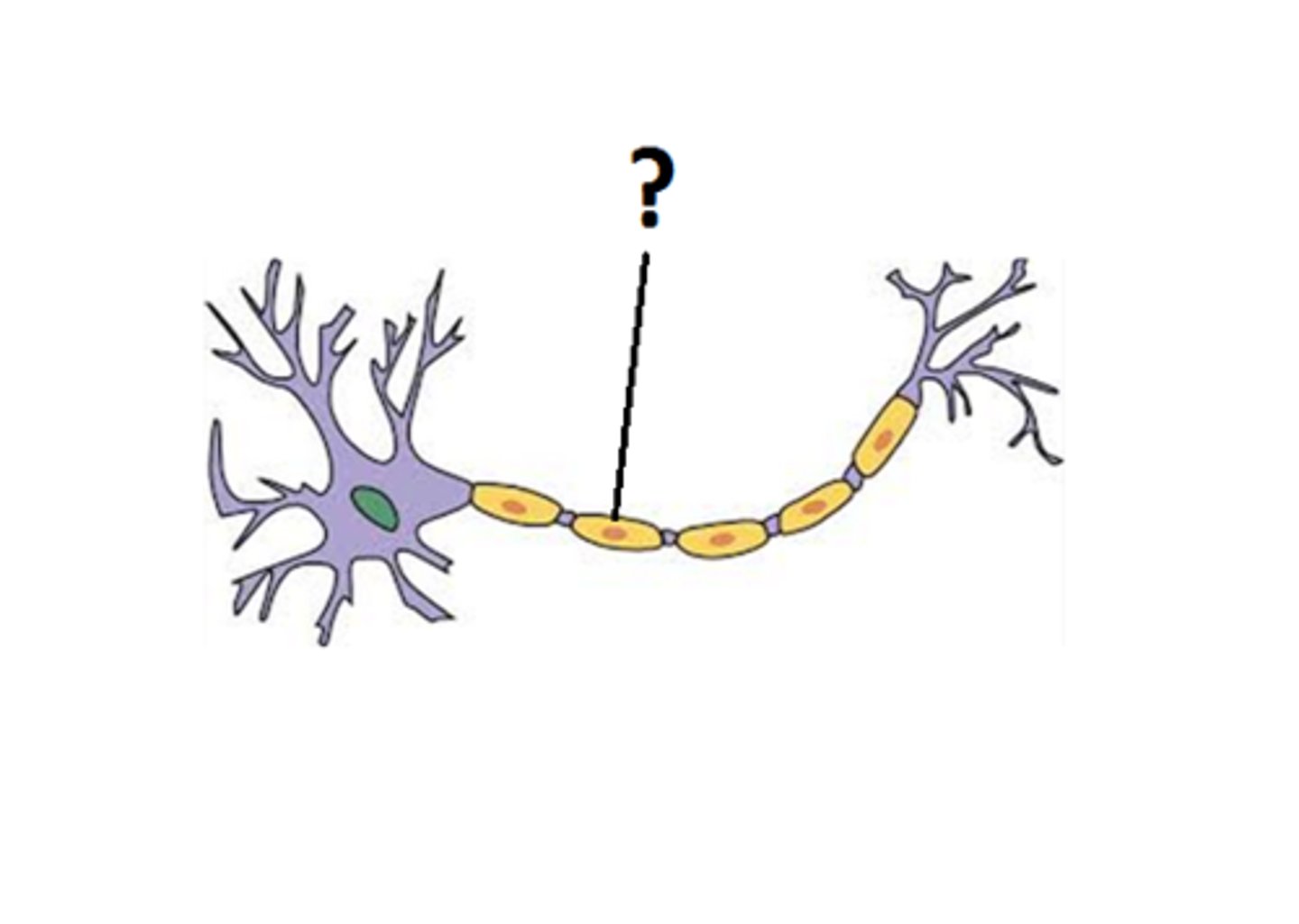
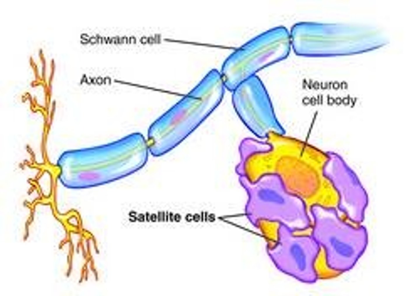
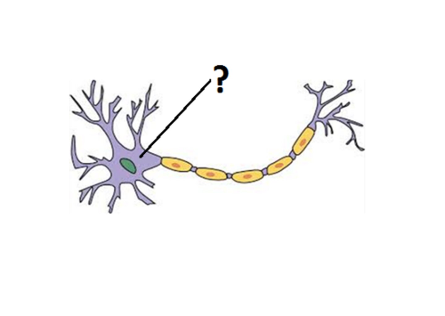
Schwann cells — PNS cells producing myelin sheaths.
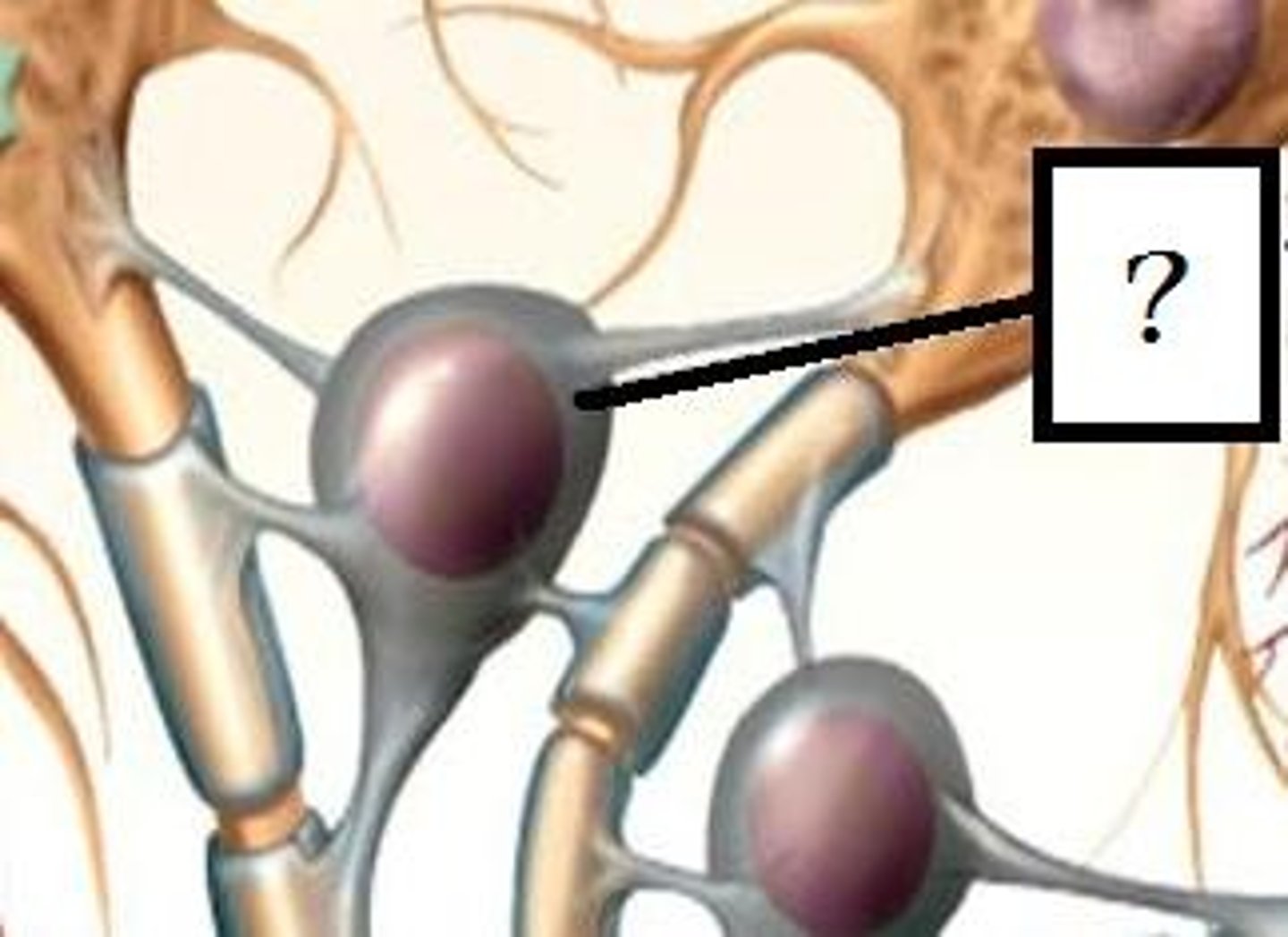
Satellite cells — PNS cells surrounding neuron cell bodies.
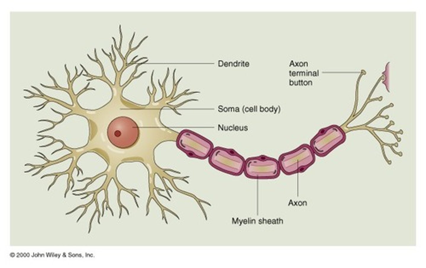
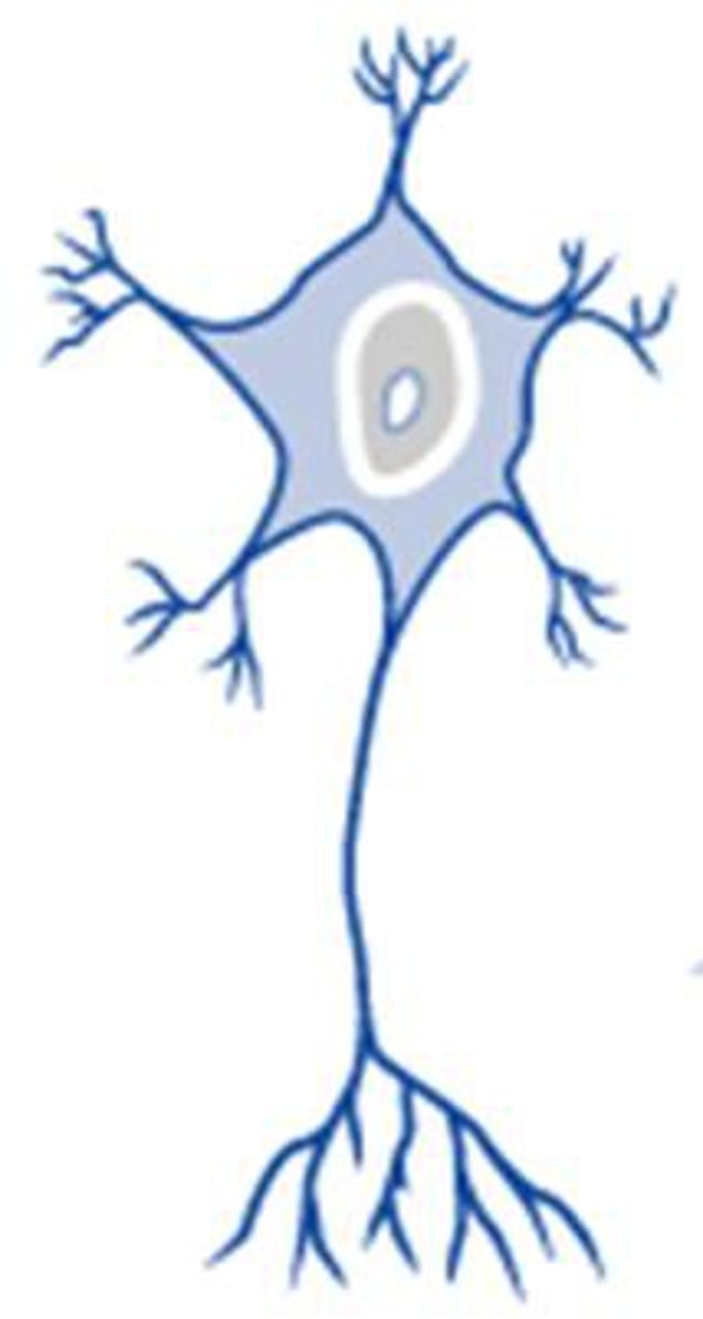
Cell body — Contains nucleus and cytoplasm of neuron.
Neurofibrils — Cytoskeletal fibers maintaining neuron shape.
Chromatophilic substance — Rough ER in neuron cell body.
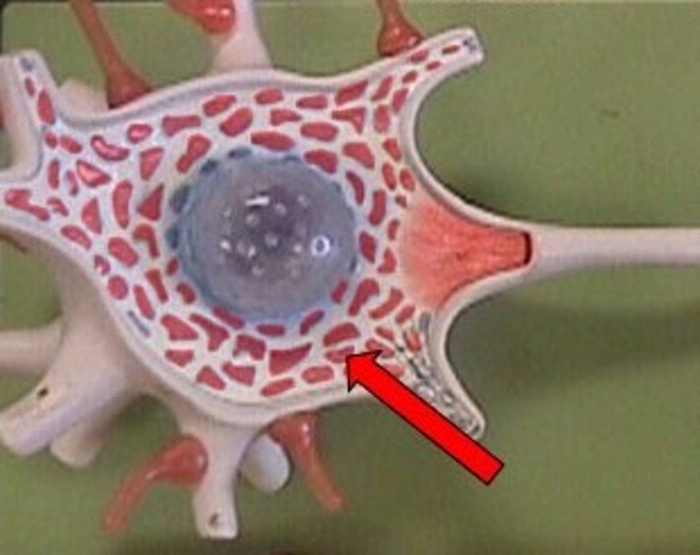
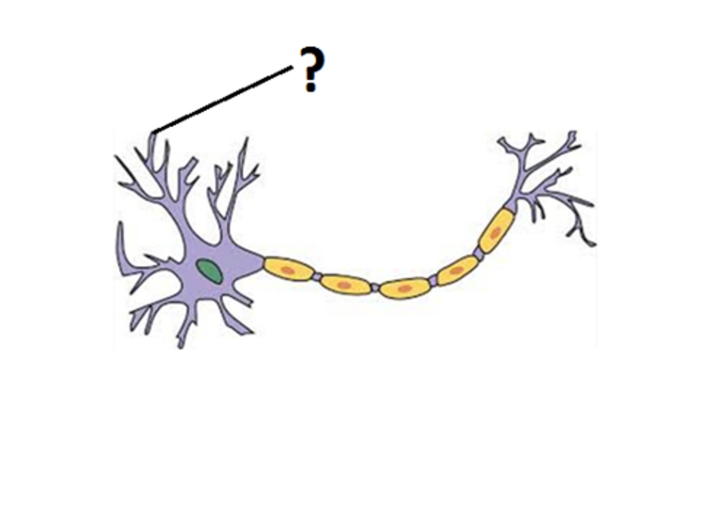
Dendrites — Structures receiving incoming signals.
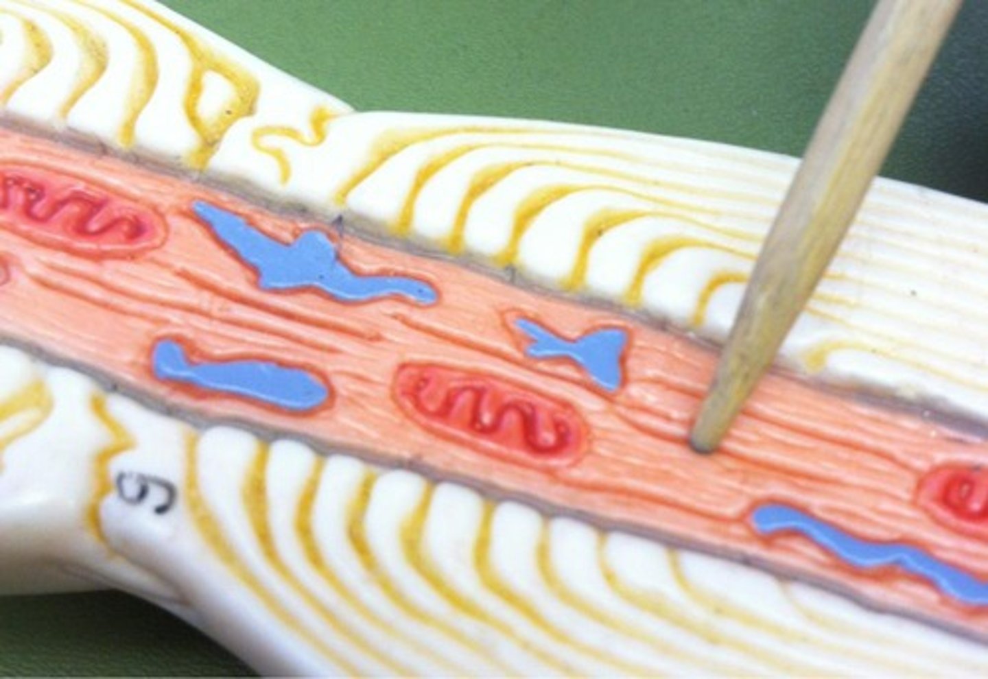
Axons — Structures transmitting impulses away from cell body.
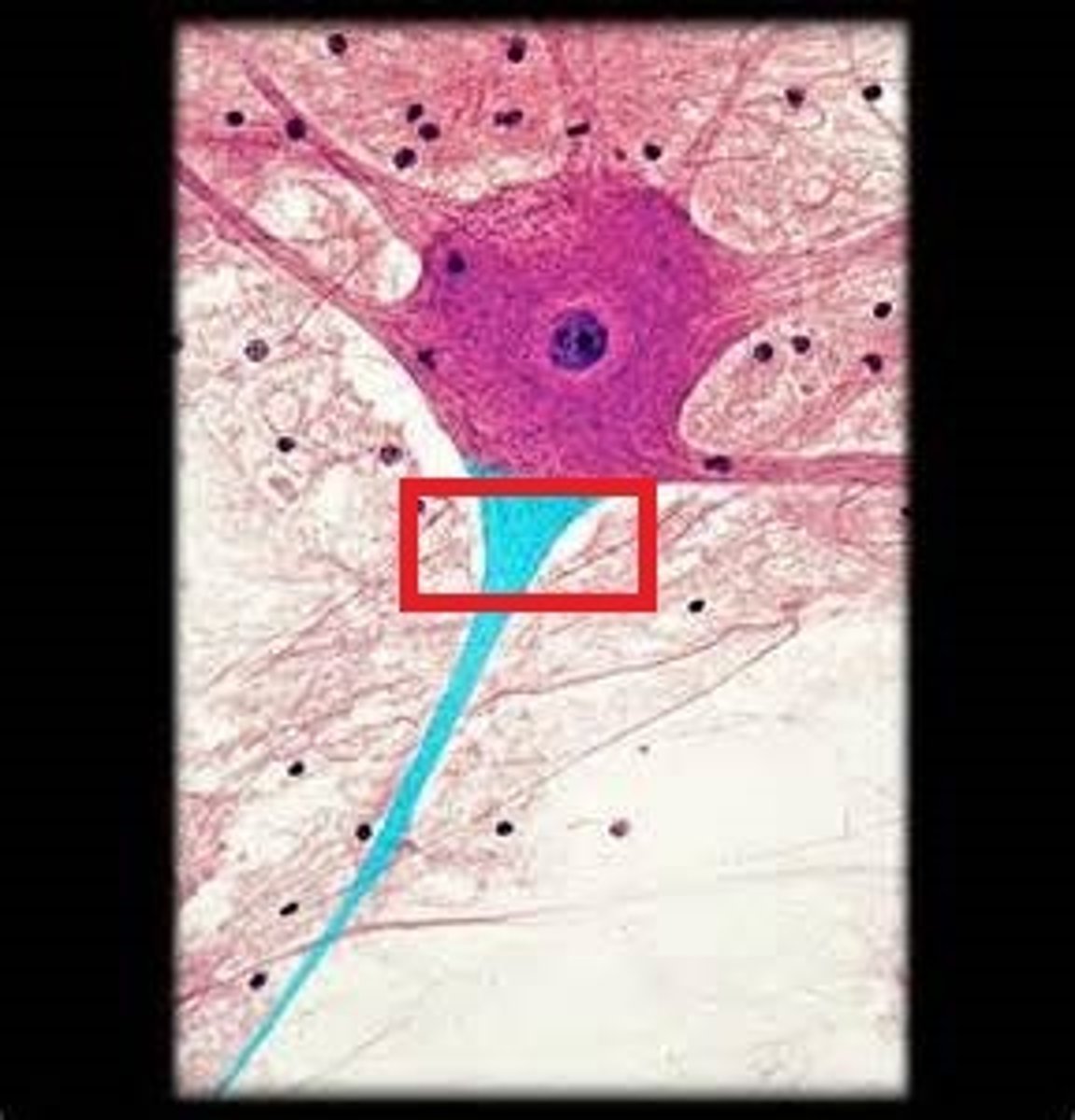
Axon hillock — Cone-shaped region where axon originates.
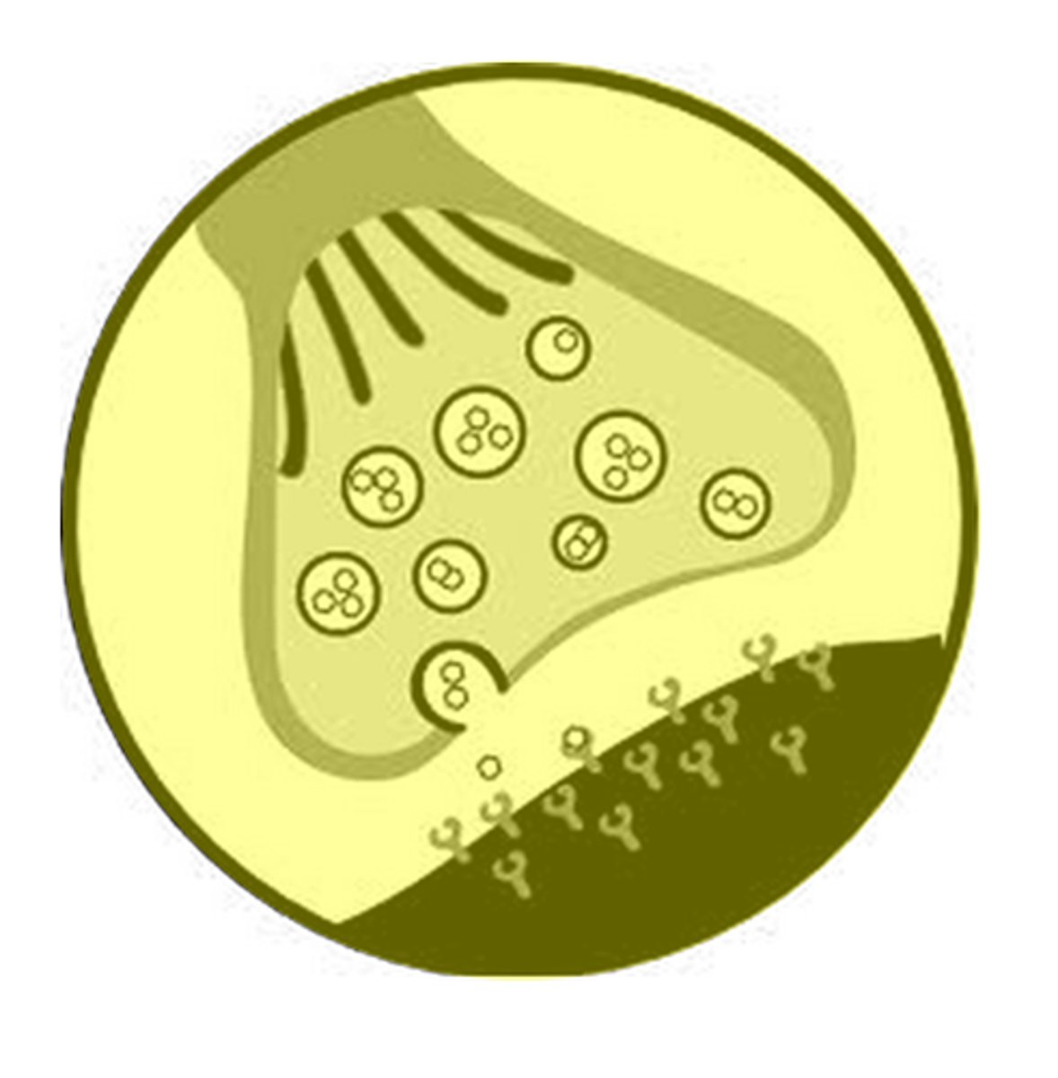
Axon terminals — Store and release neurotransmitters.
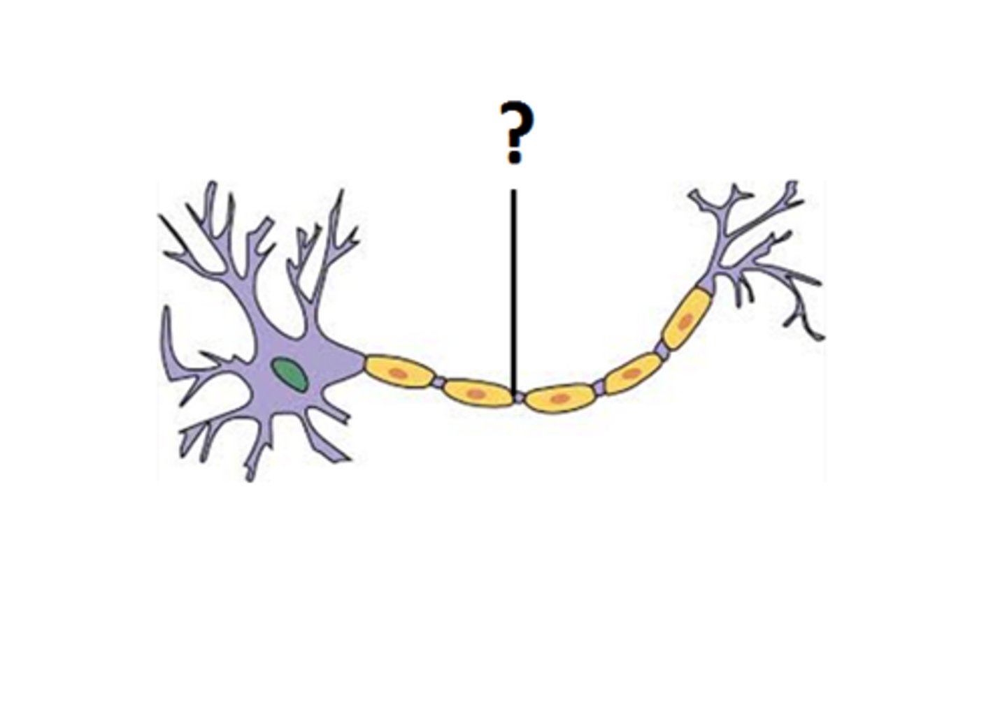
Myelin sheath — Fatty insulation increasing impulse speed.

Myelin sheath gaps (Nodes of Ranvier) — Gaps aiding rapid conduction.
Multipolar neurons — Many dendrites, one axon; most common in CNS.
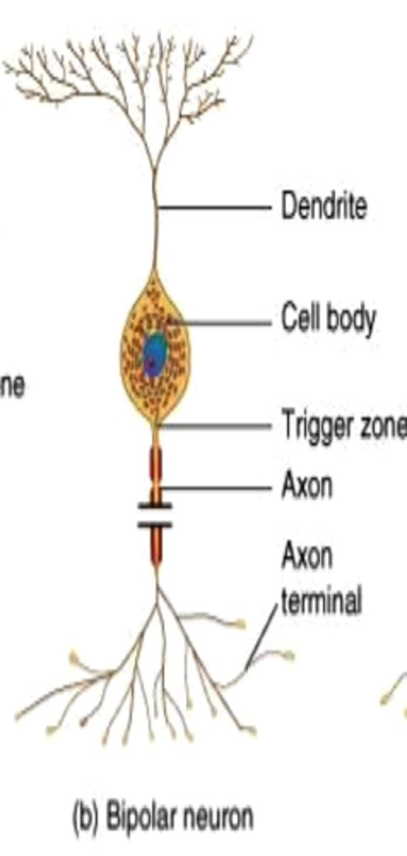
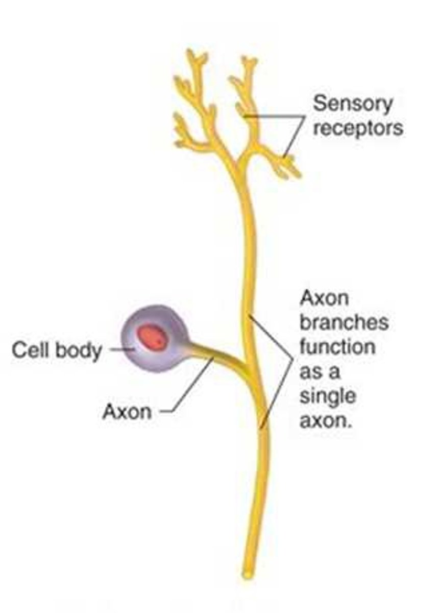
Bipolar neurons — One dendrite and one axon; found in sensory organs.

Unipolar neurons — One process branching into peripheral and central processes; common in PNS.
Sensory (afferent) neurons — Carry impulses toward CNS.
Sensory
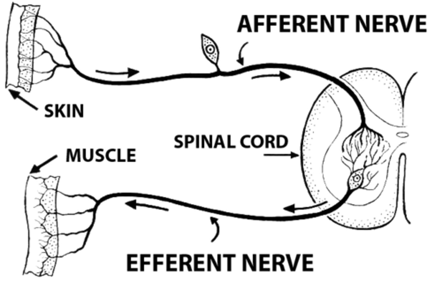
Motor (efferent) neurons — Carry impulses away from CNS.
Motor

Association neurons (interneurons) — Integrate information within CNS.
Association Neurons
Tract — Bundle of CNS axons.
Tract
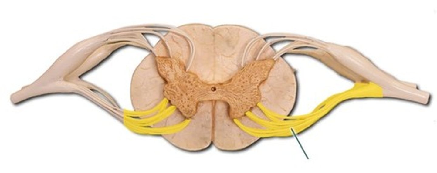
Nerve — Bundle of PNS axons.
Nerve
Mixed nerves — Contain both sensory and motor fibers.
Mixed nerves
Endoneurium — Connective tissue around one axon.
Endoneurium
Perineurium — Connective tissue surrounding fascicles.
perineurium
Epineurium — Connective tissue surrounding entire nerve.
epineurium
Cerebrum — Largest brain region responsible for conscious processes.
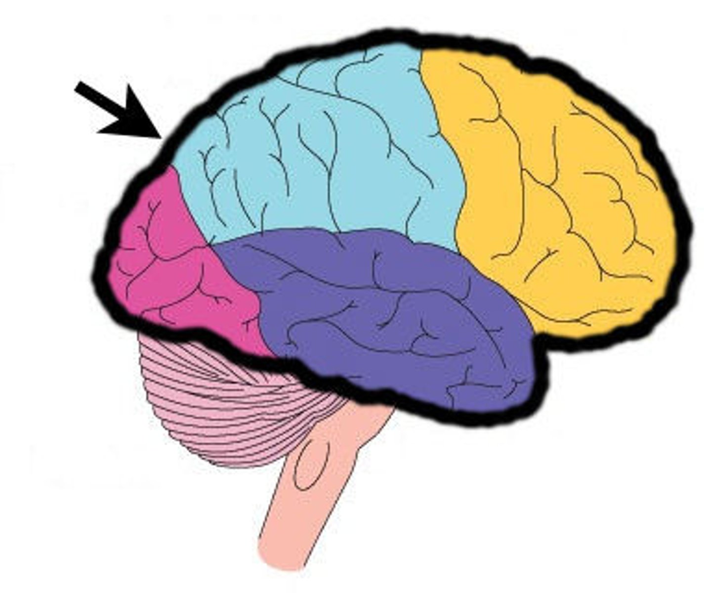
Cerebral cortex — Outer gray matter containing neuron cell bodies and dendrites.
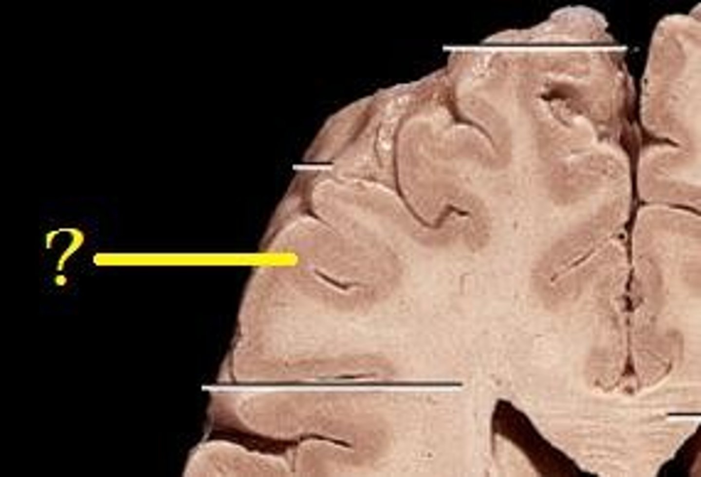
White matter (cerebrum) — Myelinated fiber tracts under cortex.
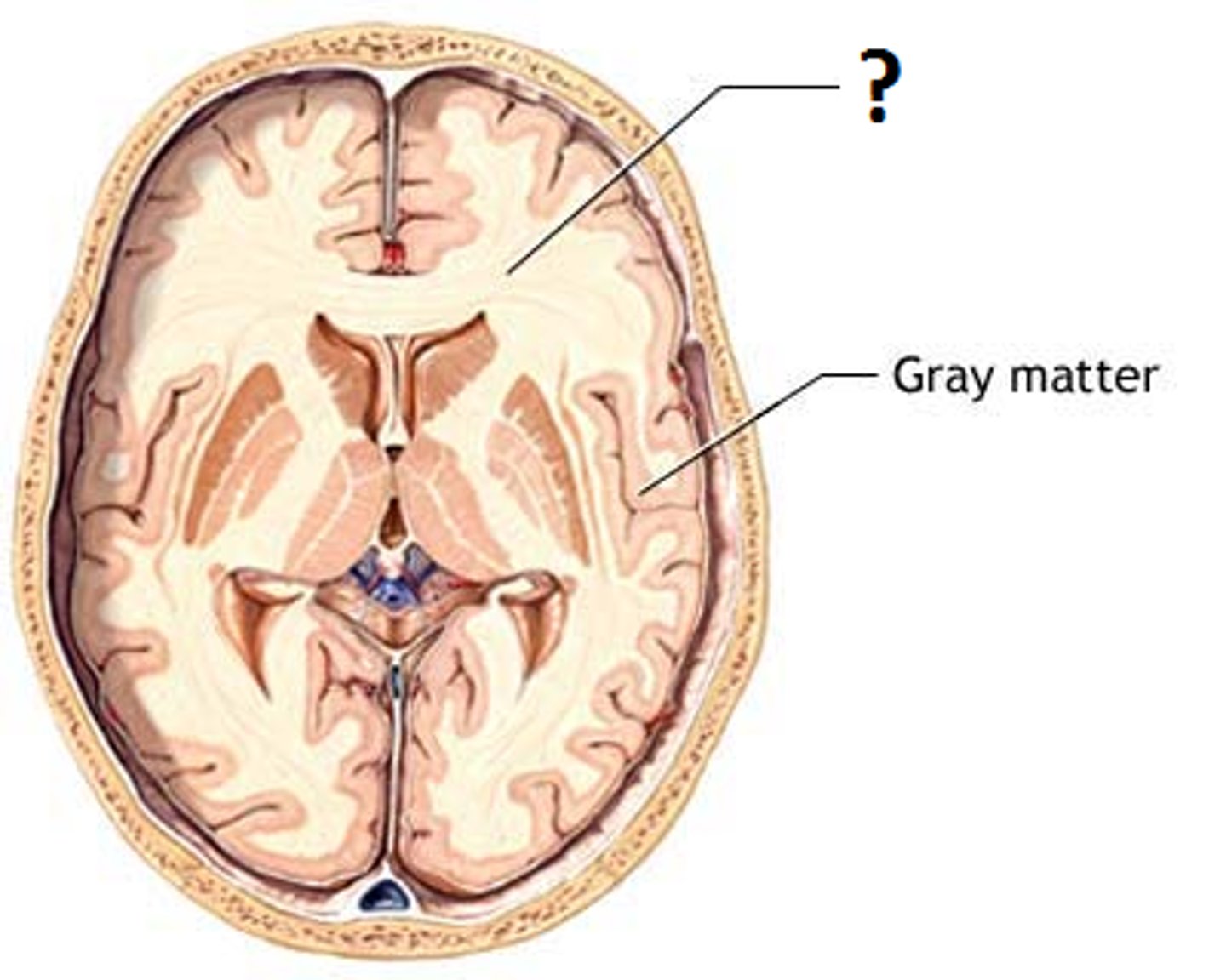
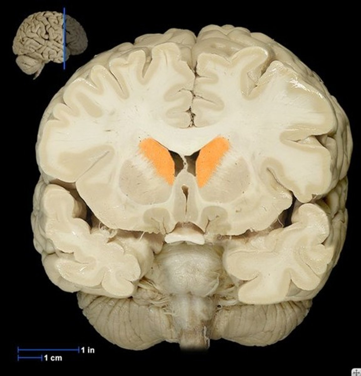
Basal nuclei — Clusters of cell bodies deep within white matter.
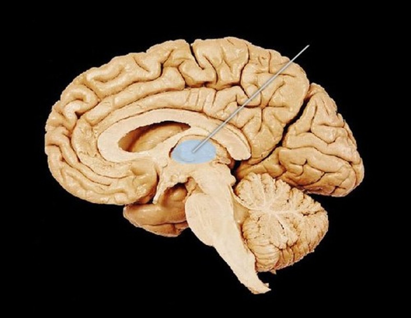
Diencephalon — Thalamus, hypothalamus, epithalamus.
What is in the diencephalon
Thalamus — Relay station for sensory information except smell.
Hypothalamus — Regulates homeostasis, hormones, emotions.
Epithalamus — Contains pineal gland; secretes melatonin.
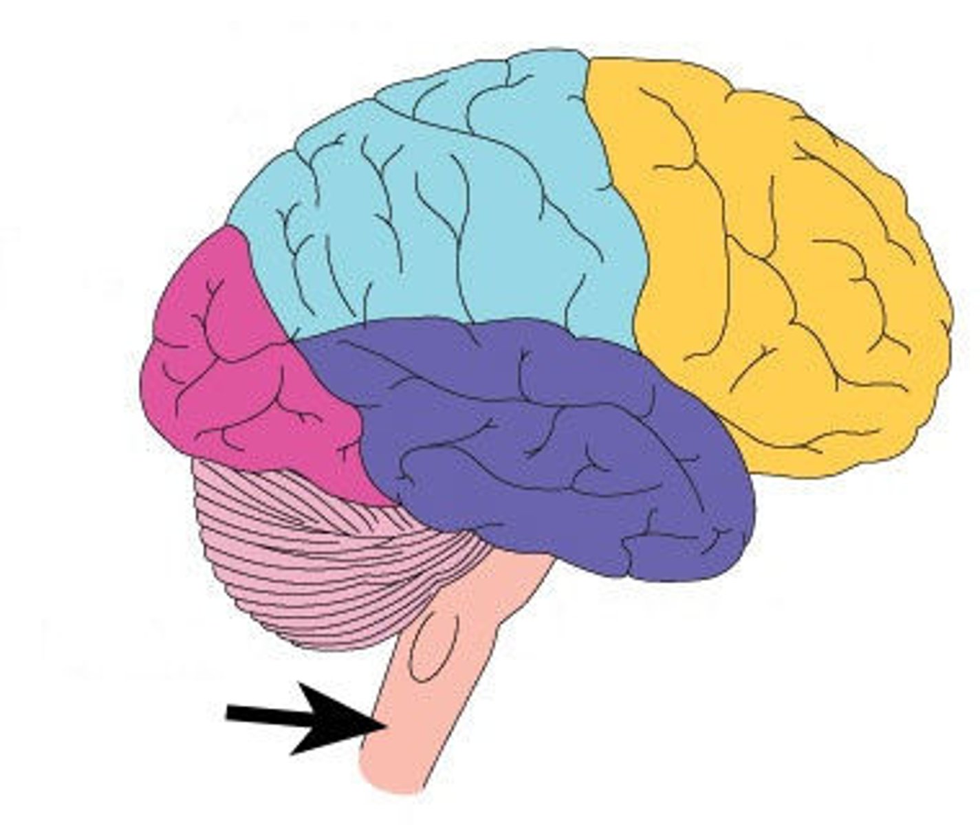
Brain stem — Midbrain, pons, medulla; controls vital functions.