Juvenile, Immature, and Developmental Bone Diseases in Small Animals
1/47
There's no tags or description
Looks like no tags are added yet.
Name | Mastery | Learn | Test | Matching | Spaced | Call with Kai |
|---|
No analytics yet
Send a link to your students to track their progress
48 Terms
What is the signalment for osteochondrosis?
Young rapidly growing large breed dogs
What causes osteochondrosis?
Failure of endochondral ossification
What are the radiographic findings of osteochondrosis?
Flattening or concavity of articular margin
Subchondral sclerosis
Mineralized flap
How does hyaline cartilage get nutrients?
Diffusion from synovial fluid
What are the predilection sites of osteochondrosis?
Caudal humeral head
Medial humeral condyle
Lateral and medial femoral condyle
Lateral and medial trochlea of talus
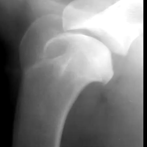
Osteochondrosis (dense subchondral bone)
What are the radiographic findings of osteochondrosis in the stife?
Flattening or subchondral defect
Subchondral sclerosis
Detached mineralized cartilage flap
DJD
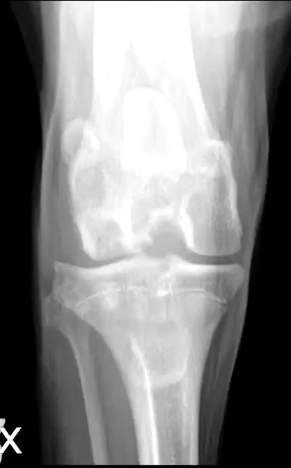
Osteochondrosis
Describe osteochondrosis in the tarsus?
Most common on medial trochlear ridge
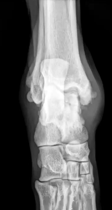
What are the radiographic findings of osteochondrosis of the tarsus?
Flattening or subchondral defect of trochlear ridge
Widening of joint space
Subchondral sclerosis
DJD
Mineralized flap
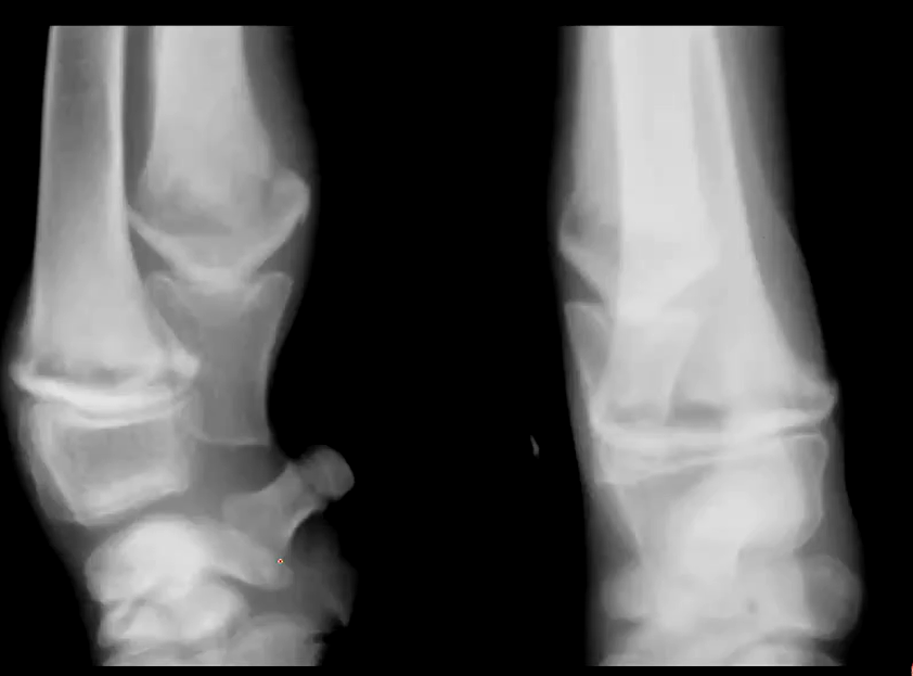
Osteochondrosis with a mineralized flap is what?
Osteochondrosis dessicans
Osteochondrosis dessicans
Describe panosteitis
Self limiting disease in large breed dogs that are 5-18 months old
What are the C/S of panosteitis?
Shifting leg lameness
What bones are affected with panosteitis?
Long bones
T/F panosteitis can occur with ED and OCD?
True
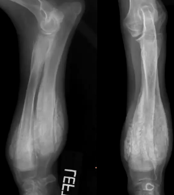
What are the early radiographic changes for panosteitis?
Increased medullary opacity
What are the middle radiographic changes of panosteitis?
Endosteal roughening, more defined opacities, mild periosteal reaction
What are the late/healed radiographic changes of panosteitis?
Hallow or lucent medullary canal, horizontal opaque bands
What are the C/S of metaphyseal osteopathy
Pyrexia
Malaise
Pain and swelling over metaphyseal region
Lameness
Can vary from mild to life threatening
What are the early radiographic changes of metaphyseal osteopathy?
Metaphysis of the long bones
Bilateral
Double physeal line
Metaphyseal lucency
Soft tissue swelling around metaphysis
Early metaphyseal osteopathy
What are the radiographic changes of late metaphyseal osteopathy?
Para-cortical/metaphyseal cuffing
Soft tissue swelling
Flaring of metaphysis
Increased metaphyseal opacity
Metaphyseal cuffing
What are the inactive radiographic changes with metaphyseal osteopathy?
Cortical cuff fuses with underlying cortex
Appearance of thickened long bones
Remodeling
What is the signalment for craniomandibular osteopathy?
Westies, scotties, Cairn, boston terriers
What are the C/S of craniomandibular osteopathy?
Mandibular swelling with pain chewing
Pyrexia
What are the radiographic signs of craniomandibular osteopathy?
Bony proliferation on mandibles, bulla petrous temporal bone, calvarium
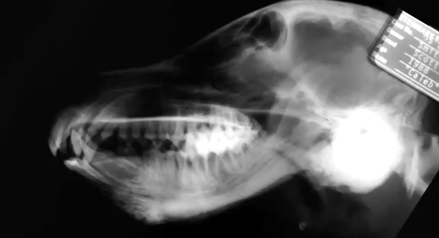
Craniomandibular osteopathy
What is the signalment for calvarial hyperostosis?
Young bull mastiffs
What are the C/S of calvarial hyperostosis?
Pain swelling
Pyrexia, depression, nasal discharge, lameness
Self limiting
Similar to CMO
What are the radiographic signs of calvarial hyperostosis?
Marked thickening of frontal and parietal bones
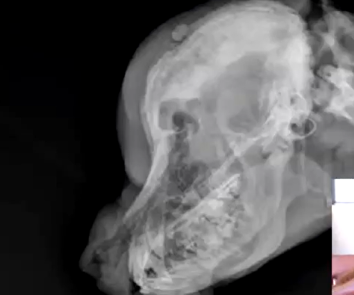
Calvarial hyperostosis
What is the signalment for avascular necrosis of the femoral head?
Small dogs
What is the pathogenesis of avascular necrosis of the femoral head?
Compromised blood supply to femoral head causes subchondral bone necrosis while cartilage continues to grow
T/F avascular necrosis of the femoral head is bilateral?
False
What is Legg-Calve perthes?
Avascular necrosis of the femoral head
What are the radiographic changes of avascular necrosis of the femoral head?
Widened joint space from thickened cartilage
Increased opacity in the epiphyseal area of femoral head
Focal bony lysis femoral head
Muscle atrophy and secondary DJD
What is the earlies radiographic change from avascular necrosis of the femoral head?
Widened joint space from thickened cartilage
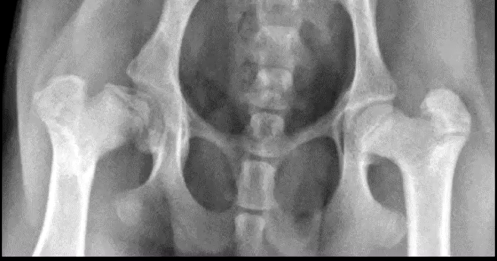
Avascular necrosis of the femoral head
What is SCFE?
Slipped capital femoral epiphyses of physeal dysplasia
What is the signalment for SCFE (sipped capital femoral epiphyses)?
Dogs and usually male cats the got neutered early
What are the C/S of physeal displasia?
Acute onset lameness, resistant to jump
Atraumatic fracture at proximal femoral physes
What is the best view to diagnose SCFE?
Frog leg view (VD flexed)
What does SCFE mimic?
Salter Harris Type 1 fractures
What is the signalment for patella fluxation?
Small breed and large breed
What are the abnormal imaging findings of a patella luxation?
Patella luxated on either side of the trochlear ridge
Shallow trochlear groove
Coxa vara/coxa valgra
Bowing at distal femur/tibia
Patella luxation