Chest wall and mediastinum
1/69
There's no tags or description
Looks like no tags are added yet.
Name | Mastery | Learn | Test | Matching | Spaced | Call with Kai |
|---|
No analytics yet
Send a link to your students to track their progress
70 Terms
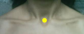
Jugular Notch
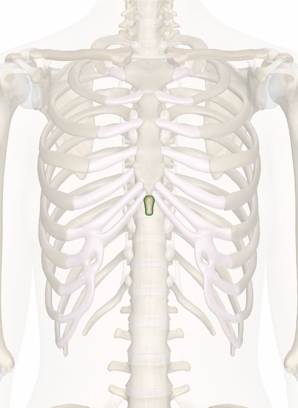
Xiphoid Process
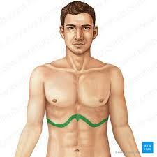
Coastal Margins
Thorax
The part of the body between the neck and the diaphragm, containing the lungs and heart, protected by the rib cage.
Suspensory retinacula
Connects the breast glands to the dermis
Lactiferous Ducts/Sinus
Carry and store milk from glands to nipple
Gland lobules
Milk producing units of the breast. Arranged like clusters of grapes, each human breast contains 15 to 20 lobes comprised of many smaller lobules. They consist of secretory sacs (alveoli) and are linked to ducts that transport milk to the nipple
Functions of lymphatic system
Collect and return lymph to blood, absorb digestive fat, production of lymphocytes (immune cells), filtration of antigens and erythrocytes
How does lymph flow
unidirectionally towards the heart
Tunica media of lymph vessels
Middle layer of lymph vessel wall, contains smooth muscle
Lymph nodes
Small, round aggregates of tissue that filter lymph using macrophages
Macrophages
Large white blood cells that act as the body's cleanup crew and frontline defenders. They patrol tissues, engulfing dead cells, cellular debris, and pathogens through a process called phagocytosis.
Superficial location of lymph nodes
Cervical, axillary, and inguinal regions
Deep locations of lymph nodes
abdominal aorta, and along illiac arteries (near groin)
Spleen
a fist-sized organ in the upper-left side of your abdomen, just beneath the rib cage. It acts as a blood filter and a core part of the immune system
Cisterna chyli
a major temporary reservoir for lymphatic fluid. It acts as the primary origin of the thoracic duct
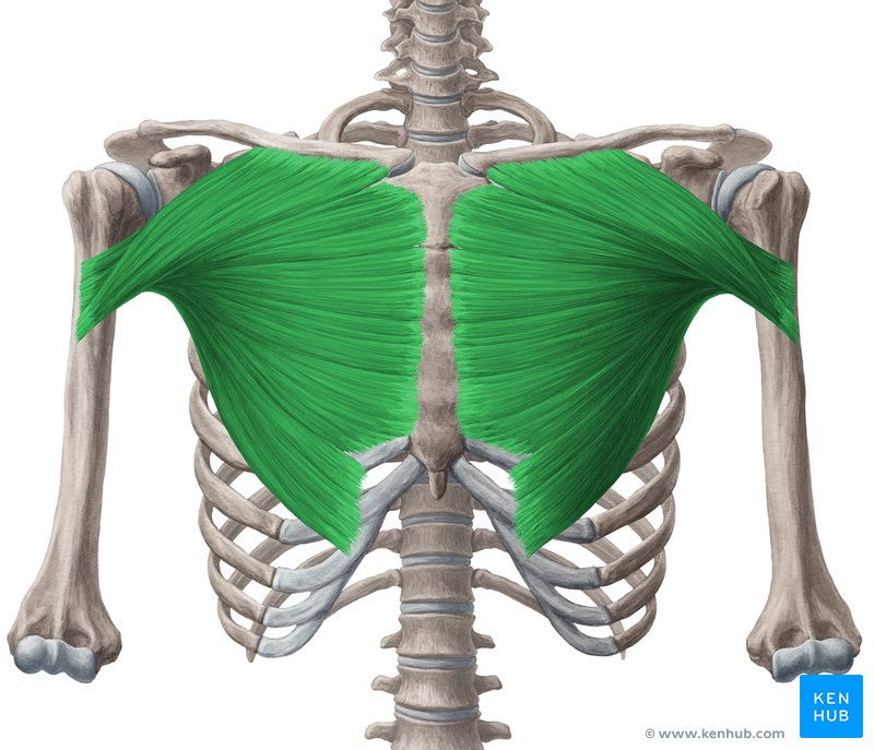
Chest wall muscles
Protect the thoracic viscera and help with limb movement
Pectoralis major
Pectoralis minor
Serratus anterior
External intercostals
responsible for active inhalation
Internal intercostals
responsible for forced exhalation; slightly deeper and face the heart
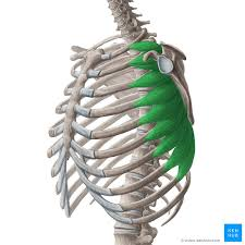
Serratus anterior
“Boxer’s muscle” responsible for pushing, punching, and lifting arms overhead
True ribs
Ribs 1-7
False ribs
Ribs 8-10
Floating ribs
Ribs 11 &12
Costal cartilage
cartilage that connects ribs to sternum
Sternum
Manubrium
Sternum body
Intercostal space
empty area between two adjacent ribs
Costochondral joint
Stiff, cartilaginous connection between the bony part of your rib and the costal cartilage
Jugular notch
U shaped depression at base of neck situated between collarbones
Clavicular notch
Connects collar bone to breastbone
Costovertebral joints
Connections between the ribs and the thoracic spine/vertebral column
Sternocostal joints
Connect the first seven ribs to the sternum
Angle of rib
Area of sharpest curvature, near the spine, near where rib is most susceptible to fracture
Tubercle
Small bony bump on posterior of rib
Rib Neck
Flattened section of rib located laterally to the spine
Rib shaft
Long flat curved main section of rib
Costal grove
Where the neurovascular structures run on the inferior aspect of ribs
Anastomosis
A connection between two tubular structures to maintain flow, supplying the same structure
Thoracic Aorta
Main and largest artery, supplies oxygenated blood to ribs
Veins
Bring deoxygenated blood back to heart
Posterior intercostal artery
Run in the intercostal groves of each rib, back side
Anterior intercostal artery
Run in the intercostal groves of each rib, front side
Internal thoracic artery
Runs vertically down the ribcage, connects to anterior intercostal arteries
Intercostal veins
Drain the thorax into the internal thoracic vein or the azygous system
Azygous vein
Runs vertically up the spine carrying deoxygenated blood to the heart, connected to interc
Intercostal thoracic vein
Runs vertical down the inside of the rib cage connecting the internal thoracic veins
Costochondral joint
Connect rib cage to cartilaginous structure
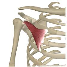
Transversus thoracis
Thin flat muscle on inner surface of anterior chest wall
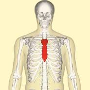
Superior vena cava
Vein that returns deoxygenated blood from the body to right atrium of heart
Mediastinum
Medical compartment of thorax, sits between the thoracic vertebrae t1-t12
Superior thorax compartment
Top of thorax, between t1 and t4
Anterior mediastinum
Where thymus is located
Middle mediastinum
Where heart, pericardium and roots of great vessels are
Posterior mediastinum
Where esophagus, azygous veins, Aorta, trachea, thoracic duct and autonomic nerves are
Diaphragm
Primary muscle of respiration, contracts inferiorly to create negative pressure
What nerves innervate the diaphragm
C3 to c5
Pericardium
Double welled sac that encloses the heart
Phrenic nerve
Paired nerves that originate in the neck from cervical spine c3-c5 and innervate diaphragm
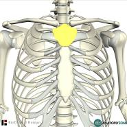
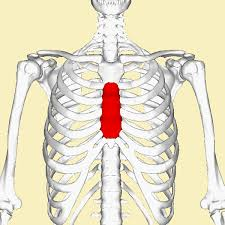
Thymus
Immune system gland that involutes and regresses into adipose with age. Two lobed. Site of t cell development and maturation.
Cortex
In thymus lobules, densely cellular
Medulla
Part of thymus, less cellular, central part
Thymic (Hassalls corpuscles)
Lamellar rings of keratinized epithelial cells in the medulla of the thymus
Thymic environment
Fibroblast (connective tissue), adipocyte, thymic epithelial cells