Vascular - Test 3 (Jo's & Mine)
1/255
There's no tags or description
Looks like no tags are added yet.
Name | Mastery | Learn | Test | Matching | Spaced | Call with Kai |
|---|
No analytics yet
Send a link to your students to track their progress
256 Terms
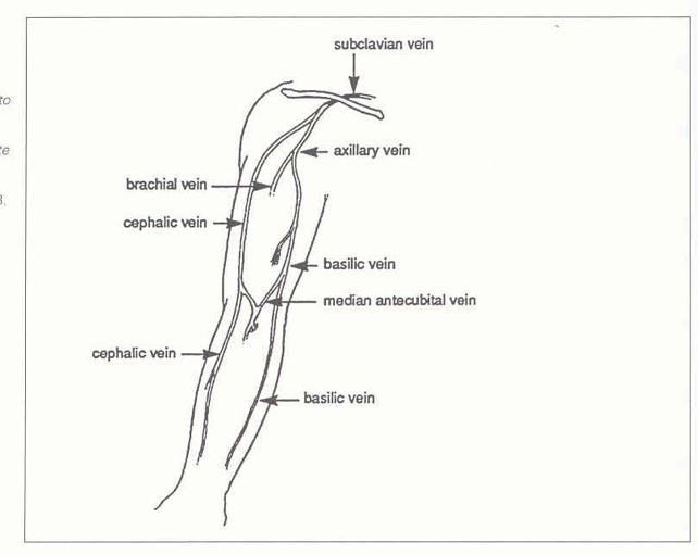
brachial and basilic veins combine to form what?
axillary vein
axillary vein and cephalic vein form what?
subclavian vein
lateral and medial veins are connected by what?
median cubital vein
transmural pressure
the difference between intraluminal (inside) pressure and interstitial (outside) pressure
determines cross sectional shape of the vessel
interstitial pressure
the pressure surrounding tissue
hydrostatic pressure
equivalent to the weight of a column of blood extending from the heart to the level where the pressure is being measured
when you do valsalva, what should happen?
you should not see flow on spectral doppler because the whole venous system stops
what does it mean if you see flow on spectral doppler during valsalva?
venous reflux/insufficiency
varicose veins
palpable, distended veins that are > 4 mm in diameter
phlegmasia alba dolens
rare complication of DVT during pregnancy where leg turns milky white
phlegmasia cerulea dolens
similar condition to “alba” except this is when severely reduced venous flow markedly reduces arterial inflow and the leg becomes cyanotic and has a bluish discoloration
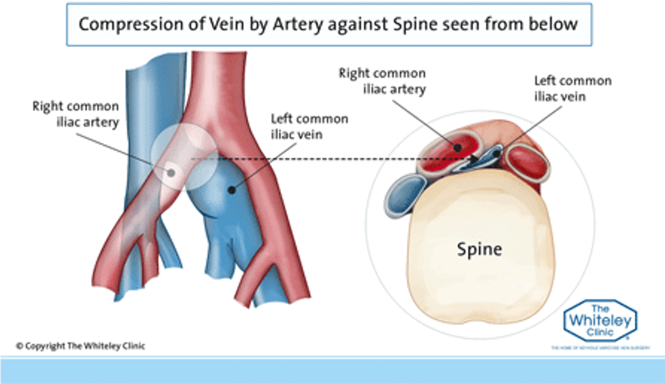
May-Thurner syndrome (MTS)
a condition in which compression of the common venous outflow tract of the left lower extremity MAY cause discomfort, swelling, pain or blood clots (DVT) in the iliofemoral veins
how many valves does GSV have?
10-12
how many valves does LSV have?
6-12
how many valves does soleal sinuses have?
none
how many valves do the perforators have?
1 each
how many valves do the calf veins have?
9-12 each
how many valves do the popliteal and SFV have?
1-3 each
how many valves does the CF have?
1
how many valves does the external iliac vein have?
has them 25% of the time
how many valves do the common and internal iliac veins have?
none
how many valves does the IVC have?
none
how many valves does the IJV have?
1
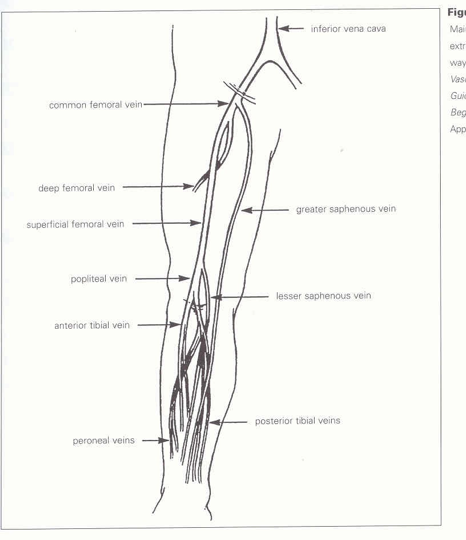
paired posterior tibial veins
empty the back of the leg and carry blood to the tibioperoneal trunk
paired anterior tibial veins
empty the front of the leg and join the tibioperoneal trunk just below the knee to form the popliteal vein
popliteal vein
becomes the superficial femoral vein at the adductor canal, turning into the common femoral vein at the inguinal ligament
lower extremity deep veins
paired posterior tibial
paired anterior tibial
popliteal
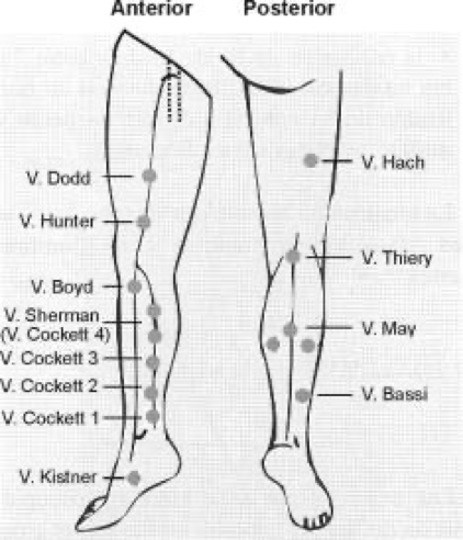
leg veins
perforators
veins that connect superficial veins to the deep veins
perforators
contain one-way valves to direct blood from superficial to deep, but never deep to superficial
perforators
Cockett
Boyd’s
Dodd’s
Hunterian
Boyd’s perforators
connect GSV to posterior tibial vein
common sites for primary varicose veins
Hunterian perforators
connect GSV to the superficial femoral vein
perforators to know
upper extremity veins
clinical findings for DVT
swelling, pain, redness (erythema), skin warmth
acute venous clinical findings
swelling, pain, redness, warmth
chronic venous clinical findings
swelling, heaviness, discoloration/ulcerations, and varicosities
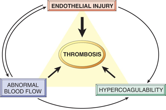
Virchow’s triad
trauma to the vessel
venous stasis
hypercoagulability
the development of venous thrombosis can be summarized based on three factors known as:
Virchow’s Triad
hypercoagulability (blood chemistry) »
Hemaglide Vitamin E & C
vessel injury (trauma) »
Hemaglide Quercetin
venous stasis (blood flow) »
Hemaglide Nattokinase, Raspberry extract
know this
examples for hypercoagulable state
malignancy
pregnancy and peri-partum period
oestrogen therapy
trauma or surgery of lower extremity, hip, abdomen or pelvis
inflammatory bowel disease
nephrotic syndrome
sepsis
thrombophilia
vascular wall injury examples
trauma or surgery
venepuncture
chemical irritation
heart valve disease or replacement
atherosclerosis
indwelling catheters
examples of circulatory stasis
atrial fibrillation
left ventricle dysfunction
immobility or paralysis
venous insufficiency or varicose veins
venous obstruction from tumor, obesity or pregnancy
venous ulcers
location: near medial malleolus
pain: none to mild
appearance: shallow, irregular
bleeding: ooze
other: brawney color, presence of variosities
arterial ulcers
location: tibial area, toes, bony structures
pain: severe
appearance: deep, regular
bleeding: little
other: shiny skin, loss of hair, thickened toenails
what causes phlegmasia alba dolens?
unclear, but edema may increase soft tissue pressure leading to ischemia and wet gangrene
phlegmasia cerulea dolens
severe form of DVT where there’s a massive iliofemoral venous thrombosis that causes almost total venous occlusion
what is phlegmasia cerulea dolens characterized by?
ischemia
marked limb swelling
extreme pain
cyanotic
VENOUS GANGRENE IF LEFT UNTREATED
what primarily triggers phlegmasia cerulea dolens?
malignancy
CFV catheterization
heparin-induced thrombocytopenia
antiphospholipid syndrome
surgery
heart failure
pregnancy
may-thurner syndrome
also known as the iliac vein compression syndrome
MTS
veins
highly compliant, explanding and contracting according to intraluminal pressure changes
when a person is supine, what is their transmural pressure?
low » a vein assumes a dumbell shape
when a person is supine, what is their hydrostatic pressure?
around 15 mmHg » rises to around 100 mmHg when patient stands
What is plethysmography?
Volume flow studies
What is venous photoplethysmography (PPG)?
Test that measures capillary volume and can evaluate presence and severity of venous reflux or insufficiency
How does photoplethysmography (PPG) work?
Photocell sensor transmits infrared light into tissues to be reflected back to sensor and converted into waveforms or analog
What is the photoplethysmography (PPG) technique?
Sensor applied above medial malleolus
Patient completes 20-25 dorsiflexions to empty veins
VRT calculated to determine need for further testing
What is a VRT?
Venous refill time
What is a normal value for VRT?
> 20 seconds
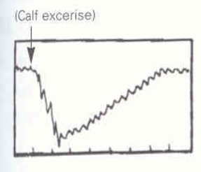
Identify this image.
Normal PPG as VRT > 20 seconds
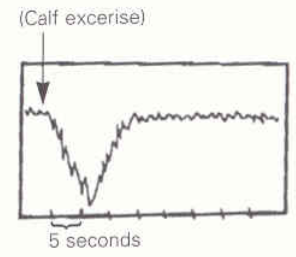
Identify this image.
Abnormal PPG as VRT < 20 seconds
What is the photoplethysmography (PPG) technique used if the initial VRT is abnormal?
BP cuff below knee to test LSV: VRT NORMALIZES to > 20 secs with inflated cuff below knee is consistent with reflux (retrograde flow) of LSV
BP cuff above knee to test GSV: VRT NORMALIZES to > 20 secs with inflated cuff above knee is consistent with reflux (retrograde flow) of GSV
Both systems: VRT < 20 seconds with AND without inflated BP cuffs is consistent with insufficiency of deep and superficial venous systems
Thigh: VRT < 20 seconds with a cuff in place on thigh indicates deep venous insufficiency or reflux
What is duplex imaging?
Combination of b-mode and PW Doppler to create images and measure blood flow
What are the advantages of duplex imaging?
Rule out or identify thrombus
Identify thrombus type (acute, subacute, etc)
Diagnose venous incompetence
Which venous characteristics should be checked when using duplex imaging for a lower extremity examination?
Compressibility: Normal veins compress
Spontaneity: Normal flow is audible and consistent
Phasicity: Normal flow is phasic with respiration
Augmentation
Valsalva Maneuver
Pulsatility
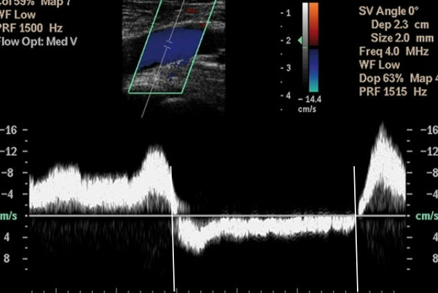
Identify this image.
Abnormal aphasic flow due to undiagnosed occlusion
What is the normal venous response with augmentation?
Normal distal augmentation: Visual and audible signal
Normal proximal augmentation: NO visual and audible signal (augmentation during proximal compression is consistent with venous reflux)
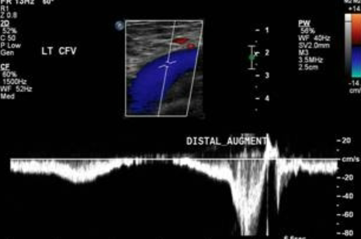
Identify this image.
Normal LEV augmentation
What is the normal venous response with valsalva maneuver?
Normal: Venous flow augmented following maneuver
Abnormal: Augmentation during maneuver is consistent with reflux
When might pulsatility be present in veins?
Fluid overload
CHF
Right heart failure
What position should the patient be in for duplex imaging of the lower extremity?
Reverse Trendelenburg’s or Semi-Fowler’s position with head higher than heart
What is reflux evaluation?
Test used to assess for DVT complications
What is the technique used for a reflux evaluation?
Rule out chronic venous outflow obstruction by assessing compressibility, phasicity, and augmentation
Evaluate vessels for reflux
Which vessels are used to evaluate for venous reflux?
CFV
Proximal femoral vein
Popliteal vein
What is the sonographic appearance of venous reflux?
Retrograde flow following augmentation seen as change in color Doppler
What is the normal response to venous reflux testing?
Cessation of flow with proximal compression
Returning flow upon release
What is the reflux duration criteria that indicates venous reflux?
Deep veins: ≥ 1 second
GSV and SSV ≥ 0.5 seconds
Perforating veins ≥ 0.35 seconds
(T/F) Longer reflux durations are seen in those in the supine position.
True
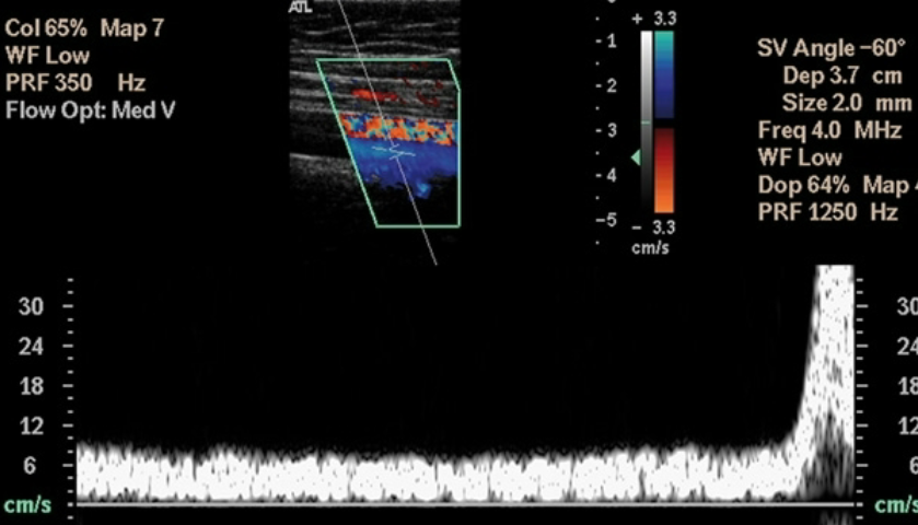
Identify this image.
Abnormal study seen as venous reflux
What is vein mapping?
Exam performed to determine if GSV or radial vein is suitable for CABG procedure
What is the criteria assessed when performing a vein mapping procedure?
Suitability
Presence or location
Patency
Size
What is the required vein diameter in order to be used in a CABG procedure?
2.5 mm
What is the gold standard of venous exams?
Contrast venography
What is contrast venography?
Invasive test that consists of injecting contrast into veins and x-ray shows any filling defects
(T/F) Heparin dissolves clots.
False; Heparin PREVENTS GROWTH of clots
What is radiofrequency ablation?
Minimally invasive procedure that uses catheter-based techniques to heat and destroy varicose veins
What is vein stripping?
Invasive procedure performed under general anesthesia in which small incisions allow removal of individual varicose vein clusters
What is sclerotherapy?
Minimally invasive procedure performed while patient is standing in which a salty solution is injected into varicose veins to scar and close them
What is a thrombectomy?
Procedure that removes a blood clot or thrombus from a vessel by using a catheter-based device and a stent to restore blood flow
Which conditions could a thrombectomy resolve?
Acute cerebral ischemic stroke
Pulmonary embolism (PE)
Acute myocardial infarction (MI)
What is endovenous ablation therapy?
Non-surgical procedure that uses heat, chemicals, glue, or a wire to damage and close off varicose veins
What is laser light therapy (EVLA)?
Procedure used to compliment or replace vein stripping that uses a light beam on varicose vein to seal it off, causing vein to dissolve from heat
What is laser therapy?
Procedure in which a laser fiber is inserted through skin, directly into
varicose vein to damage, collapse, shrink, and reabsorb it back into body
What is a microphlebectomy?
Minimally invasive procedure in conjunction with sclerotherapy to remove varicose veins
The venous puncture for introducing contrast in venography to assess for deep venous thrombosis is done at what level?
Dorsal vein on foot