2.2 Cells - electron microscopy
1/22
There's no tags or description
Looks like no tags are added yet.
Name | Mastery | Learn | Test | Matching | Spaced | Call with Kai |
|---|
No analytics yet
Send a link to your students to track their progress
23 Terms
What is electron microscopy?
Electron microscopy is a technique that uses a beam of electrons instead of light to form highly magnified and high-resolution images of cell structures, allowing detailed observation of sub-cellular organelles.
Why do electron microscopes have higher resolution than light microscopes?
Electron microscopes use electrons with a much shorter wavelength (~1 nm) compared to visible light (400–700 nm), giving a much higher resolving power (0.5 nm), allowing finer cellular detail to be seen.
Define resolution (electron microscopy context)
Resolution is the ability to distinguish two points as separate; in electron microscopy it is extremely high (about 0.5 nm), allowing organelles and large molecules to be seen clearly.
What is a transmission electron microscope (TEM)?
A TEM passes electrons through very thin sections of a specimen to form a detailed image of internal cell structures, which is recorded on a screen or camera.
Why must specimens be thin in electron microscopy?
Electron beams cannot penetrate thick specimens, so samples must be cut into very thin sections (about 100–500 nm) to allow electrons to pass through and form a clear image.
Why are electron microscopes used in vacuum conditions?
Air molecules would scatter the electron beam, so a vacuum is used to ensure electrons travel in a straight path without interference.
Differences between light and electron microscopes
Light microscope: uses light, lower resolution (200 nm), max magnification ~×1000, can view living cells
Electron microscope: uses electrons, higher resolution (0.5 nm), magnification up to ~×250,000, only dead specimens
Why can light microscopes view living cells but electron microscopes cannot?
Light microscopy does not require fixation or vacuum conditions, so cells remain alive. Electron microscopy requires fixation, dehydration, and vacuum, which kill the specimen.
Why do electron microscope images show artefacts?
Chemical fixation and heavy metal staining can distort structures, creating artificial features that are not present in living cells.
Advantage of light microscopy over electron microscopy
Light microscopes allow observation of living processes such as cell movement, mitosis, and cytoplasmic streaming, which electron microscopes cannot show.
Advantages of electron microscopy
Much higher resolution (0.5 nm)
Much higher magnification (up to ×250,000+)
Can view organelles and large molecules not visible with light microscopes (e.g. ribosomes)
Provides detailed ultrastructure of cells
What is meant by cell ultrastructure?
Ultrastructure refers to the fine detail of cell organelles and internal structures visible only with an electron microscope.
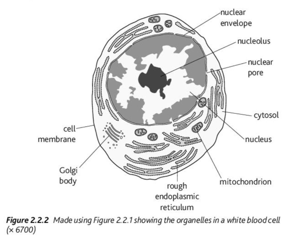
Organelles visible in a white blood cell (electron micrograph)
Nucleus (with nuclear envelope, pores, nucleolus)
Rough endoplasmic reticulum
Golgi apparatus
Mitochondria
Cell membrane and cytosol
Function of mitochondria in white blood cells
Mitochondria produce ATP through respiration to provide energy for active processes such as phagocytosis.
Function of rough endoplasmic reticulum
RER is involved in protein synthesis and transport, especially in cells producing enzymes or antibodies like white blood cells.
Differences between plant and animal cells (electron microscope view)
Plant cells: cell wall, chloroplasts, large vacuole, regular shape
Animal cells: no cell wall or chloroplasts, smaller vacuoles, irregular shape
Why are chloroplasts not found in animal cells?
Animal cells do not perform photosynthesis, so they do not require chloroplasts, which are responsible for light energy conversion in plant cells.
Why is TEM better than light microscopy for cell structure?
TEM has much higher resolution, allowing detailed observation of organelles like ribosomes, membranes, and internal structures that are too small for light microscopy.
Why are electron microscope images black and white?
Electrons are not detected in colour; images are formed based on electron density, and colour is sometimes added artificially using computer software.
Why is fixation required in electron microscopy?
Fixation preserves cell structure by stabilising proteins and lipids, preventing decay during preparation, but it kills the cell.
When is a light microscope better than an electron microscope?
Light microscopes are better when studying living cells or dynamic processes such as mitosis, movement, or cytoplasmic streaming.
When is an electron microscope better than a light microscope?
Electron microscopes are better for studying fine cell structure (ultrastructure), including organelles like ribosomes, membranes, and mitochondria.
Why are ribosomes only visible in electron microscopes?
Ribosomes are about 25–30 nm in size, which is below the 200 nm resolution limit of light microscopes.