Health science
 Call Kai
 Learn
Learn Practice Test
 Spaced Repetition
 Match
Match Flashcards
 Knowt Play
1/47

There's no tags or description
Looks like no tags are added yet.
Name | Mastery | Learn | Test | Matching | Spaced | Call with Kai |
|---|
No analytics yet
Send a link to your students to track their progress
48 Terms
What is the cardiovascular system?
The cardiovascular (cardio = heart, vascular = veins, arteries and other vessels) system is what pumps and supplies blood around the body. It transports nutrients, waste products, gases and hormones throughout the body, whilst also playing an important role in the immune system and regulating body temperature. It consists of the heart, all the associated blood vessels, blood and our lymphatic system
Describe the heart
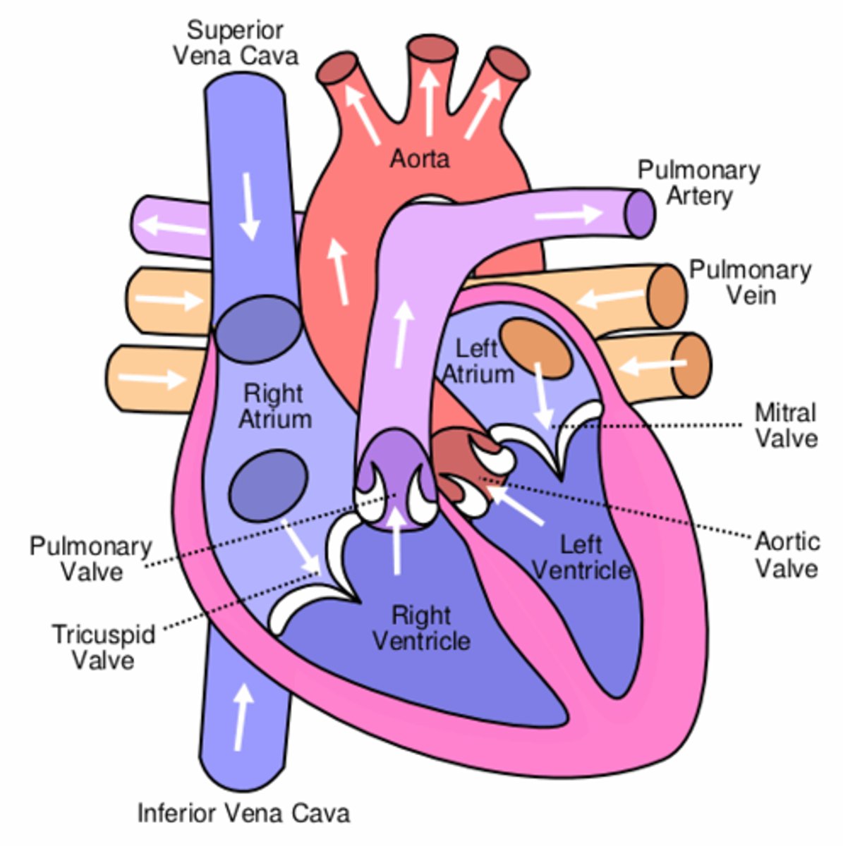
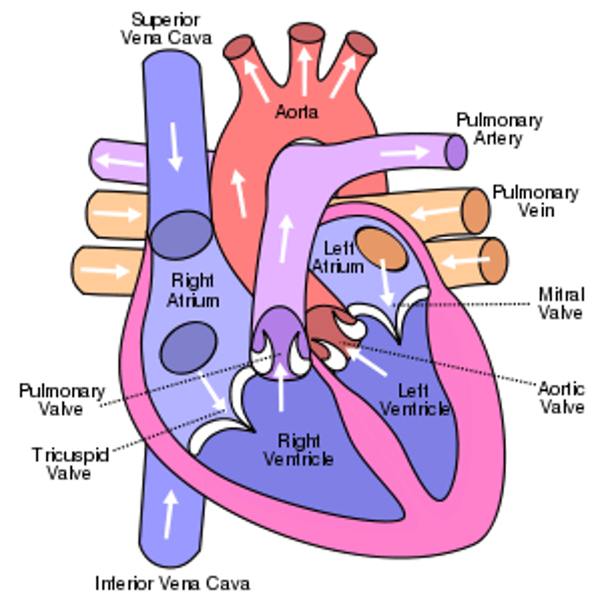
A muscular organ about the size of your clenched fist and can be considered two pumps in one. It is a four chambered pump divided into the left and the right side of the heart.
Draw the anatomy of the heart.
What are the chambers of the heart?
The top two chambers are called atria (plural of atrium) and the bottom two chambers are called ventricles (Marieb, 2012). There are valves between the atria and ventricles known as the left and right atrioventricular valves.
Describe the transport of blood through the heart.
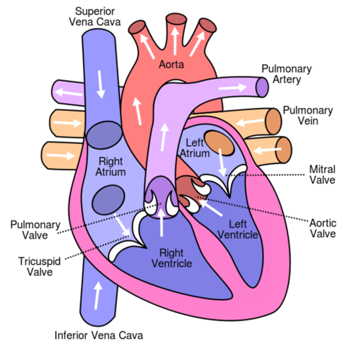
The right side of the heart takes deoxygenated blood returning from the body and pushes it to the lungs, where the blood can get rid of carbon dioxide and get more oxygen from the air we breathe in. The left hand side of the heart accepts oxygenated blood from the lungs and pushes it out to the body
How does the vena cava and aorta transport blood?
The superior vena cava (SVC) receives deoxygenated blood from the head and upper body whilst the inferior vena cava (IVC) returns blood to the heart from the lower body. These two structures are veins and they both empty into the right atrium. The Aorta is the largest artery in the human body and carries oxygenated blood out to the body. It is good to remember that veins always carry blood towards the heart and arteries always carry blood away from the heart. However arteries do not always carry oxygenated blood and veins do not always carry deoxygenated blood, exceptions are: Pulmonary arteries (going AWAY from heart to lung) carry deoxygenated blood! Pulmonary veins (going TOWARDS the heart from the lungs) carry oxygenated blood. Other exception is the umbilical artery and vein in pregnancy.
Describe the heart's walls.
As the left ventricle needs to pump blood the furthest distance and at the greatest pressure - its walls are thickest. The right ventricle only deals with blood going to the lungs, so its walls are thinner.
What is the systemic circulation?
Blood enters the right atrium from the systemic circulation, which is returning all the deoxygenated blood from all the tissues of the body.
What is pulmonary circulation?
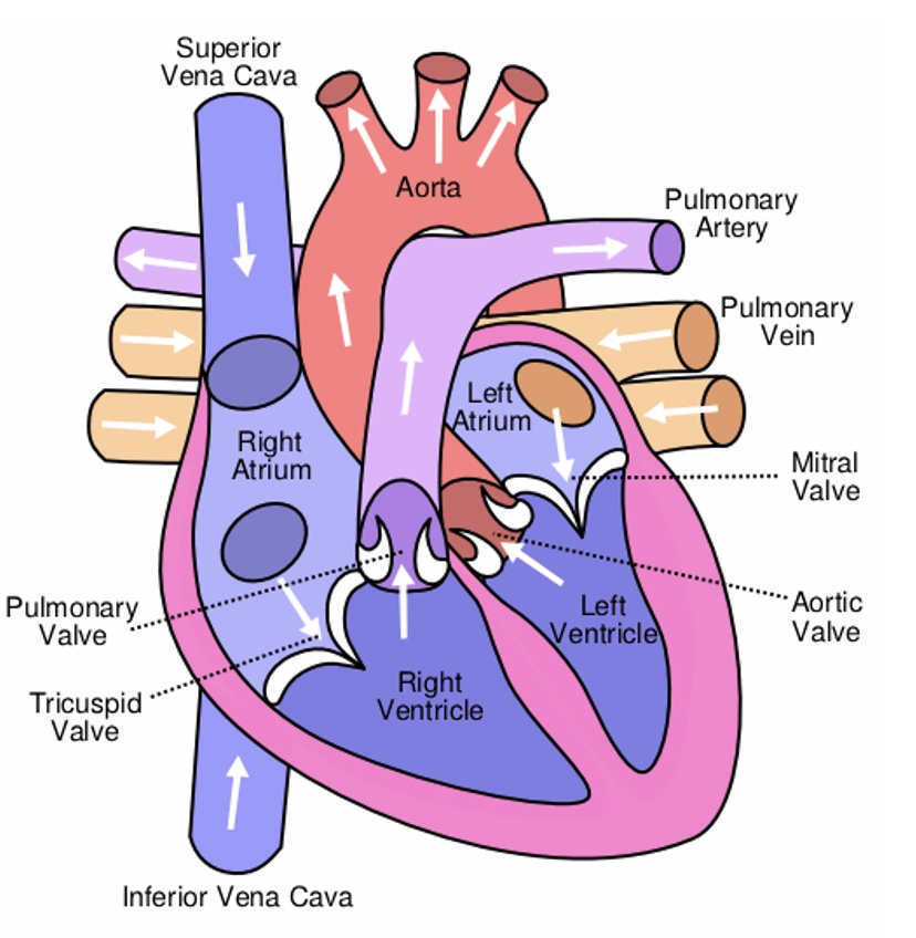
Blood then travels to the lungs (pulmonary circulation) to receive oxygen, which then enters the left atrium, following into the left ventricle and out the aorta to the rest of the body.
Give a summary of blood flow through the heart
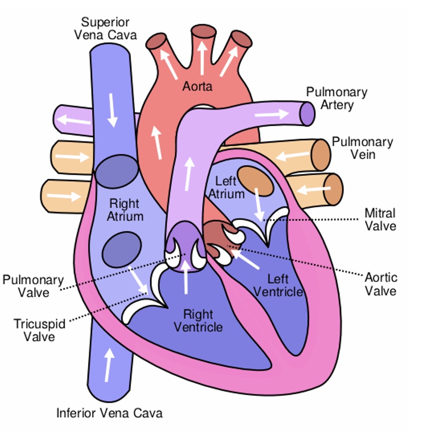
Deoxygenated blood returns from the body, draining via the vena cavae into the right side of the heart (right atrium). Blood is then pushed through the tricuspid valve to the right ventricle and through the pulmonary semilunar valve into the pulmonary trunk and arteries, to the lungs. In the lungs, the blood dumps its CO2 and picks up O2. This occurs at the respiratory membrane where tiny capillaries meet the alveoli. Remember the right side of the heart has relatively smaller and thinner walls (Pulmonary circulation) - just going to the lungs (Marieb, 2012).
Oxygenated blood returns from the lungs via pulmonary veins into the left atrium then through the bicuspid valve (also known as mitral valve) to left ventricle. Blood is then pumped to the entire body through the aortic semilunar valve and aorta. Remember the left ventricle has bigger/thicker walls (Systemic Circulation) - going to the rest of the body (VanPutte et al., 2014). Once through the aorta, flow of blood is as follows:
Elastic arteries → muscular arteries → arterioles → capillaries → venules → veins → vena cavae then back into the right atrium of the heart (Marieb, 2012).
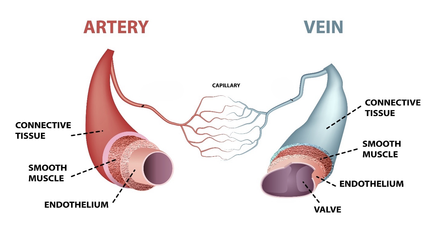
Describe arteries and draw structure
Arteries are elastic to cope with surging of blood.
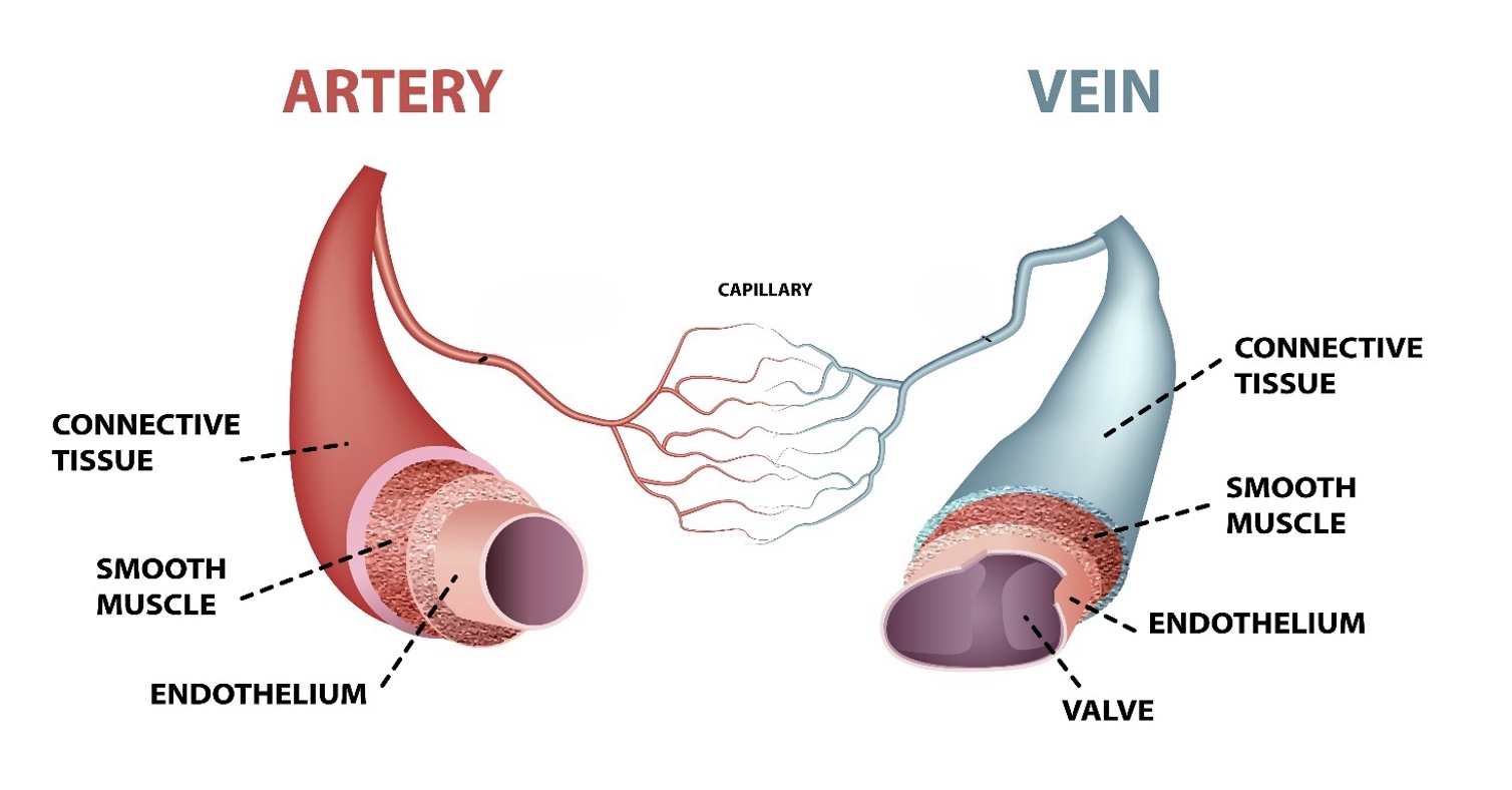
Describe veins and draw structure
Veins don't need to be elastic as they have valves to prevent back flow and deal with constant flow of blood, which ensures one-way flow.
What is blood pressure?
Blood pressure is the pressure of the blood in the arteries as the heart pumps it around the body, it’s the pressure the blood exerts against the inner walls of the blood vessels, and it is the force that keeps blood circulating continuously even between heartbeats. The term blood pressure in this discussion is understood to mean the pressure within the large systemic arteries near the heart. Blood pressure involves the off-and-on flow of blood into the arteries as the heart alternately contracts and relaxes, causing the blood pressure to rise and fall during each beat. Thus, two arterial blood pressures are usually measured: systolic pressure, the pressure in the arteries at the peak of ventricular contraction, and diastolic pressure, the pressure when the ventricles are relaxing. Blood pressures are reported in millimetres of mercury (mm Hg), with the higher systolic pressure written first—120/80 (read “120 over 80”) translates to a systolic pressure of 120 mm Hg and a diastolic pressure of 80 mm Hg. High blood pressure can result in increased risks of stroke, heart disease, kidney disease, eye disease and nerve damage.
What are diastole’s characteristics?
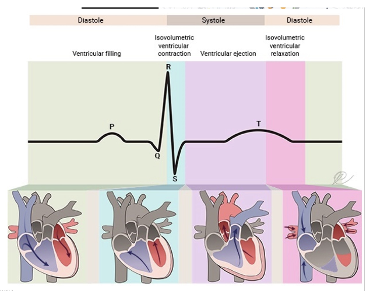
Diastole is when the heart muscle relaxes.
What are systole’s characteristics?
It’s when the heart contracts, during systole a person’s blood pressure increases.
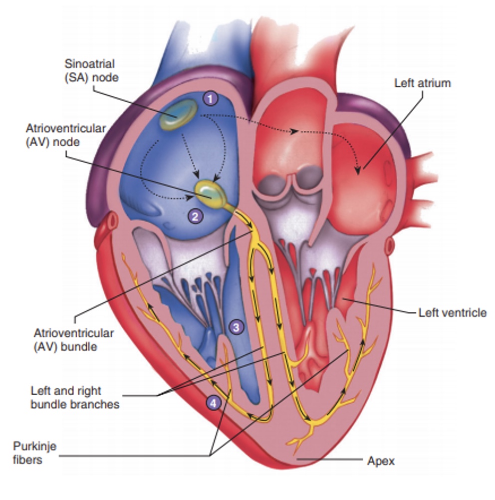
Annotate the conducting systems of the heart.
1. Action potentials originate in the sinoatrial (SA) node (the pacemaker)
2. Then pass through the AV node and along the atrioventricular (AV) bundle
3. The AV bundle divides into right and left bundle branches.
4. Purkinje fibers
What does the autonomic nervous system do in relation to the heart?
Act like brakes and gas pedals to decrease or increase the heart rate, depending on which division is activated.
What does the intrinsic conduction (or nodal system) system do to the heart?
Sets its basic rhythm, it’s composed of a special tissue found nowhere else in the body; it is much like a cross between muscle and nervous tissue. This system causes heart muscle depolarisation in only one direction from the atria to the ventricles. In addition, the intrinsic conduction system enforces a contraction rate of approximately 75 beats per minute on the heart; thus, the heart beats as a coordinated unit.
What are the parts of intrinsic conduction system?
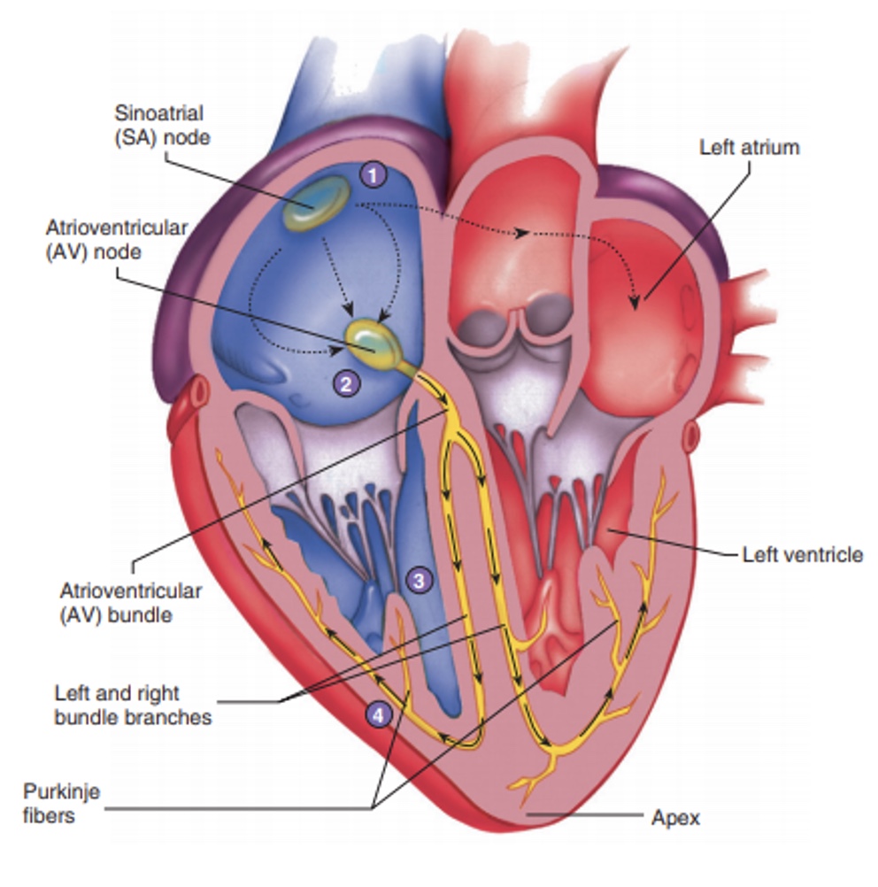
A crescent-shaped node of tissue called the sinoatrial (SA) node, located in the right atrium. Other components include the atrioventricular (AV) node at the junction of the atria and ventricles, the atrioventricular (AV) bundle and the right and left bundle branches located in the interventricular septum, and finally the Purkinje fibers, which spread within the myocardium of the ventricle walls.
What is the Sinoatrial (SA) node?
A tiny cell mass with a mammoth job. Because it has the highest rate of depolarisation (a.k.a. action potential) in the whole system, it starts each heartbeat and sets the pace for the whole heart. Consequently, the SA node is often called the pacemaker.
Where does the impulse spread from the SA node?
Spreads through the atria to the AV node, and then the atria contract. At the AV node, the impulse is delayed briefly to give the atria time to finish contracting. It then passes rapidly through the AV bundle, the bundle branches, and the Purkinje fibers, resulting in a “wringing” contraction of the ventricles that begins at the heart apex and moves toward the atria. This contraction effectively ejects blood superiorly into the large arteries leaving the heart
What is electrocardiogram (ECG)?
Record contractions of cardiac smooth muscle produces electric activity resulting in a series of waves on the ECG.
What are the functions of blood?
1. Transport dissolved gases bringing oxygen from the lungs to the tissues and carrying carbon dioxide from the tissues to the lungs.
2. Distribute nutrients absorbed from the digestive tract or released from storage in adipose tissue or the liver.
3. Transport metabolic wastes from peripheral tissues to sites of excretion, especially the kidneys.
4. Deliver enzymes and hormones to specific target tissues.
5. Stabilise the pH and electrolyte composition of interstitial fluids throughout the body.
6. Prevent fluid losses through damaged vessels or at other injury sites.
7. Defend against toxins and pathogens.
8. Stabilise body temperature by absorbing and redistributing heat.
Expand on blood’s stabilise the pH and electrolyte composition of interstitial fluids throughout the body.
By absorbing, transporting, and releasing ions as it circulates, blood helps prevent regional variations in the ion concentrations of body tissues. An extensive array of buffers enables the bloodstream to deal with the acids generated by
Explain blood’s Prevent fluid losses
The clotting reaction seals the breaks in the vessel walls, preventing changes in blood volume that could seriously affect blood pressure and cardiovascular function.
Explain blood’s Defence against toxins and pathogens.
Blood transports white blood cells (specialised cells that migrate into peripheral tissues) to fight infections or remove debris, and delivers antibodies, special proteins that attack invading organisms or foreign compounds. The blood also collects toxins, such as those produced by infection, and delivers them to the liver and kidneys, where they can be inactivated or excreted.
Explain blood’s Stabilising body temperature
Active skeletal muscles and other tissues generate heat, and the bloodstream carries it away. When body temperature is too high, blood flow to the skin increases, as does the rate of heat loss across the skin surface. When body temperature is too low, warm blood is directed to the most temperature-sensitive organs. These changes in circulatory flow are controlled and coordinated by the cardiovascular centres in the medulla oblongata.
What is the composition of blood?
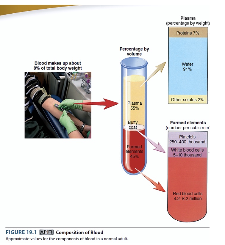
Blood consists of plasma and formed elements (red blood cells, white blood cells and platelets).
What is plasma?
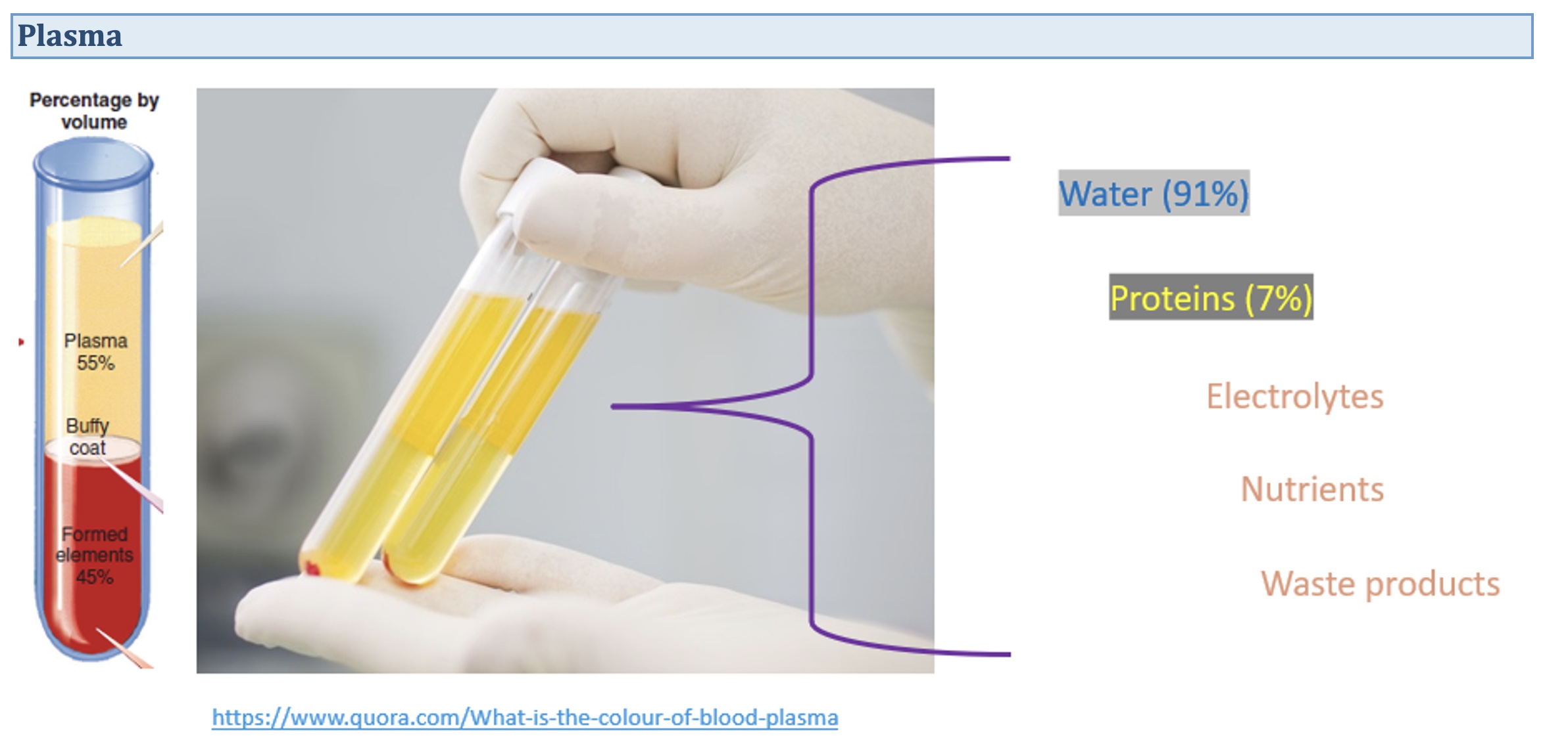
Plasma is the liquid component of blood. It has a density only slightly greater than water. Plasma contains dissolved proteins and other solutes such as nutrients, electrolytes, and wastes.
What are formed elements of blood cells?
Formed elements are blood cells (red blood cells and white blood cells) and cell fragments (platelets) suspended in the plasma. Red blood cells (RBCs) transport oxygen and carbon dioxide. White blood cells (WBCs) are components of the immune system and are less numerous than RBCs. Platelets are small, membrane-enclosed packets of cytoplasm containing enzymes and clotting factors, proteins that play a role in blood clotting.
What determines the viscosity of blood?
Its components can be separated, or fractionated, for clinical purposes. Whole blood is sticky, cohesive, and resistant to flow. Solutions are compared with pure water, which has a viscosity of 1.0. Plasma has a viscosity of 1.5, but whole blood is 5 times as viscous as water. Its high viscosity results from interactions among dissolved proteins, formed elements, and water molecules in plasma.
What is blood made up of?
Plasma is approximately 55% of the volume of whole blood. It is a pale-yellow fluid that consists of about 91% water and 9% other substances, such as proteins, ions, nutrients, gases, waste products, and regulatory substances. Plasma is a colloid (e.g. suspension), which is a liquid containing suspended substances that do not settle out of solution. Serum is plasma without the clotting factors.
What is hematopoiesis?
The process of blood cell production. In the embryo and fetus, this process occurs in tissues such as the yolk sac, liver, thymus, spleen, lymph nodes, and red bone marrow. After birth, hematopoiesis is confined primarily to red bone marrow, with some lymphatic tissue helping produce lymphocyte
What are the characteristics of Red Blood Cells (Erythrocyte)?
· Biconcave: concave on both sides
· Without a nucleus, mitochondria or ribosomes
· Red colour due to presence of haemoglobin molecules
What are the functions of Red Blood Cells?
· Transport oxygen from lungs to tissues, and carbon dioxide from tissues to lungs
What is the life cycle of Red Blood Cells?
· 120- day life expectancy, do not divide once mature, therefore they must be continually replaced to ensure the body functions efficiently. Red blood cells travel vast distances during their 120 day lifespan and are constantly exposed to severe stresses, such as squeezing in and out of capillaries
· Recycled and re-used during erythropoiesais through the spleen and liver
Explain Erythrocyte
The most abundant cells in the blood. RBCs make up roughly one-third of all cells in the human body. A standard blood test reports the number of RBCs per microliter (μL) of whole blood as the red blood cell count. A single drop of whole blood contains approximately 260 million RBCs.
Describe the structure of red blood cells.
Red blood cells do not have a nucleus and are absent of most organelles, such as mitochondria and ribosomes, leaving space for large amounts of haemoglobin (Hb) which gives red colour to the cell. Haemoglobin bind oxygen in the lungs and carry it to tissues throughout the body where it is exchanged for the waste product carbon dioxide. They are thin, disc-shaped cells, about 7.5μm in diameter, with a depression in the middle on both sides. This biconcavity increases the surface area to allow efficient diffusion of gases. The cells are small and flexible enough to squeeze through tiny capillaries.
Explain what are White blood cells/leukocytes.
Blood is separated from each other, white blood cells (WBCs), or leukocytes, form a thin, white layer of cells between the plasma and the red blood cells. This layer is often referred to as the buffy coat. White blood cells lack hemoglobin but have a nucleus. White blood cells function as part of our body defense against foreign substances and pathogens entering the body, and the removal of cellular debris.
Where are Leukocytes?
Leukocytes, are found circulating within the blood vessels of the cardiovascular system and scattered throughout peripheral tissues. Most of the leukocytes are found in peripheral tissues. White blood cells defend the body against invasion by pathogens and remove toxins, wastes, and abnormal or damaged cells. All WBCs are as large as or larger than RBCs.
What are the characteristics of white blood cells?
With a nucleus and other organelles
What are functions of white blood cells?
Against invasion by pathogens and remove toxins, waste, and abnormal or damaged cells.
What is the life cycle of white blood cells?
Vary depending on which type of leukocytes, e.g. neutrophils surviving minutes to days
What are characteristics of platelets (Thrombocytes)?
· Cytoplasmic fragments
· Without a nucleus
What are functions of platelets (Thrombocytes)?
· Clump together and stick to vessel wall
· Control blood loss
What is the life cycle of platelets (Thrombocytes)?
· 5-9 days life expectancy
What are platelets?
Minute fragments of cells consisting of a small amount of cytoplasm surrounded by a plasma membrane. Cytoplasmic fragments allow platelets to attach to other molecules, such as collagen in connective tissue.
What are platelets roles?
(1) forming platelet plugs that seal holes in small vessels and
(2) promoting the formation and contraction of clots that help seal off larger wounds in the vessels.