BIOCH 200 Unit 4 - Protein Function (Questions)
1/26
There's no tags or description
Looks like no tags are added yet.
Name | Mastery | Learn | Test | Matching | Spaced | Call with Kai |
|---|
No analytics yet
Send a link to your students to track their progress
27 Terms
What are the functions of myoglobin?
Acts as a local reserve of O2 during intense exercise
Stores O2 in aquatic animals
Faciliates O2 diffusion through muscle tissue when needed in the muscles
Inactivates NO and scavenges reactive O2 species
Myoglobin/hemoglobin (are/are not) stabilized by disulphide bonds. Why?
Not, cytoplasmic proteins → cytoplasm is reducing environment so cystine cannot form
Why does O2 need a vessel to transport it?
O2 is a nonpolar molecule and is thus insoluble in water and can’t be transported through water-based bodily fluids. It requires something that can bind to it in order to move through the body
The greater the affinity of a protein X for a ligand Y, the (more/less) the concentration of ligand binded to protein (XY) will exist.
More
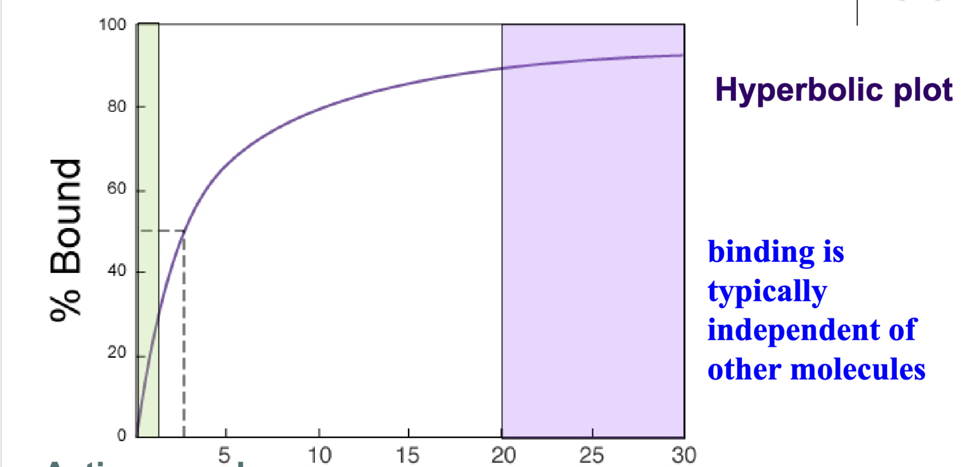
Describe a ligand binding curve. How can it be changed?
Hyperbola shape, with Kd at 50% midpoint. Once peak 100% is reached it plateaus and does not change as all binding sites are full and adding more ligand does not increase binding.
Ligands with a higher affinity for a specific protein will have a lower Kd, which shows as the midpoint of the graph being further back. Changing the concentration of protein will increase or decrease the 100% ceiling by adding or removing binding sites to the solution
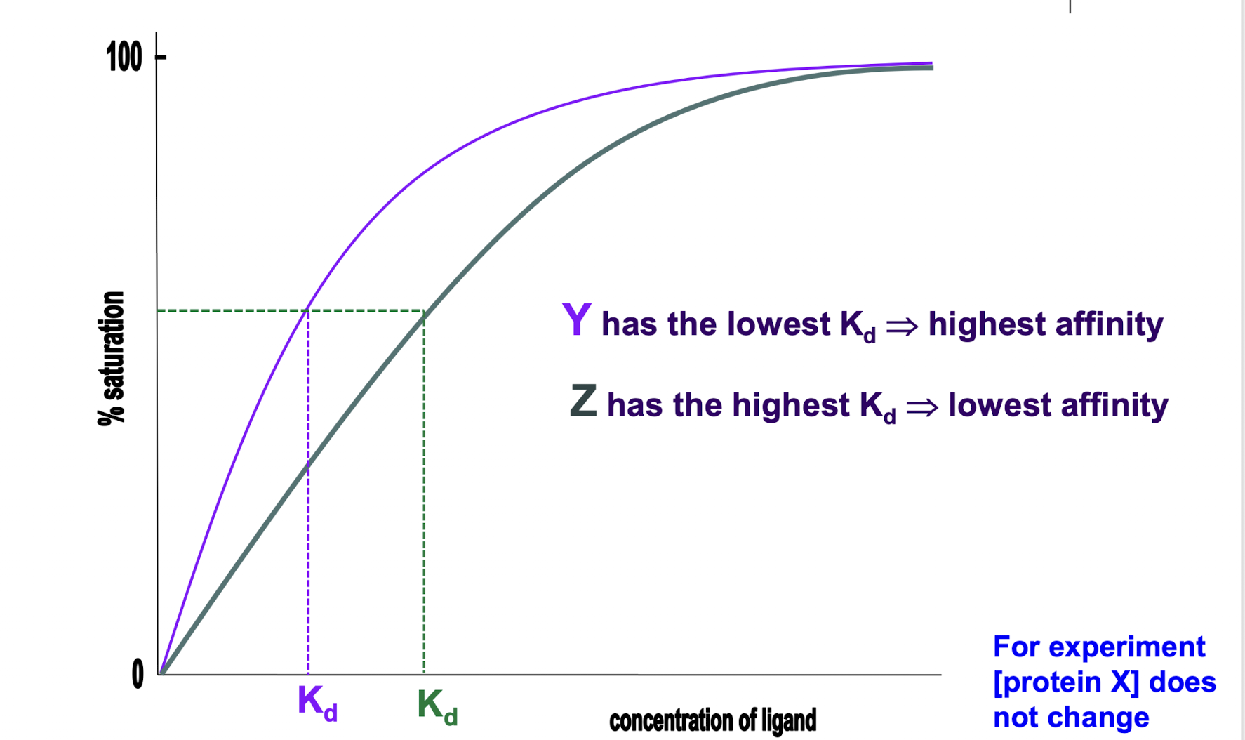
What concentrations determine Kd? What does a greater Kd represent?
Greater Kd = lower [XY] = less affinity. More ligand needed to reach Kd = lower affinity
![<p>Greater Kd = lower [XY] = less affinity. More ligand needed to reach Kd = lower affinity</p>](https://assets.knowt.com/user-attachments/67723e3e-db1e-4250-8f49-8e8eaf3798dd.png)
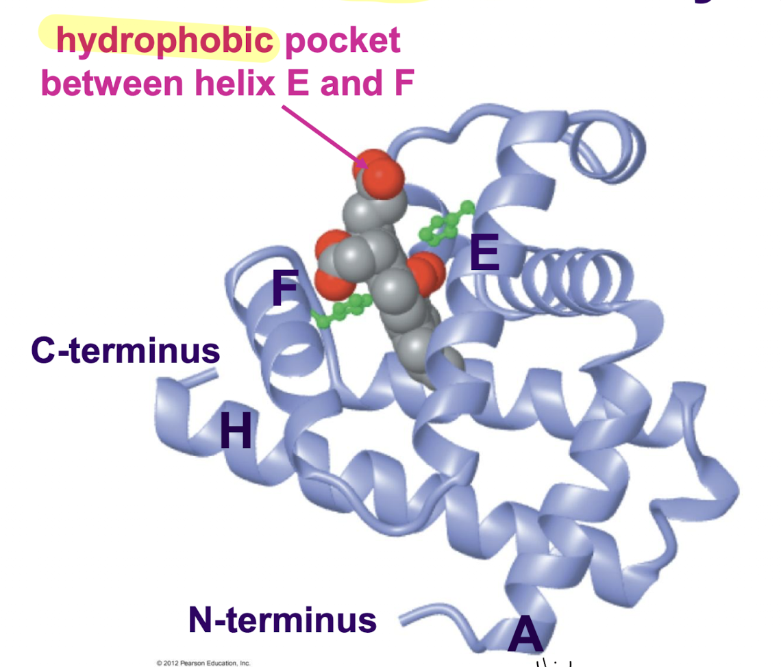
Describe the structure of myoglobin, including secondary structures, prosthetic groups, hydrophobic/hydrophilic areas, and all noteworthy sections. How does this mirror other globin-type proteins?
1 domain that consists of 8 alpha helices held together by loops. Heme prosthetic group attached to globin protein. Hydrophobic pocket between alpha helix E and F where heme resides
Commonly, all have 8 alpha helices and loops, with hydrophobic pocket between E and F where heme slots
Describe the structure of heme, including shape, prosthetic groups, hydrophobic/hydrophilic areas, and all noteworthy sections.
Circular and planar nonprotein. Porphyrin ring (Fe2+ coordinated between 4 N) located at center. Aliphatic ring section that branches out from ring is hydrophobic, two propionyl groups at the bottom of the ring are polar
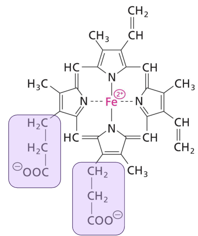
What is the purpose of the nonpolar and polar sections of heme?
Nonpolar aliphatic sections: ensures heme is able to slot into the hydrophobic pocket in myoglobin
Polar propionyl groups: will stay on the surface of the hydrophobic pocket, ensuring the heme slots into the pocket in the same orientation every time
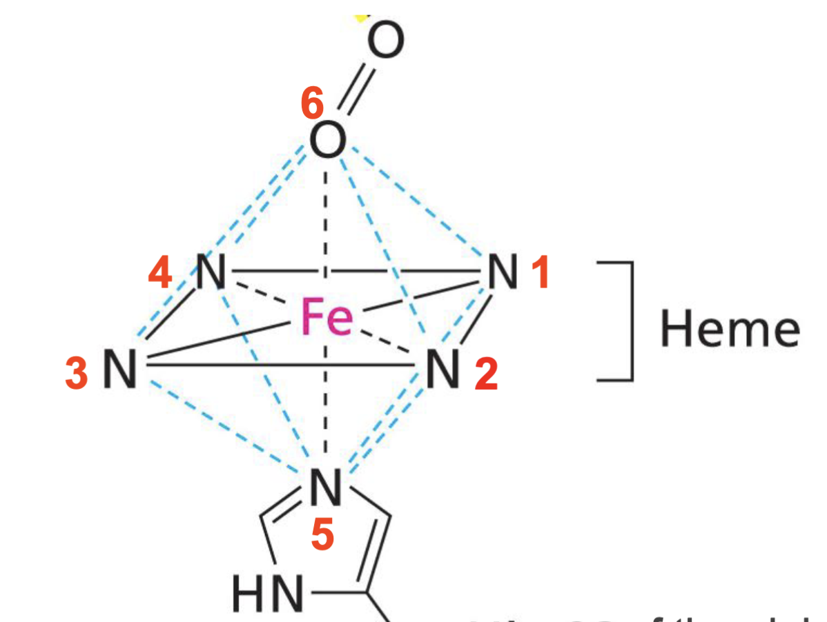
The Fe2+ ion in heme can form ___ coordination bonds. Describe each one and why they are there.
6 coord bonds:
4 coordination bonds with nitrogen that form propionyl ring
5th coordination position is with the HisF8/proximal his residue of myoglobin at the bottom of the ring, which is the major force that holds the porphyrin ring in place (permanently attaches heme to globin protein) along with hydrophobic interactions
6th coordination position is with O2, which is needed to load and offload protein as heme’s role
Can heme bind to O2 by itself? Why or why not?
No, heme binding to a polypeptide prevents the oxidation of Fe2+ to Fe3+. The oxidized form of the iron ion does not bind to O2 and thus heme cannot carry O2 on its own
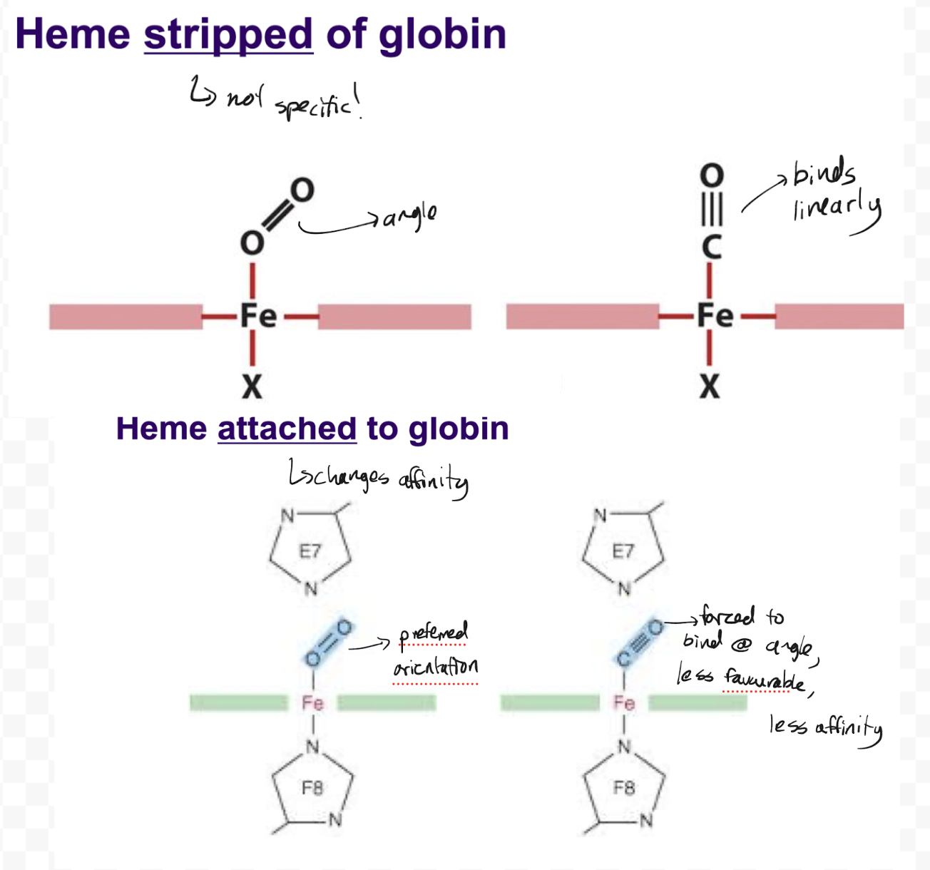
Explain how heme optimizes specificity and affinity of its O2 binding site in myoglobin.
Heme stripped of globin is unspecific. O2 binds to it at an angle while linear molecules such as cyanide and carbon monoxide bind linearly, and thus the binding of these linear molecules is more energetically favoured. CO will bind to heme with 20 000 times the affinity of O2, thus holding onto any CO it finds and never letting go. O2 has no chance to bind as just heme.
Heme attached to globin forces all molecules to bind at an angle due to being blocked by distal histidine. This doesn’t change the affinity of O2, but lowers the affinity of CO and other linear molecules to only 200 times the affinity of O2. Since the body produces a high amount of O2 and only a small amount of CO, heme in myoglobin is more specific towards O2 and will be able pick up O2 without issue
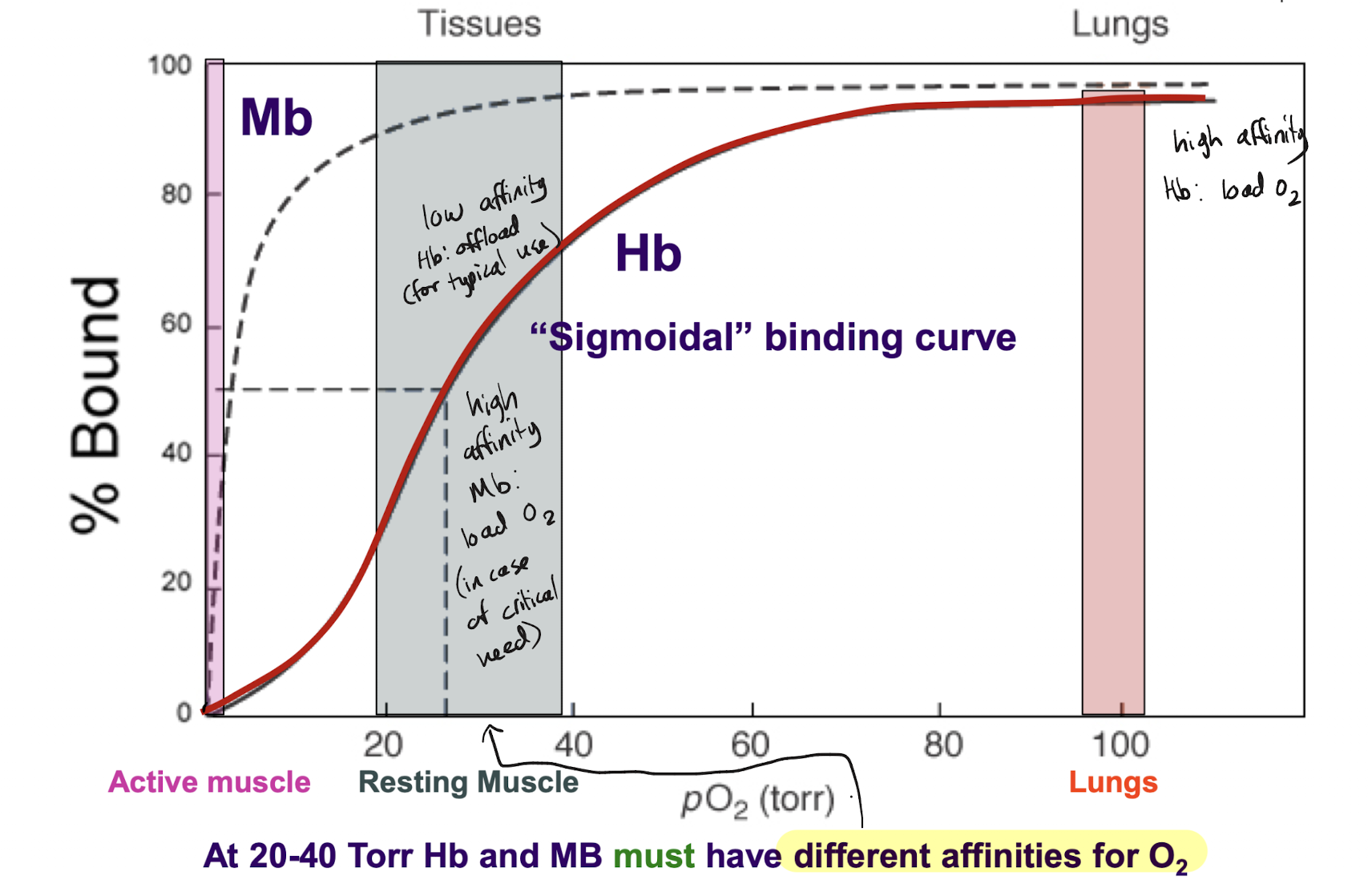
Describe the binding curve of oxygen to myoglobin in terms of shape and its implications for independence and affinity. Include the stages in which myoglobin is in active and resting muscle.
Hyperbolic plot with one line: binding is independent of other molecules, high affinity but reversible.
Start of the plot: active muscle (low oxygen due to being used. Myoglobin is offloading it to the muscle → low binding). End of the plot: resting muscle (relatively higher oxygen due to lack of usage. Myoglobin is loading oxygen → high binding)
Describe the structure of hemoglobin, including subunits, prosthetic groups, binding sites and all noteworthy sections.
Heterotetramer (4 polypeptide chains) with 2 types of globin: alpha globin and beta globin.
2 identical alpha chain subunits, 2 identical beta chain subunits. Each subunit has a heme prosthetic group, meaning there are 4 oxygen binding sites (one on each subunit), and 8 alpha helices + loops
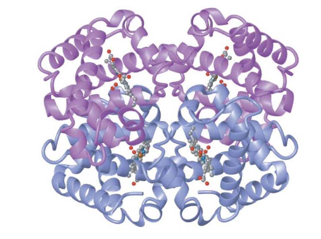
Hemoglobin binds ___ O2, while myoglobin binds ___ O2.
4, 1
Commonly, what kind of substitutions are conservative and critical?
Conservative: substitutions of the same class, similar sized groups/side chains, isomerism. Ex. Leu → Ile, Thr → Ser
Critical: swapping of class, differing functional groups, amino acids at reactive sites. Ex. Ser → Val, His → Lys if Histidine is crucial to a reactive site
Compare the structures of alpha globin, beta globin, and myoglobin. How does this extend to other homologous proteins?
All have 8 alpha helices connected by loops, with a hydrophobic heme binding pocket between helices E and F and a heme prosthetic group. Heme binds oxygen at exact same manner as Mb in alpha and beta globin
Alpha and beta subunits are ~40% identical, subunits are ~18% identical to Mb in primary sequence. Substitutions are mainly conservative
All homologous proteins have very similar secondary and tertiary structure
Which residues in heme-binded proteins (alpha/beta globin and Mb) are invariant? What does this mean in terms of their importance?
HisF8 and HisE7 are invariant across all types because they are critical to the main function of the protein
On a ligand binding curve, a hyperbolic curve is indicative of ___ binding affinity, commonly seen in (Hb/Mb). A sigmoidal curve is indicative of ___ binding affinity, commonly seen in (Hb/Mb). Why does this trend occur?
Constant, Mb (monomers). Cooperative, Hb (oligomers). Monomers have 1 binding site and thus don’t often change ligand affinity, but oligomers have multiple and thus they can affect one another
Describe the binding curve of oxygen to hemoglobin in terms of shape and its implications for independence and affinity. Compare this to myoglobin’s curve, what are their affinities in comparison to each other?. Include active and resting muscle as well as the lungs for both proteins.
Start of the plot: active muscle (critically low oxygen due to being used during strenuous activity) Myoglobin and hemoglobin offloading oxygen to the muscle → low binding, low affinity state.
Middle of plot: resting muscle (relative level of oxygen being used during typical metabolistic activities) Myoglobin will load oxygen in case of critical need → high binding, high affinity. Hemoglobin will offload the oxygen needed to perform daily metabolism → low binding, low affinity. At this stage Hb and Mb must have different affinities for oxygen in order to fulfill different needs
End of the plot: lungs (very high oxygen). Myoglobin, though not present in the lungs, would still load oxygen with same affinity as resting muscle. Hemoglobin will load oxygen for daily metabolism → high binding, high affinity state.
What are the different conformations of hemoglobin? Describe them structurally.
In T state (deoxy), His residue on the beta subunit fits between a Pro and Thr on the alpha subunit. Once oxygenated, the hemoglobin changes shape to R state so that the His residue on the beta subunit now fits between two Thr on the alpha subunit.
T state will have a larger central cavity and more salt bridges than R state
What type of curve is characteristic of a homoallosteric relationship?
Sigmoidal
Proteins generally require what kind of structure for allostery, and why?
Quaternary, need more than 1 subunit (more than 1 binding site)
What type of allosteric relationship do oxygen and hemoglobin have? Explain.
Homoallosteric activation: the binding of O2 to one subunit will cause O2 affinity to increase at other subunits
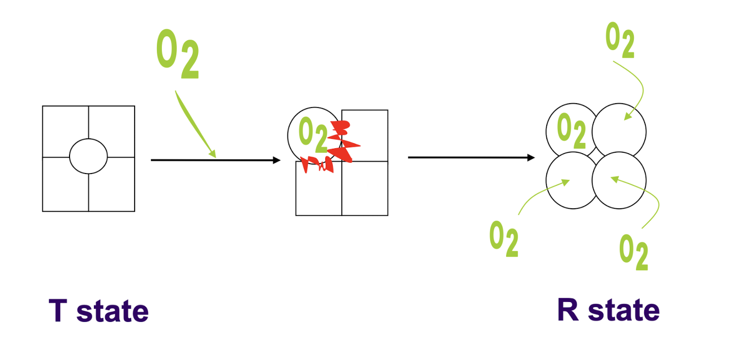
How does hemoglobin change its affinity for oxygen? Describe this process.
Conformational change in structure from T state to R state
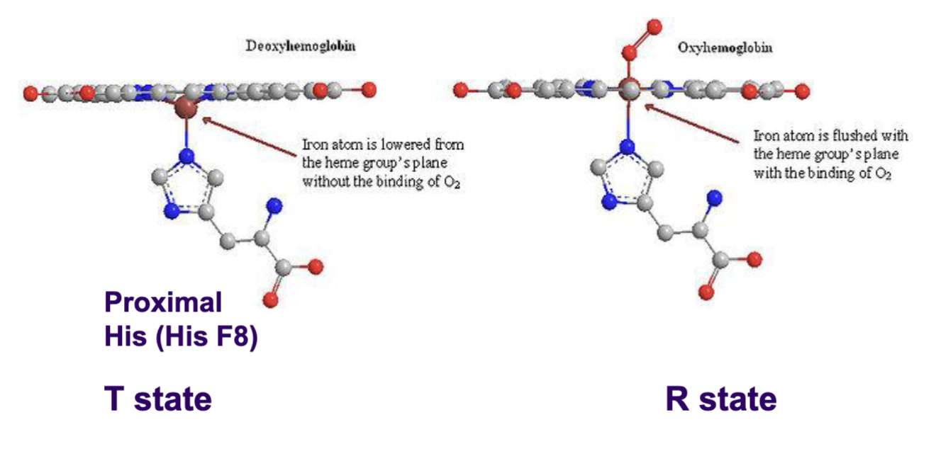
In T state with no oxygen bound, the Fe2+ ion of the heme group of each subunit lies below the plane of the heme ring. Thus, the proximal His attached to the ion lies lower and oxygen binds with lower affinity to the Fe2+ due to its position.
Once O2 binds to a subunit, the iron ion will move into the plane of the heme. This pulls up this proximal histidine, moving the F helix as a whole and changing the subunit interface. This will trigger a chain reaction in all other subunits, who will all undergo iron ion, F helix, and subunit interface movement.
Once this conformational change has occurred, the oxygen binding sites are high affinity (R state), with iron in the plane of the heme ring. Oxygen binds more readily to these sites.
From the following allosteric inhibitors, which favour the T state and which favour the R state? What type of effectors does this make them? O2, BPG, H+, CO2
O2 favours R state (high oxygen/lungs): positive effector/activator
BPG, H+, CO2 favour T state (low oxygen/tissues): negative effectors/inhibitors
How does BPG work as an effector in oxygen binding to hemoglobin? Explain, using hemoglobin states and any bonds/interactions.